

Methods for identifying inhibitors
a technology of inhibitors and vif proteins, applied in the field of methods for identifying inhibitors, can solve the problems of accumulating immunologic damage, unable to counteract the homologous antiviral factors of agms or more distantly related species, and unable to elucidate the function of vif proteins, so as to prevent its incorporation into hiv-1 virions, rapid degradation, and elimination of cells
- Summary
- Abstract
- Description
- Claims
- Application Information
AI Technical Summary
Benefits of technology
Problems solved by technology
Method used
Image

Examples
example 1
HIV-1 Vif Protein Binds the Editing Enzyme APOBEC3G and Induces its Degradation
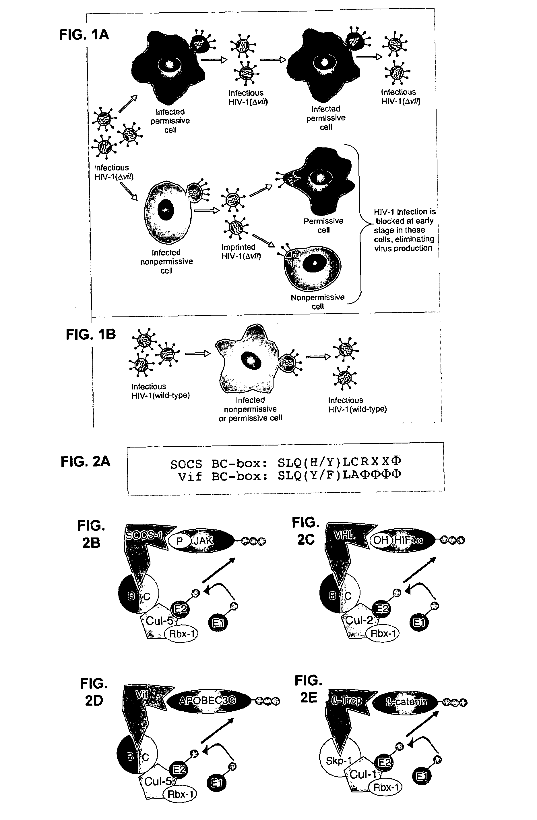
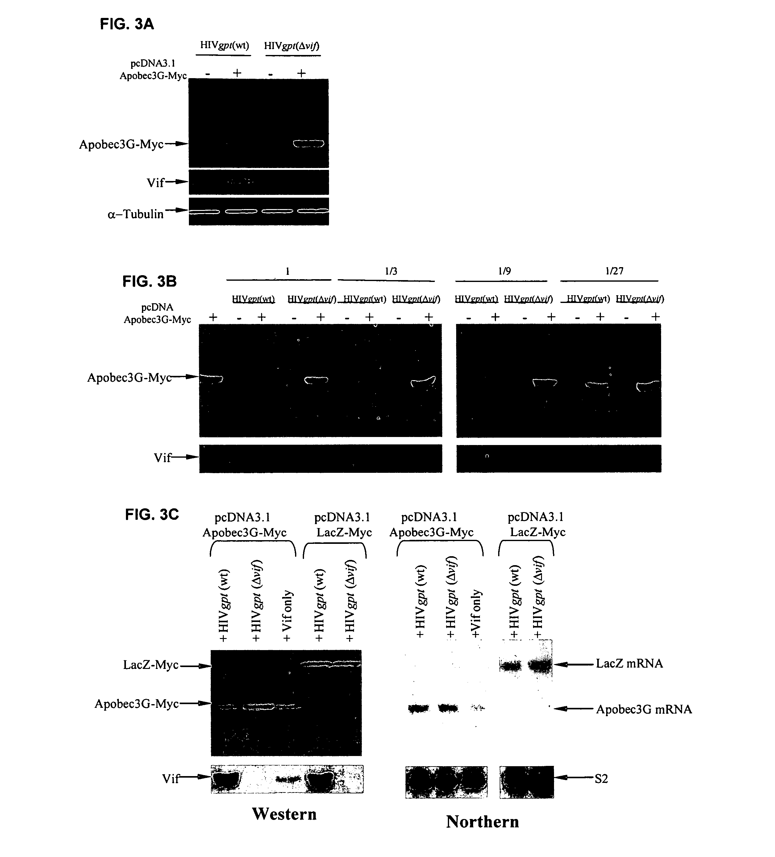
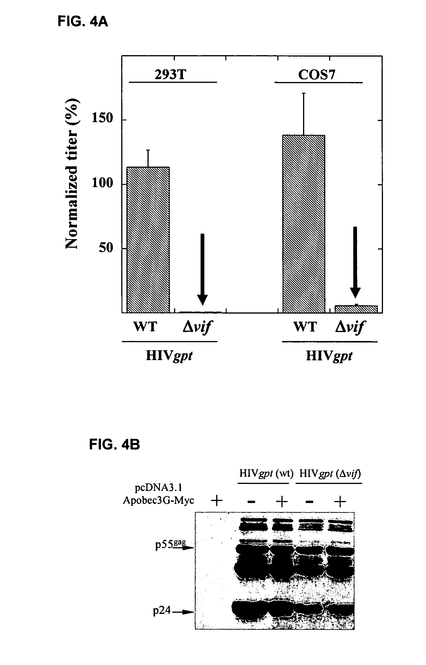
[0284]This example demonstrates that Vif binds to APOBEC3G and induces its rapid degradation, thus eliminating it from cells and preventing its incorporation into HIV-1 virions (Marin et al., Nat. Med. 9:1398–1403, 2003).
[0285]Studies of Vif mutants imply that it contains two domains, one that binds APOBEC3G and another with a conserved SLQ(Y / F)LA motif that mediates APOBEC3G degradation by a proteasome-dependent pathway.
Methods
[0286]Expression vectors: pHIV-gpt(wt) and its derivative pHIV-gpt(Δvif) were previously described (Madani & Kabat, J. Virol. 72, 10251–10255, 1998; Page et al., J Virol 64, 5270–5276, 1990; Madani & Kabat, J. Virol. 74, 5982–5987, 2000). pcDNA3.1-Vif was donated by D. Gabuzda (Dana-Farber Cancer Institute, Harvard, Boston, Mass.). Rev-dependent plasmids for expression of the Vif mutants Δ2, Δ5, Δ6, Δ7, Δ9, Δ10, Δ12 and Δ13 (Simon et al., J. Virol. 73, 2675–2681, 1999) were provide...
example 2
Cell-Based System Capable of Identifying Inhibitors of Vif-Mediated APOGEC3G Degradation
[0308]This example provides an additional APOBEC3G fusion protein, YFP-APOBEC3G (SEQ ID NO: 4), and demonstrates its use in a cell-based system for use in methods of identifying inhibitors of Vif-mediated APOBEC3G degradation.
[0309]Using methods essentially similar to those described in Example 1, a vector was constructed that encodes a chimeric protein containing the yellow fluorescent protein (YFP) at the amino terminus and APOBEC3G at the carboxyl terminus (YFP-APOBEC3G; SEQ ID NOs: 3 and 4). Cotransfection of plasmids that encode this chimera and Vif into human 293T cell cultures results in only a few fluorescent cells, consistent with a Vif-induced degradation (FIG. 13A, top two panels). In contrast, cotransfection with a vector lacking Vif results in a much larger fraction of fluorescent cells. Moreover, treatment of the culture that contains Vif and YFP-APOBEC3G with a proteasome inhibitor...
example 3
Rapid Binding Assay to Detect Vif-APOBEC3G Association
[0311]This example provides a description of one method for detecting the binding or association of Vif with APOBEC3G.
[0312]APOBEC3G-Myc attached onto beads (results shown) or other surfaces (results not shown) actively adsorbs Vif from solution. FIG. 13 shows the results of using this as a rapid binding assay to detect Vif-APOBEC3G association. 293T cultures were transiently transfected or cotransfected with either pcDNA3.1APOBEC3G-Myc-His, pHIVgptWT, or pHIVgptΔvif and pcDNA3 Δ12 Vif. Thirty-six hours post-transfection, cells transiently expressing APOBEC3G-Myc-His were lysed in RIPA buffer (50 mM Tris-Cl (pH 7.4), 1% Nonidet P40, 0.1% sodium deoxycholate, 150 mM NaCl) and 350 μl of lysate was combined with 70 μl of Ni-NTA agarose (Qiagen, Chatsworth, Calif.) and rotated at 4° C. for 4 hours. APOBEC3G-Myc-His bound to Ni-NTA agarose was washed three times in 300 μl RIPA buffer to remove all unbound proteins. Cultures transientl...
PUM
| Property | Measurement | Unit |
|---|---|---|
| molar ratio | aaaaa | aaaaa |
| temperature | aaaaa | aaaaa |
| thermal melting point Tm | aaaaa | aaaaa |
Abstract
Description
Claims
Application Information