Screening method of anti-HIV drug

A technology of animal cells and cells, applied in biochemical equipment and methods, measurement/inspection of microorganisms, cells modified by introducing foreign genetic material, etc., can solve the problems of rapid mutation and drug resistance of viruses

Inactive Publication Date: 2009-12-02
MEDICINE & BIOENG INST OF CHINESE ACAD OF MEDICAL SCI
View PDF0 Cites 4 Cited by
  • Summary
  • Abstract
  • Description
  • Claims
  • Application Information

AI Technical Summary

Problems solved by technology

The pathogenic virus is naturally highly mutated, and under the selection pressure

Method used


Image

  • Screening method of anti-HIV drug
  • Screening method of anti-HIV drug
  • Screening method of anti-HIV drug

Examples


Embodiment 1

[0061] Example 1 Cell Culture

[0062] 293T cells were taken for culture. After the cells filled the culture flask, the old medium was discarded and digested with a digestion solution containing 0.25% trypsin and 0.02% EDTA. When the cells become round, the digestion solution is discarded, and high-sugar DMEM medium (HyClone) containing 10% FBS (purchased from Biological industries) is immediately added, and the bottom of the bottle is gently blown with a straw to completely separate the cells from the bottom of the bottle and disperse them as single cell suspension. After counting on a hemocytometer, adjust the cell concentration to 2.2 × 10 with medium 5 cells / ml, take 15ml of cell suspension and inoculate it on a 10cm culture dish, and use it for cell transfection after 12 hours (the cell abundance is about 70%).

Embodiment 2

[0063] Example 2 Construction of expression vector

[0064] The whole gene of hA3G (GeneID: 60489) was amplified by PCR. The template is human cDNA, and the primers used are: upstream primer 5′-GCC AGA ATT CAA GGA TGA AGCCTC ACT TCA G, downstream primer 5′-TAG AAG CTC GAG GTT TTC CTGATT CTG GAG AAT GG,

[0065] reaction system:

[0066]

[0067] Reaction conditions: 98°C for 5min; 94°C / 1min, 55°C / 1min, 72°C / 1min, a total of 30 cycles; 72°C for 10min.

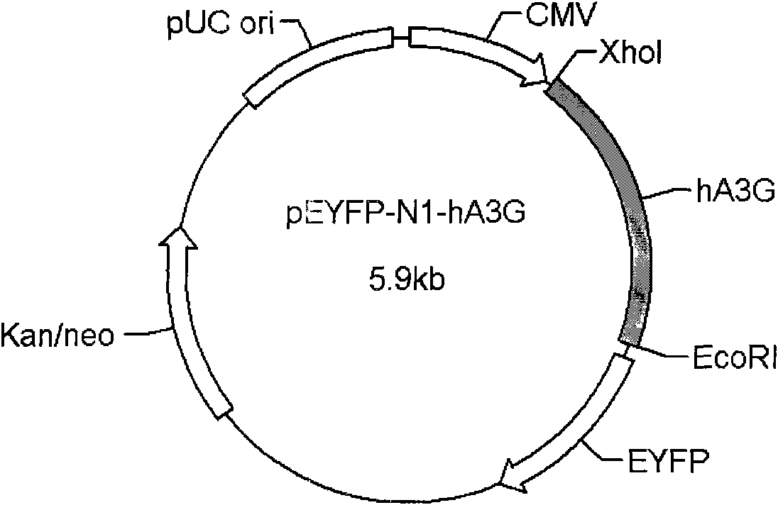
[0068] After the amplified product was verified, it was inserted into the multiple cloning site (Xho1 and EcoR1) of the eukaryotic cell expression vector pEYFP-N1 (Clontech) to obtain the plasmid pEYFP-N1-APOBEC3G. pEYFP-N1-APOBEC3G expresses the fusion protein of hA3G and YFP.

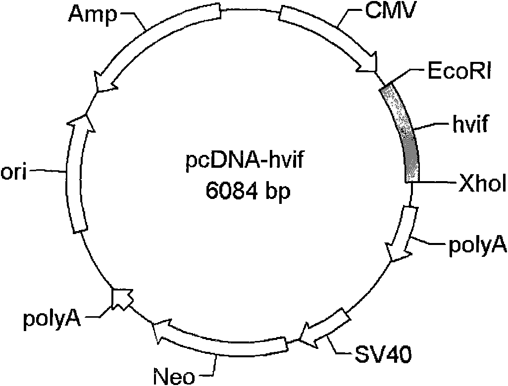
[0069] The full-length Vif gene (GeneID: 326389) was amplified by PCR from wild-type HIV-1 strain pSVC21.BH10 (NIH AIDS Research & Reference Reagent Program). Primers used:

[0070] Upstream primer 5′-TAG AAG GAA TTC ATG GAA AAC AGA TGC

[0071...

Embodiment 3

[0076] Example 3 Cell Transfection

[0077] The transfection step was carried out according to the instructions of Lipofectamine 2000 (Invitrogene). The specific operation is as follows: mix the plasmid used for transfection with 1.5ml high-sugar DMEM medium, and then mix gently. Positive group: co-transfected with 12 μg pEYFP-N1-APOBEC3G and 12 μg control empty vector pcDNA3.1, experimental group: co-transfected with 12 μg pEYFP-N1-APOBEC3G and 12 μg pcDNA-hvif; blank control group: transfected with 24 μg control empty vector pcDNA3 .1.

[0078] Dilute 60 μl Lipofectamine 2000 with 1.5 ml high glucose DMEM medium. Incubate at room temperature for 5 minutes, gently mix the medium containing the transfection plasmid and Lipofectamine 2000 (total volume is 3ml), incubate at room temperature for 20 minutes, and add to the cell culture supernatant in a 10cm culture dish.

[0079] After transfection, 37 °C, 5% CO 2 Cultivate for 10h. Then suck out the old culture medium, diges...


PUM


Abstract

The invention discloses a screening method of an anti-HIV drug, which comprises the following steps: a sample to be tested is added to a fusion protein which expresses hA3G and reporter gene and a Vif cell; the blocking effect of the sample on Vif pathway is judged by investigating the quantity of the encoding products of the reporter gene; whether the initially chosen sample is an inhibitor in the pathway of proteasome degradation is further judged to screen out the anti-HIV drug. In the method provided by the invention, a hA3G is taken as a target point, solves the problem of drug resistance caused by high mutation of viruses and has no cross resistance with the anti-HIV-1 drug used in clinical application for the moment.

Description

technical field [0001] The present invention relates to a method for screening drugs, in particular to a method for screening anti-HIV drugs. Background technique [0002] AIDS, caused by human immunodeficiency virus (HIV-1), is a major infectious disease that my country focuses on. At present, the number of HIV-1 infected people in my country has reached 1 million, and the number of infected people has entered a period of rapid growth. AIDS has not only become a serious health problem in my country, but also causes hundreds of billions of direct economic losses every year. According to the latest report by the Ministry of Health of China at the end of 2006, nearly 200,000 cases of AIDS have been reported nationwide over the years. [0003] At present, the prevention and treatment of AIDS mainly relies on drug treatment. Clinically used anti-HIV-1 drugs can be mainly divided into four categories: nucleoside and non-nucleoside reverse transcriptase inhibitors, protease inh...

Claims


Application Information

Patent Timeline
IPC IPC(8): C12N5/10C12N15/85C12Q1/68C12Q1/02
Inventor 岑山余利岩蒋建东徐建李卓荣彭宗根
Owner MEDICINE & BIOENG INST OF CHINESE ACAD OF MEDICAL SCI
Why Eureka
Try Eureka