Method and apparatus for monitoring glucose levels in a biological tissue

a biological tissue and glucose level technology, applied in the field of biological tissue glucose level monitoring methods and apparatuses, can solve the problems of serious medical complications, low compliance, and low level of compliance, and achieve the effects of reducing noise or speckle, and measurable differences in scattering properties

Active Publication Date: 2007-08-07
MASIMO CORP
View PDF100 Cites 799 Cited by
  • Summary
  • Abstract
  • Description
  • Claims
  • Application Information

AI Technical Summary

Benefits of technology

[0011]An apparatus for measuring the blood glucose level in biological tissue comprises one or more light sources to provide light. Optical fiber or lens-directed paths direct the light onto the tissue, an area scanner continuously scans the light over a two-dimensional area of the tissue and an interferometer effectively scans the tissue in depth. The interferometer also collects, analyzes and measures the light reflected within the tissue. A processor responsive to the light measurements then calculates the glucose level of blood-profused regions of the tissue. Advantageously, the apparatus uses low coherence light sources (light emitting diodes (LEDs) or super luminescent diodes (SLEDs), a low coherence interferometer (LCI), and beam focusing optics. The continuous scanning over a two-dimensional area and the depth scanning reduce noise or speckle and optimize the amount of blood-profused tissue scanned.

Problems solved by technology

But the inconvenience of this procedure—specifically the blood extraction and the use and disposition of test strips—has led to a low level of compliance.
Such low compliance can lead to serious medical complications.
Despite significant efforts, these techniques have shortcomings such as low sensitivity, low accuracy (less than current invasive home monitors) and insufficient specificity of glucose concentration measurement within the relevant physiological range (4-30 mM or 72-540 mg / dL).
There are, however, major drawbacks to the use of OCT for glucose monitoring.
First, the OCT process requires lengthy scanning to reduce optical noise (“speckle”).
However this approach in OCT requires a time period impractically long for a home monitor, and even then speckle in OCT remains problematic for achieving a sufficiently accurate measurement of glucose level.
A second drawback of OCT is that it requires complex processing to form an image and even further processing to analyze the image data to determine glucose levels.
A third drawback is that OCT requires expensive, bulky, precision equipment neither suitable for transport or for use outside the laboratory.

Method used


Image

  • Method and apparatus for monitoring glucose levels in a biological tissue
  • Method and apparatus for monitoring glucose levels in a biological tissue
  • Method and apparatus for monitoring glucose levels in a biological tissue

Examples


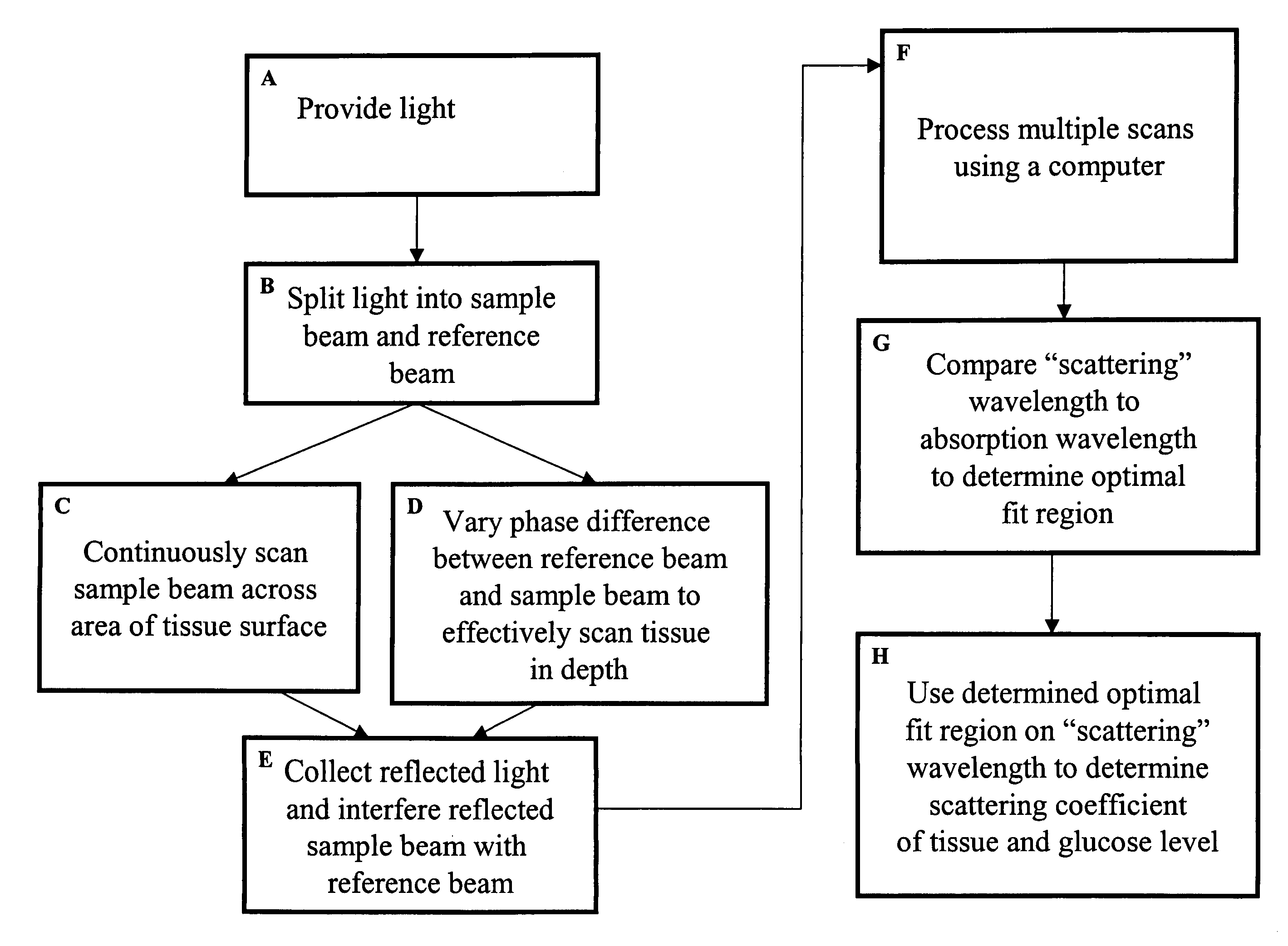
Embodiment Construction

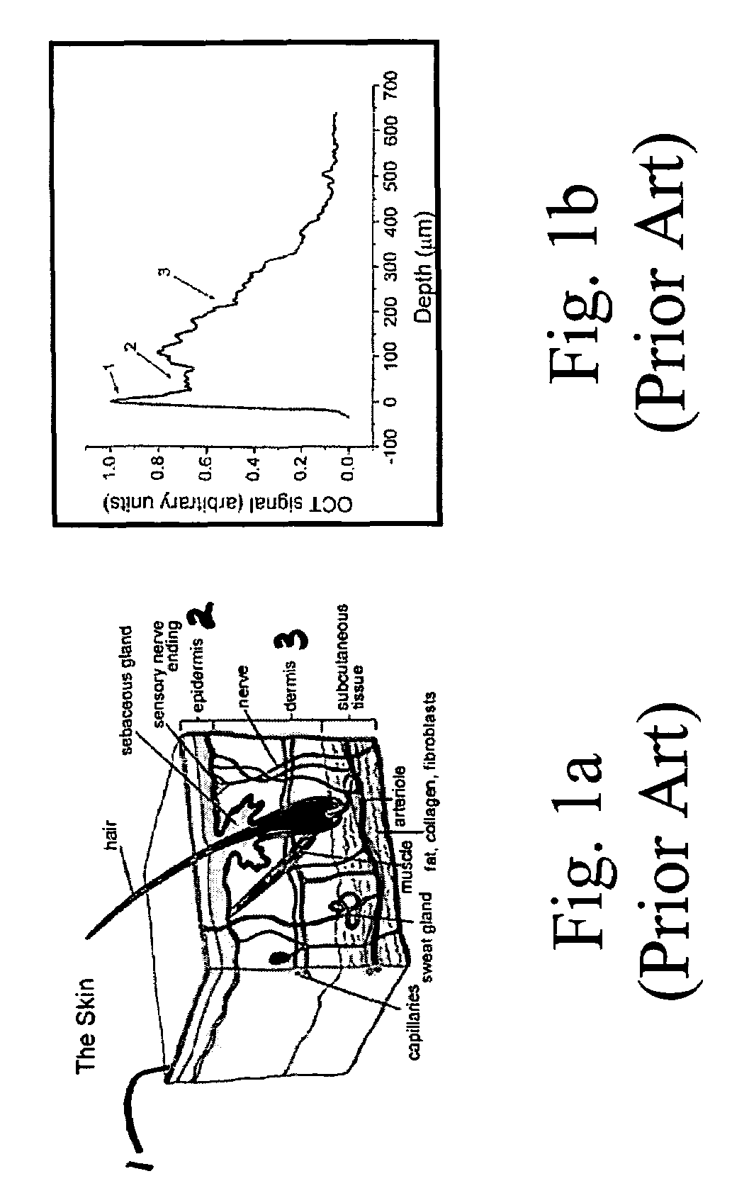
[0018]FIG. 1a is a schematic cross section of the skin showing the skin surface 1, the epidermis layer 2 and the dermis layer 3. Various additional structures are labeled. Some structures, such as hair and its associated follicles, scatter light in a manner unrelated to glucose concentration. Other regions, such as regions in the dermis near the capillaries are profused with blood and IF (blood profused tissues) and light scattered from these regions is highly correlated with glucose concentration.

[0019]FIG. 1b graphically illustrates light scattered within the skin as a function of depth. Region 1 of the curve corresponds to light reflected from the skin surface. Region 2 shows light scattered within the epidermis, and Region 3 is light scattered from the dermis. As we will show, slope of the curve at depths where the tissue is profused with blood (e.g. near the capillaries) is highly correlated with blood glucose levels.

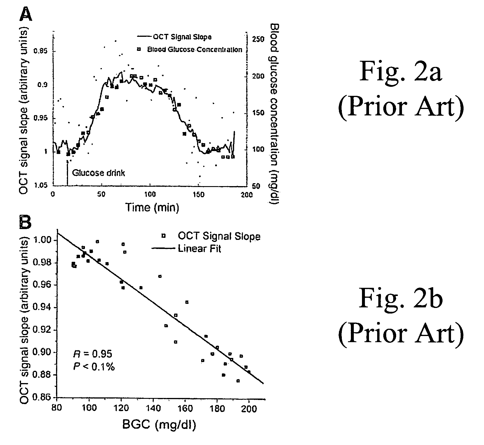
[0020]FIG. 2a graphically plots the slope of the optical sign...


PUM


Abstract

In accordance with the invention, a low coherence interferometer is used to non-invasively monitor the concentration of glucose in blood by shining a light over a surface area of human or animal tissue, continuously scanning the light over a two dimensional area of the surface, collecting the reflected light from within the tissue and constructively interfering this reflected light with light reflected along a reference path to scan the tissue in depth. Since the reflection spectrum is sensitive to glucose concentration at particular wavelengths, measurement and analysis of the reflected light provides a measure of the level of glucose in the blood. The measurement of glucose is taken from multiple depths within blood-profused tissue, and sensitivity is preferably enhanced by the use of multiple wavelengths. Noise or speckle associated with this technique is minimized by continuously scanning the illuminated tissue in area and depth.

Description

FIELD OF THE INVENTION[0001]This invention pertains to methods and apparatus for monitoring the blood glucose level in biological tissues. It is especially useful for non-invasive monitoring of blood glucose in human or animal diabetics. Specifically, it relates to methods and apparatus for non-invasive monitoring of blood glucose using optical coherence interferometry with continuous area scanning and simultaneous depth scanning to reduce the effect of speckle (noise).BACKGROUND OF THE INVENTION[0002]Monitoring of blood glucose (blood sugar) concentration levels has long been critical to the treatment of diabetes in humans. Current blood glucose monitors involve a chemical reaction between blood serum and a test strip, requiring an invasive extraction of blood via a lancet or pinprick. Small handheld monitors have been developed to enable a patient to perform this procedure anywhere, at any time. But the inconvenience of this procedure—specifically the blood extraction and the use ...

Claims


Application Information

Patent Timeline
Patent Type & Authority Patents(United States)
IPC IPC(8): A61B5/00
CPCA61B5/0064A61B5/441A61B5/14532A61B5/0066
Inventor SCHURMAN, MATTHEW J.SHAKESPEARE, WALTER J.
Owner MASIMO CORP
Why Eureka
Try Eureka