
Method for generating T1-weighted magnetic resonance images and quantitative T1 maps

a magnetic resonance image and quantitative technology, applied in the field of medical imaging systems, can solve the problems of poor signal-to-noise ratio (snr), long scan time, and inconvenient use of fetal brain long scans

Active Publication Date: 2007-10-02
THE BOARD OF TRUSTEES OF THE LELAND STANFORD JUNIOR UNIV +1
View PDF7 Cites 16 Cited by
  • Summary
  • Abstract
  • Description
  • Claims
  • Application Information

AI Technical Summary

Benefits of technology

"The patent text describes a method and system for generating T1-weighted images using magnetic resonance imaging (MRI). The method involves acquiring two sets of MR signals and using them to generate the T1-weighted image. The technical effect of this invention is the ability to produce high-quality T1-weighted images with improved accuracy and reliability."

Problems solved by technology

The inversion recovery technique utilizes a long repetition time and thus results in a long scan time.
The low signals result in a poor signal-to-noise ratio (SNR).
Further, long scans are not useful for the fetal brain because of motion of the fetal brain.
However, the invasive procedures are prone to movement of the object and contamination from the environment in which the invasive procedures are performed.
However, obtaining measurements of fluid T1 in order to estimate pO2 has not previously been demonstrated in vivo.

Method used


Image

  • Method for generating T1-weighted magnetic resonance images and quantitative T1 maps
  • Method for generating T1-weighted magnetic resonance images and quantitative T1 maps
  • Method for generating T1-weighted magnetic resonance images and quantitative T1 maps

Examples


Embodiment Construction

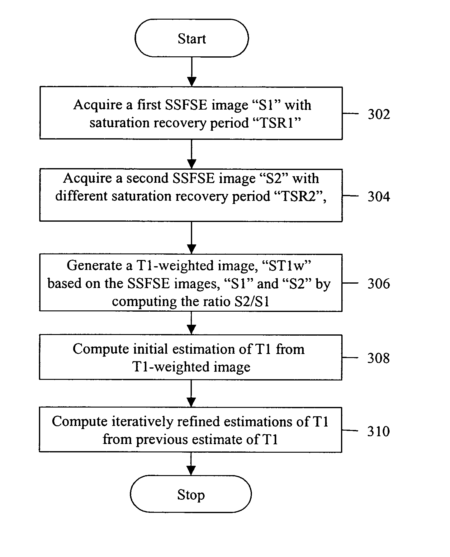
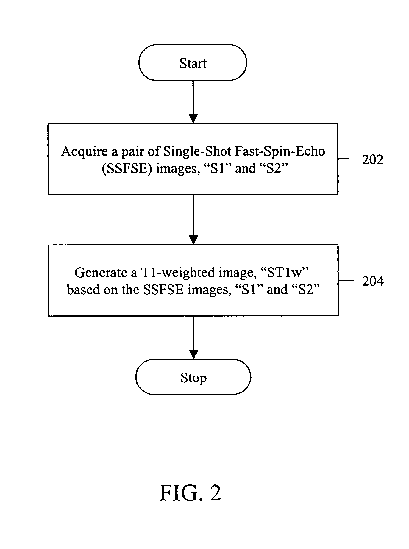
[0015]Various embodiments of the invention provide a method and system for generating T1-weighted Magnetic Resonance (MR) images and quantitative T1 maps. The method is performed by acquiring a pair of Single-Shot Fast-Spin-Echo (SSFSE) images.

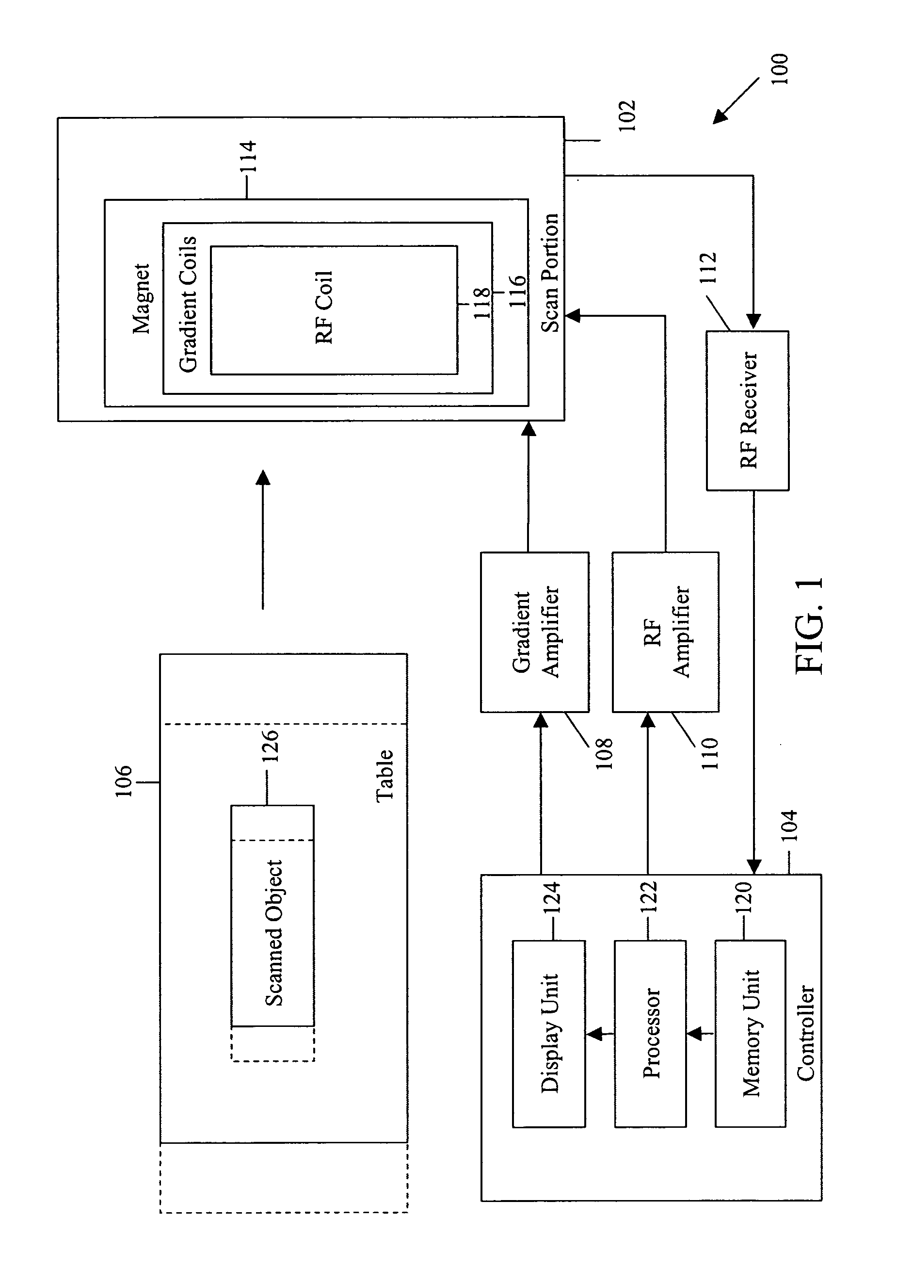
[0016]FIG. 1 is a block diagram of an MRI system 100, in which various embodiments of the invention can be implemented. MRI system 100 generally includes a scan portion 102, a controller 104, a table 106, a gradient amplifier 108, an RF amplifier 110, and an RF receiver 112. Scan portion 102 includes a magnet 114, a set of gradient coils 116, and an RF coil 118. Controller 104 includes a memory unit 120, a processor 122 and a display unit 124.

[0017]In an embodiment of the invention, an object 126 (e.g., a patient) to be scanned is placed on table 106. MRI data of object 126 is obtained by scan portion 102. This is achieved through the application of a magnetic field generated by magnet 114, a plurality of magnetic gradient fields, along x, y, ...


PUM


Abstract

Methods and systems for generating T1-weighted images are provided. The method includes acquiring a pair of single-shot fast-spin-echo (SSFSE) images S1 and S2. The method further includes generating a T1-weighted image ST1w based on S1 and S2.

Description

BACKGROUND OF THE INVENTION[0001]This invention relates generally to medical imaging systems, and more particularly, to methods and systems for generating purely T1-weighted images using data from two or more acquisitions.[0002]Magnetic Resonance Imaging (MRI) generates an image based on a plurality of properties specific to a material of an object being imaged. One of the properties is a longitudinal relaxation time T1 and another one of the properties is a transverse relaxation time T2. Known imaging methods generate images by applying a sequence of Radio Frequency (RF) pulses to the object. This sequence is repeated periodically after a time interval TR. The signal produced by the object after applying RF pulses depends on the material of the object and the T1 of that material. MRI employs the property of different T1 relaxation rates for the material of the object to obtain contrast within the image. The image obtained by emphasizing T1 and de-emphasizing T2 of the material is r...

Claims


Application Information

Patent Timeline
Patent Type & Authority Patents(United States)
IPC IPC(8): G01V3/00
CPCG01R33/50G01R33/5617G01R33/5615
Inventor BUSSE, REED F.PAULY, JOHN M.ZAHARCHUK, GREG
Owner THE BOARD OF TRUSTEES OF THE LELAND STANFORD JUNIOR UNIV
Learn More