
Method, device and software for separating the individual subjects of an anatomical structure segmented from 3D datasets of medical examination procedures

a three-dimensional character and anatomical structure technology, applied in the field of medical image processing of three-dimensional character datasets, can solve the problems of insufficient merely conveying a spatial impression of the displayed body region to the medical practitioner, insufficient form of presentation of examination results, and inability to achieve the effect of preparing for an intervention to treat a complex multiple fractur

Inactive Publication Date: 2007-11-06
SIEMENS HEALTHCARE GMBH
View PDF6 Cites 3 Cited by
  • Summary
  • Abstract
  • Description
  • Claims
  • Application Information

AI Technical Summary

Benefits of technology

[0010]Time-consuming procedures thus can be shifted from the operation into the planning phase. The precise planning of an intervention in the preparatory phase of the operation not only shortens the duration of the intervention and, thus, produces less stress on the patient, but also enables more precise and safer work for the medical practitioner during the operation. Also advantageously, the use of suitable aids such as, for example, screws, nails, etc., can already be tested before the operation. Likewise advantageously, implants can now be pre-shaped according to the identifiable conditions on the basis of the present invention and no longer need be intuitively designed during the course of the operation.
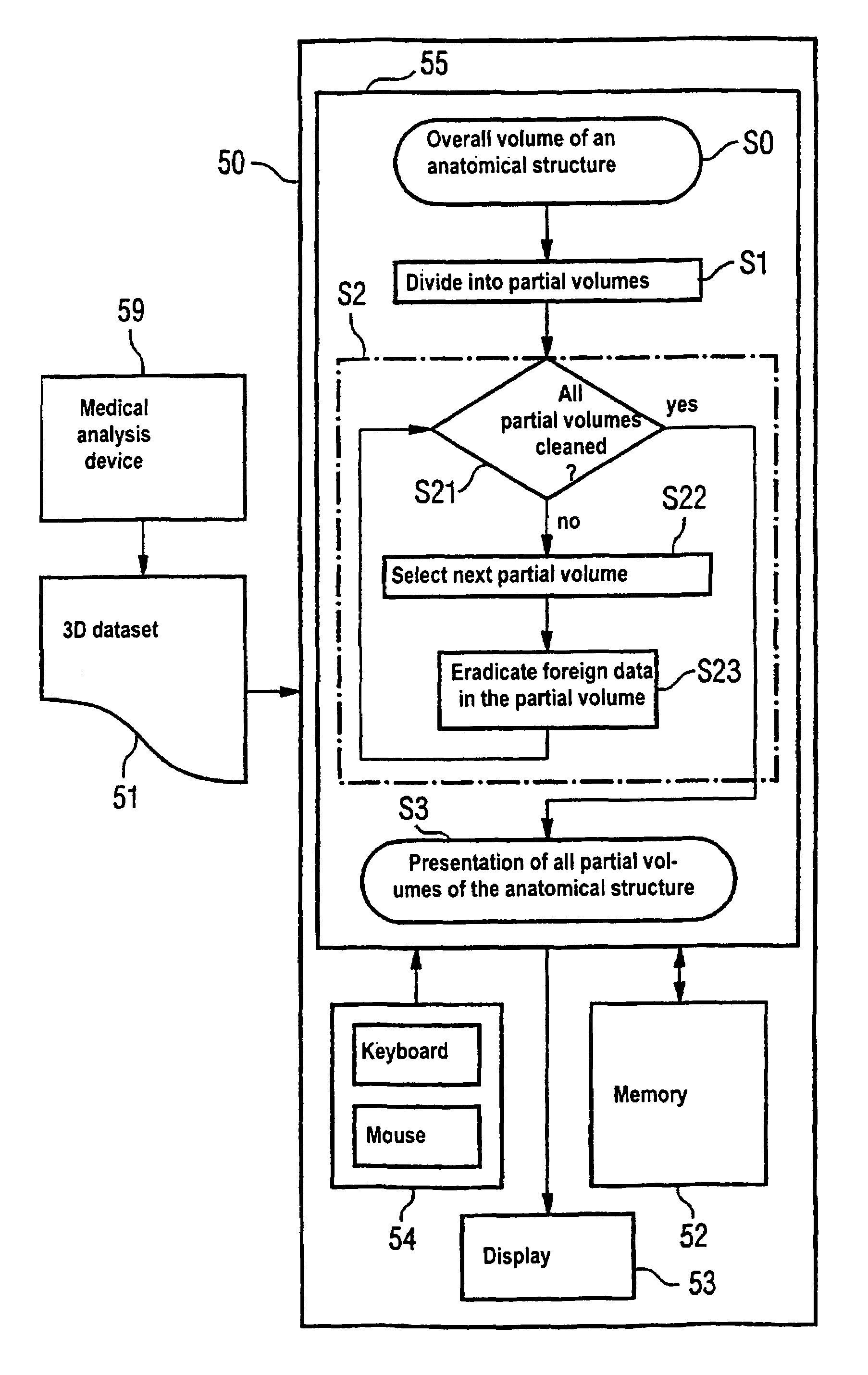
[0011]In an embodiment of the invention, the partial volumes have the shape of a cuboid, as a result of which the individual volumes can be easily joined. Together with the data of each partial volume, the appertaining coordinate information are also stored. The editing of 3D computer tomography datasets is particularly advantageous, so the selection of a bone structure as a structure of interest, with the individual bones and / or bone fragments as individual subjects of the structure to be segmented, is especially advantageous.

Problems solved by technology

In general, however, it is not adequate to merely convey a spatial impression of the displayed body region to the medical practitioner.
For planning a surgical intervention, for example after an accident resulting in a complicated fracture, however, this form of presentation of the examination results is often inadequate since although it reflects the actual condition of the structure of interest it does not allow a manipulation of the data with respect to measures to be undertaken; the individual subjects within each and every structure remain fixed in their geometrical arrangement relative to one another in known segmenting methods.
The preparation for an intervention to treat a complex multiple fracture therefore requires an enormous mental effort on the part of the orthopedist since he or she cannot verify the allocation of the various fracture surfaces and thus bone fragments to one another at the picture screen.

Method used


Image

  • Method, device and software for separating the individual subjects of an anatomical structure segmented from 3D datasets of medical examination procedures
  • Method, device and software for separating the individual subjects of an anatomical structure segmented from 3D datasets of medical examination procedures
  • Method, device and software for separating the individual subjects of an anatomical structure segmented from 3D datasets of medical examination procedures

Examples


Embodiment Construction

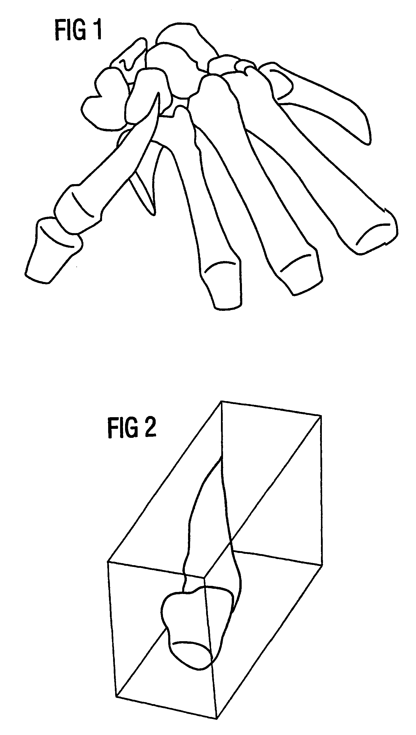
[0017]FIG. 1 provides a conceptualization of the graphic representation of a computer-tomographic 3D dataset as is currently available to a surgeon. In practice, the data are visualized by means of a method referred to as volume rendering, wherein the surface of the identified osseous structure is presented with a suitably calculated distribution of light and shadow so that it conveys a plastic impression of the underlying spatial structure to the viewer or the surgeon.

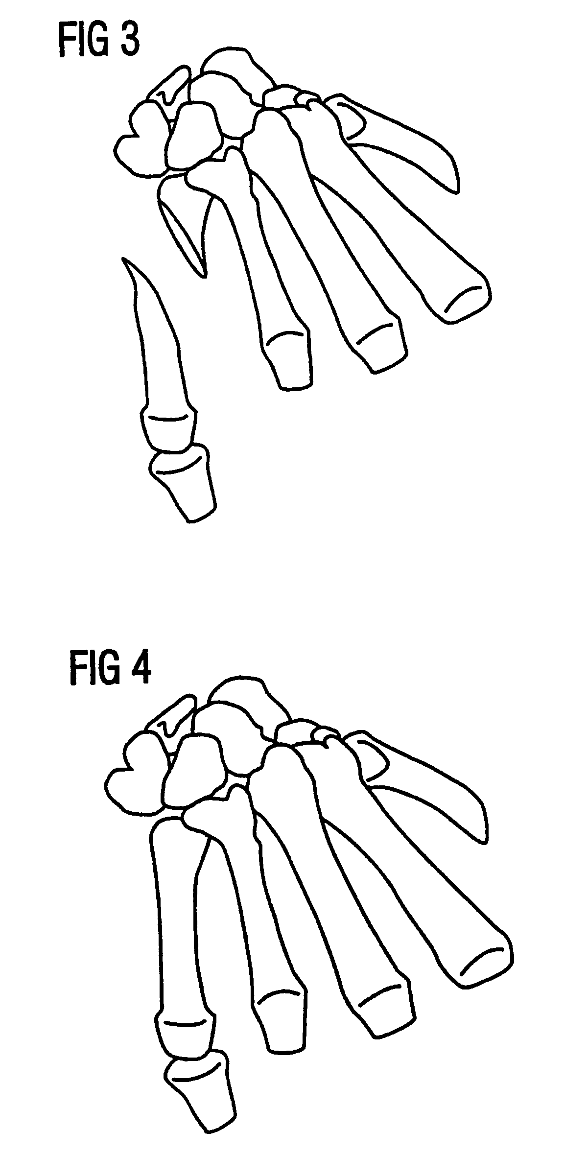
[0018]A bone fracture with a dislocated fragment of the os metacarpal V can be clearly recognized in the left half of FIG. 1. The relative position of this bone fragment with respect to the other subjects of the graphically visualized structure cannot be modified at this initial stage S0 of the inventive method since the structure for a single, interconnected subject in this stage as established by the standard methods of medical image processing. Although a viewer has the possibility of examining details such as, for...


PUM


Abstract

The invention relates to a method for carrying out the imaging processing of data sets of three-dimensional characters obtained from medical examination methods. According to the invention, a separation of the data of relevant anatomic structures as well as of other structures is carried out based on data provided in the form of volume elements. In addition, a spatial representation of at least one structure of interest ensues, and the individual objects of the structure of interest are separated from one another in such a manner that enables them to be repositioned in the spatial representation.

Description

BACKGROUND OF THE INVENTION[0001]1. Field of the Invention[0002]The invention is directed to a method, a device and a software product for the medical image processing of datasets of a three-dimensional character acquired from medical examination procedures, of the type wherein a separation of the data of relevant anatomical structures as well as of other structures is undertaken proceeding from data in the form of volume element, and a spatial presentation of at least one structure of interest ensues.[0003]2. Description of the Prior Art[0004]The human ability to conceptualize quickly reaches it limits in the spatial interpretation of the information from X-ray exposures or from tomograms as acquired with modern tomographic methods. Modern methods of medical image processing therefore convert the information of a three-dimensional character acquired in medical examinations into spatial impressions of the inside of the body. In general, however, it is not adequate to merely convey a...

Claims


Application Information

Patent Timeline
Patent Type & Authority Patents(United States)
IPC IPC(8): G06K9/00A61B5/00A61B6/03G06T17/40G06T19/00
CPCG06T19/20G06T2219/2004G06T2210/41
Inventor SEISSLER, LEGAL REPRESENTATIVE, INGEBORGSEISSLER, LEGAL REPRESENTATIVE, PAULSEISSLER, LEGAL REPRESENTATIVE, JAKOBSEISSLER, LEGAL REPRESENTATIVE, ANKESCHRAMM, LEGAL REPRESENTATIVE, SANDRASEISSLER, DECEASED, WOLFGANG GEORG
Owner SIEMENS HEALTHCARE GMBH
Learn More