

Cardiac imaging system and method for quantification of desynchrony of ventricles for biventricular pacing
a biventricular pacing and imaging system technology, applied in the field of cardiac rhythm management systems, can solve the problems of delayed left ventricular ejection, different regions of the left ventricle not contracting in a coordinated fashion, and ineffective contraction of the left ventricl
- Summary
- Abstract
- Description
- Claims
- Application Information
AI Technical Summary
Benefits of technology
Problems solved by technology
Method used
Image

Examples
Embodiment Construction
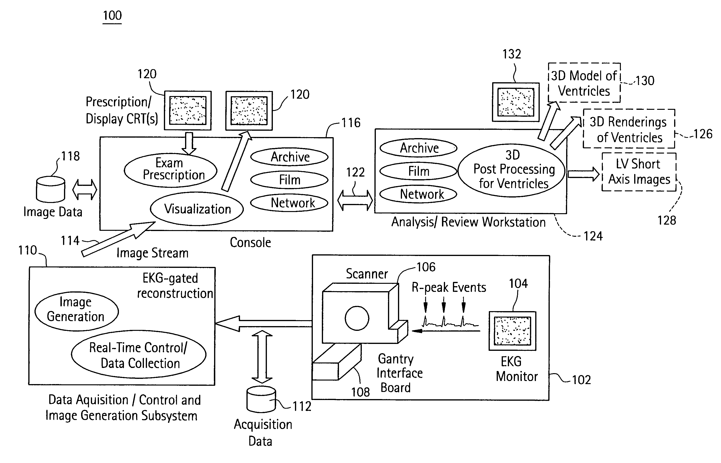
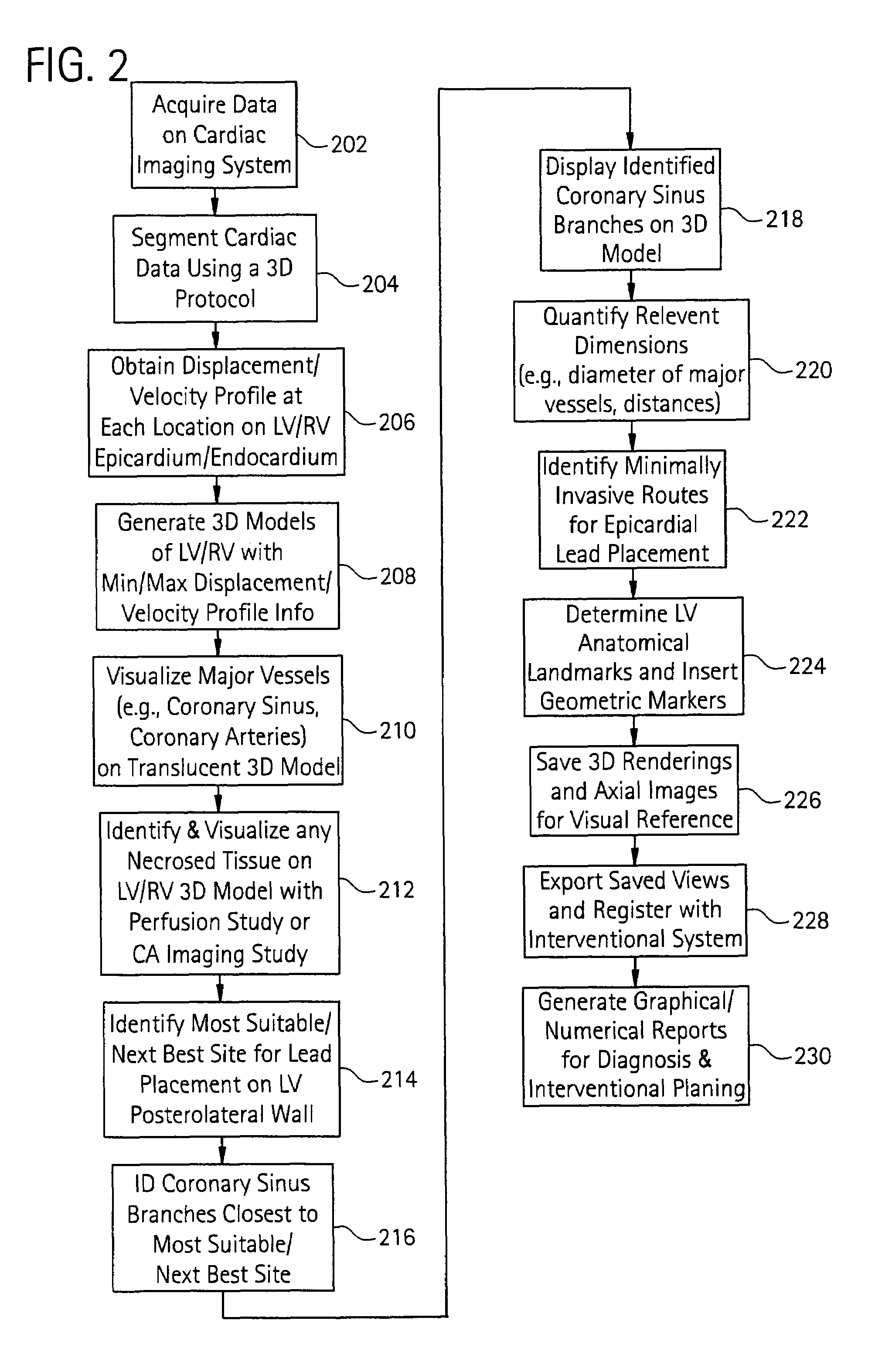
[0026]Disclosed herein is a cardiac imaging system and method for quantifying the desynchrony of the right and left ventricles so as to provide information for planning biventricular pacing interventional procedures enabling the practitioner (e.g., electrophysiologist, cardiologist, surgeon) to plan in advance the approach to take for the procedure. LV contractility can be visualized to identify the best location for placement of the LV epicardial pacing lead by, for example, determining the LV region that is the “last to contract” (i.e., a time-based contraction parameter). As used hereinafter, the term “last to contract” may be quantified as either last to start contraction or last to complete contraction by the exemplary method described in greater detail below. Alternatively, LV contractility can be visualized by determining the LV region that exhibits the most / least amount of displacement with respect to a starting position at a given point in the cardiac cycle (i.e., a displac...
PUM
 Abstract
Description
Claims
Application Information