Apparatus and method for compensating for respiratory and patient motion during treatment

a technology of respiratory and patient motion and apparatus, which is applied in the field of apparatus for improving the accuracy and efficacy of surgical treatments, can solve the problems of difficult tracking of respiratory motion using external sensors, focusing radiation, and tumor movement during treatmen

Inactive Publication Date: 2008-01-15
ACCURAY
View PDF36 Cites 266 Cited by
  • Summary
  • Abstract
  • Description
  • Claims
  • Application Information

AI Technical Summary

Problems solved by technology

The problem is that the tumor may move during treatment, especially due to the breathing motion of the patient.
Such respiratory motion is difficult to track using external sensors, since the extent and direction of the internal breathing motion of the patient cannot be seen with traditional imaging devices.
The breathing and other motion of the patient means that it is more difficult to focus the radiation on the tumor which means that the treatment may be less effective and healthy tissue may be unnecessarily damaged.
Although radiosurgery has been applied with dramatic success to brain tumors, the extension of this technique to tumors outside the head or neck areas has eluded easy solutions.
The main reason for this difficulty has been the problem of accurate target localization (i.e., accurate tracking of the motion of the target).
In particular, breathing motion and other organ and patient motion make it difficult to track the target tumor with high precision.
Thus, in the presence of breathing motion, for example, it is difficult to achieve the goal of providing a high dose of radiation to the tumor while protecting surrounding healthy tissue.
Conventional systems can only move the radiation beam along circular arcs in space so that irregular breathing motions cannot be easily followed since these breathing motions may not occur along the axis of the circular arcs traced by the radiation beam.
This method relates to finding fixed targets, such as a brain tumor, but does not address tracking the motion of a target organ, such as lung due to breathing, with respect to the skin surface, or tracking the motion of internal abdominal organs with respect to externally visible motion.
This fiducial implant permits internal structures of the human body to be analyzed, but does not attempt to compensate for motion of a target organ which moves throughout the respiratory cycle.

Method used


Image

  • Apparatus and method for compensating for respiratory and patient motion during treatment
  • Apparatus and method for compensating for respiratory and patient motion during treatment
  • Apparatus and method for compensating for respiratory and patient motion during treatment

Examples


first embodiment

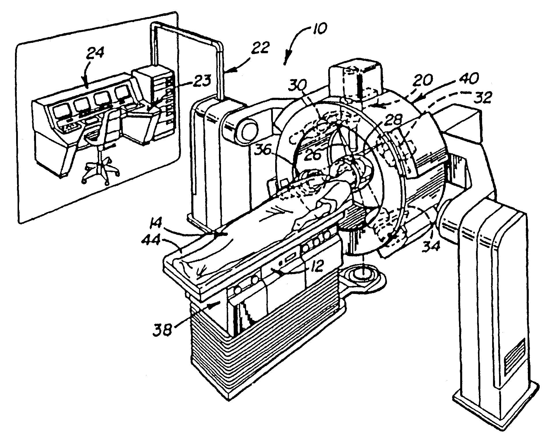
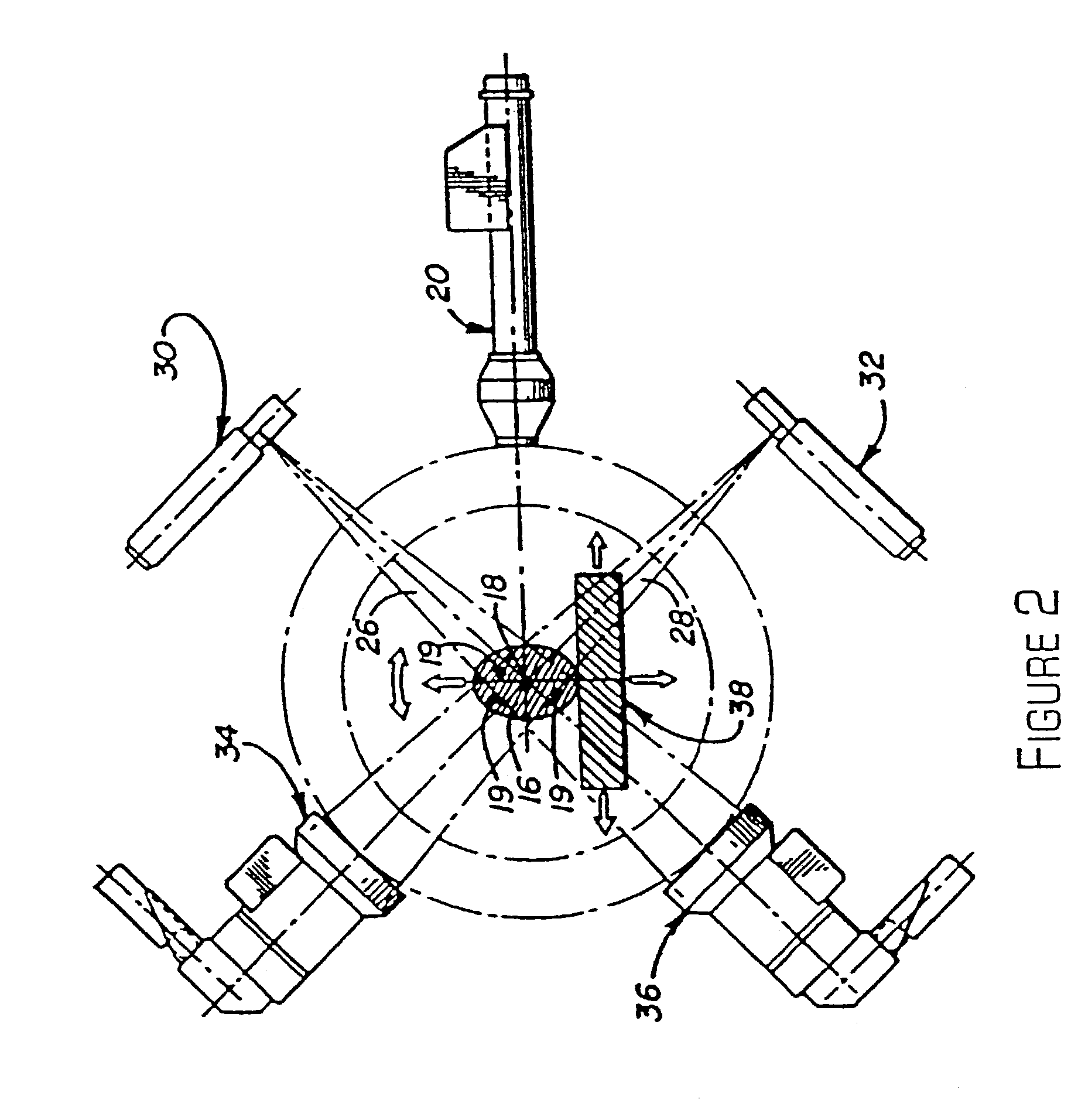
[0048]Now, various other embodiments of the method for compensating for breathing and other patient motion in accordance with the invention will be described. In all of these embodiments, the respiratory and diaphragmatic excursion may be limited and minimized by binding the abdomen or compressing the abdomen. In a first embodiment, one or more small metal markers (also known as landmarks) are attached to the target organ before treatment. There may be three or four metal markers with possibly distinct shapes or sizes, which may be, for example, small gold beads. The exact position of these internal markers is determined by two x-ray cameras, which acquire a stereo image of the target site. There may also be one or more infra-red probes which are attached to the patient's skin surface. The infra-red probes give a very accurate and high speed position reading, but they only show the surface of the patient's body. In this embodiment, internal imaging of the internal markers and extern...

second embodiment

[0049]During the actual operation, it is difficult to obtain x-ray images more than once every predetermined number of seconds due to concerns about exposing the patient to too much radiation and due to the fact that the treatment beam cannot operate when x-ray imaging is being done. The x-ray imaging alone would therefore be too slow to follow breathing motion with high precision. Therefore, the external landmarks on the skin surface, as seen by the infrared system, are used for intra-operative localization, where we continuously reference the previously computed model of relative motions of the internal and external markers. This allows the exact placement of the internal landmarks (gold beads) to be predicted at time points where no x-ray images are available. Now, the method will be described.

[0050]In a second embodiment, there are situations in which it is desirable to compensate for respiratory motion as well as to compensate for pulsation effects. These pulsation effects may ...

third embodiment

[0051]In the method in accordance with the invention, no internal landmarks attached to the target organ are used. Instead, an ultra-sound camera is used to acquire the pre-operative image series, again in combination with an infra-red tracking system. The infra-red system in this embodiment establishes both the position of the external landmarks and the position of the (movable) ultra-sound camera, which must be moved by a human operator during this pre-operative phase. During the pre-operative phase, the ultra-sound images may be analyzed manually or semi-automatically in order to locate the target. During treatment, the external landmarks (infra-red probes) are used to compensate for the motion of the target organ since the motion model we have established allows the determination of the position of the internal target organ from the position of the external markers.

[0052]Furthermore, there are situations, where the attachment of markers to an anatomic target is difficult, undesi...


PUM


Abstract

An apparatus and method for performing treatment on an internal target region while compensating for breathing and other motion of the patient is provided in which the apparatus comprises a first imaging device for periodically generating positional data about the internal target region and a second imaging device for continuously generating positional data about one or more external markers attached to the patient's body or any external sensor such as a device for measuring air flow. The apparatus further comprises a processor that receives the positional data about the internal target region and the external markers in order to generate a correspondence between the position of the internal target region and the external markers and a treatment device that directs the treatment towards the position of the target region of the patient based on the positional data of the external markers.

Description

RELATED APPLICATION[0001]This application is a continuation application of U.S. patent application Ser. No. 09 / 657,771 filed Sep. 8, 2000 now U.S. Pat. No. 6,501,981 which is in turn is a continuation-in-part application of U.S. patent application Ser. No. 09 / 270,404 now U.S. Pat. No. 6,144,875 filed on Mar. 16, 1999. Both of these applications are incorporated herein by reference.BACKGROUND OF THE INVENTION[0002]This invention relates generally to an apparatus for improving the accuracy and efficacy of surgical treatments and more particularly to locating a target region to be treated and tracking the motion of the target region due to respiratory and other patient motions during the treatment.[0003]Various different treatments may accurately track the motion of a target region in order to apply the treatment to the target region. In radiation therapy and radiosurgery, for example, a tumor may be destroyed by a beam of ionizing radiation which kills the cells in the tumor. The prob...

Claims


Application Information

Patent Timeline
Patent Type & Authority Patents(United States)
IPC IPC(8): A61B6/00A61B8/00G01B11/00A61B5/055A61B5/06A61B6/03A61B6/08A61B6/12A61B10/00A61B10/02A61B17/00A61B17/34A61B18/00A61B18/20A61B19/00A61N5/06A61N5/10A61N7/02G01B15/00G01B21/00G01R33/48
CPCA61B6/08A61B6/12A61B6/527A61B19/20A61N5/1049A61B10/0233A61B18/20A61B19/5244A61B19/54A61B19/56A61B2017/00694A61B2017/00699A61B2019/505A61B2019/5255A61B2019/5272A61B2019/5276A61N5/1037A61N5/1067A61N5/107A61N7/02A61N2005/1058A61N2005/1061A61B34/20A61B34/25A61B90/10A61B90/39A61B2034/105A61B2034/2055A61B2034/2072A61B2090/378
Inventor SCHWEIKARD, ACHIMADLER, JOHN R.
Owner ACCURAY
Why Eureka
Try Eureka