Detection and analysis of lesions in contact with a structural boundary

a structural boundary and lesions technology, applied in the field of lesions detection and analysis in digital images, can solve the problems of inflexible methods in dealing with the size variability of nodules, difficulty in optimizing the size of the disk filter and controlling the spacing, and processing speed and accuracy

Inactive Publication Date: 2008-04-15
MEVIS MEDICAL SOLUTIONS
View PDF16 Cites 27 Cited by
  • Summary
  • Abstract
  • Description
  • Claims
  • Application Information

AI Technical Summary

Benefits of technology

[0026]An especially useful application of the invention is in the detection of pleural nodules and the invention will be described in that context.

Problems solved by technology

Key issues in digital image processing are speed and accuracy.
This method suffers from inflexibility in dealing with the size variability of nodules.
Such a technique has difficulty optimizing the disk filter size and in controlling the spacing between test points on the pleura.
It also has limitations in its extension to 3-D, and therefore does not fully exploit the smoothness of lung shapes.

Method used


Image

  • Detection and analysis of lesions in contact with a structural boundary
  • Detection and analysis of lesions in contact with a structural boundary
  • Detection and analysis of lesions in contact with a structural boundary

Examples


Embodiment Construction

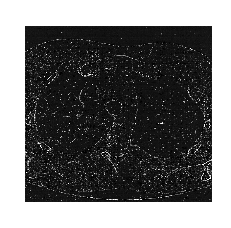
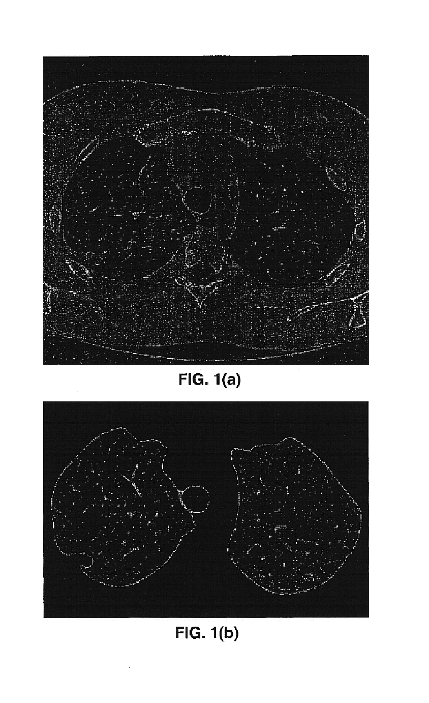
[0041]The present invention is preferably performed on a computer system, such as a Pentium™-class personal computer, running computer software that implements the algorithm of the present invention. The computer includes a processor, a memory and various input / output means. A series of CT axial or other digital images representative of a portion of the body are input to the computer. Illustratively, the portion of the body that is of interest is the thoracic volume; and examples of digital images or sections of the thoracic volume are shown in FIGS. 1(a) and 1(b). The terms “digital” and “digitized” as used herein will refer to images or volumes, as appropriate, in a digital or digitized format acquired via a digital acquisition system or via conversion from an analog image.

[0042]The present invention provides for systems and methods capable of effective and accurate lesion detection from 2-D and 3-D digital images. The digital image sections to be processed, rendered, displayed or...


PUM


Abstract

An algorithm is disclosed that detects and analyzes possible lesions that are left in contact with a structural boundary. A morphological closing with a structuring element is performed along the boundary to detect lesions within the threshold of the structuring element. A deformable surface-based analysis is performed on distinctive surfaces of the structure for the identification of lesions. The integrated use of a deformable surface model and chamfer distance potential enables explicit representation of regularized, or smoothed, surfaces from which lesion candidates may be detected.

Description

CROSS-REFERENCE TO RELATED APPLICATIONS[0001]This application is a continuation-in-part of application Ser. No. 09 / 993,789, filed Nov. 23, 2001.[0002]Related applications are:[0003]“Density Nodule Detection in 3-Dimensional Medical Images,” Ser. No. 09 / 993,792 filed Nov. 23, 2001;[0004]“Method and System for the Display of Regions of Interest in Medical Images,” Ser. No. 09 / 990,508, filed Nov. 21, 2001;[0005]“Vessel Segmentation with Nodule Detection,” Ser. No. 09 / 993,791 filed Nov. 23, 2001;[0006]“Automated Registration of 3-D Medical Scans of Similar Anatomical Structures,” Ser. No. 09 / 993,790, filed Nov. 23, 2001;[0007]“Lung Field Segmentation From CT Thoracic Images,” Ser. No. 09 / 993,793, filed Nov. 23, 2001; and[0008]“Graphical User Interface for Display of Anatomical Information, ” Ser. No. 09 / 990,511, filed Nov. 21, 2001, claiming priority from Ser. No. 60 / 252743 filed Nov. 22, 2000 and from Ser No. 60 / 314,582 filed Aug. 24, 2001.[0009]“Region Growing in Anatomical Images,” S...

Claims


Application Information

Patent Timeline
Patent Type & Authority Patents(United States)
IPC IPC(8): G06K9/62A61B10/00G06T7/00
CPCG06T7/0012G06T2207/30004Y10S128/922
Inventor ZENG, XIAOLANZHANG, WEI
Owner MEVIS MEDICAL SOLUTIONS
Why Eureka
Try Eureka