Drug delivery to the inner ear and methods of using same
a technology of therapeutic agents and inner ear, which is applied in the direction of goggles, viruses, peptide/protein ingredients, etc., can solve the problems of high-frequency hearing loss, and achieve the effect of protecting against potential hearing loss
- Summary
- Abstract
- Description
- Claims
- Application Information
AI Technical Summary
Benefits of technology
Problems solved by technology
Method used
Image
Examples
example 1
Round Window Approach
[0059]Adult CBA mice were anesthetized using intraperitoneal Avertin (2,2,2 Tribromoethanol, Sigma-Aldrich, T4,840-2). A dorsal postauricular incision was made and the bone medial to the tympanic ring was exposed. Using a No. 18 needle, a hole was drilled exposing the middle ear space medial to the tympanic ring. The round window niche and the bone overhanging the niche were both identified. The bone was scraped away revealing the round window membrane. The adenoviral vector with the E1 / E3 / E4 deletions expressing gfp driven by a CMV promoter was used (Ad11D) for all experiments (see Kanzaki et al. supra).
[0060]Vector injections consisted of 108 i.p. / μL of vector in 0.5-4.0 μL of volume (n=5 for each condition). Vector injections were carried out using a Hamilton microsyringe with 0.25 μL graduations. A 32-gauge needle was used to puncture the round window membrane using a micromanipulator (Singer Instruments, UK) while the animals were immobilized to minimize in...
example 2
Semicircular Canal Approach
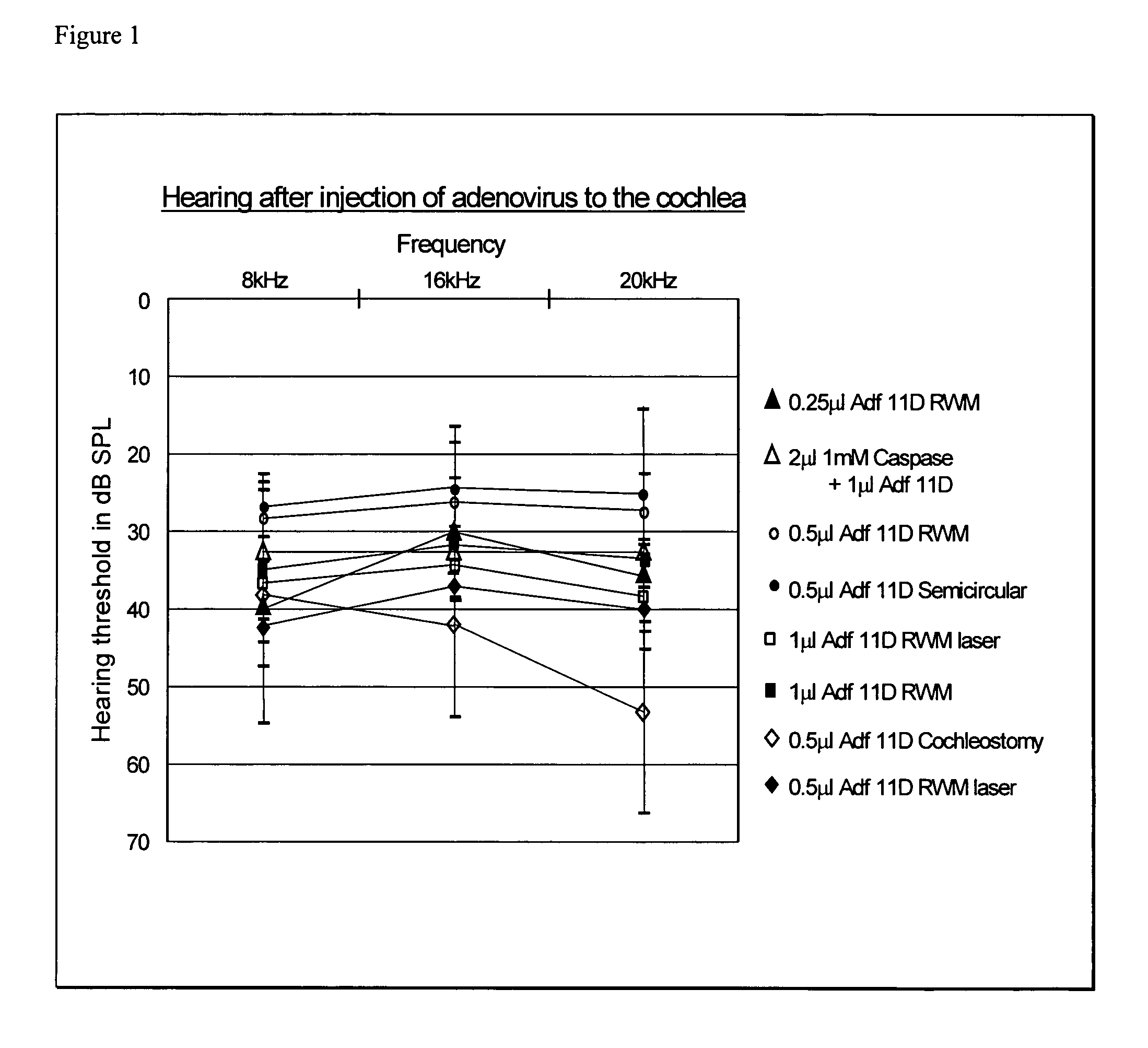
[0064]After surgically exposing the temporal bone, the superior semicircular canal was identified and marked with India ink. An argon laser was used to create an opening in the bone with a single burst of 100 W / 0.2 s. Injection of vector and data acquisition was carried out as described in Example 1. Injection of vector into the semicircular canal did not result in any hearing loss (FIG. 1).
example 3
Cochleostomy Approach
[0065]Animals were prepared as previously described in Example 1. The promontory overlying the basal turn of the cochlea was identified and marked with India ink. An argon laser was used to create a 200 μm cochleostomy just anterior to the round window using settings of 100 W / 0.2 s. Injection of vector was then carried out as in Example 1. The cochleostomy was sealed with a piece of muscle tissue, and the animal was allowed to recover. Use of a basal turn cochleostomy with a low volume of vector injection (1 μL) having approximately 1×10¹³ infectious particles per mL resulted in hearing loss at 20 kHz, suggesting that this approach is more damaging than direct injection through the round window membrane (FIG. 1).
PUM
| Property | Measurement | Unit |
|---|---|---|
| volume | aaaaa | aaaaa |
| volume | aaaaa | aaaaa |
| volumes | aaaaa | aaaaa |
Abstract
Description
Claims
Application Information
 - R&D
- Intellectual Property
- Life Sciences
- Materials
- Tech Scout
- Unparalleled Data Quality
- Higher Quality Content
- 60% Fewer Hallucinations
Browse by: Latest US Patents, China's latest patents, Technical Efficacy Thesaurus, Application Domain, Technology Topic, Popular Technical Reports.
© 2025 PatSnap. All rights reserved.Legal|Privacy policy|Modern Slavery Act Transparency Statement|Sitemap|About US| Contact US: help@patsnap.com


