Endovascular graft with separable sensors

a technology of endovascular graft and sensor, which is applied in the field of body lumen treatment, can solve the problems of high morbidity and mortality rate of abdominal surgery, and the associated implantation of endovascular grafts, and achieve the effects of improving the potential for diagnosis and treatment, reducing the cost of implantation, and facilitating confirmation of a successful implant procedur

Inactive Publication Date: 2008-07-15
LIFESHIELD SCI
View PDF58 Cites 52 Cited by
  • Summary
  • Abstract
  • Description
  • Claims
  • Application Information

AI Technical Summary

Benefits of technology

[0012]Briefly and in general terms, the present invention is embodied in an endovascular graft with sensors attached thereto. The endovascular graft has the ability to be delivered endovascularly and measure pertinent parameters within the lumen in which it is implanted. The endovascular graft has the ability to transmit data about intra-lumen parameters to an external monitoring device. Confirmation of a successful implant procedure is quickly and easily obtained. Patient follow-up is less costly (conducted in the physician office), non-invasive, and more accurate, allowing prompt intervention in those patients most at risk for acute AAA rupture. The invention would also allow for more frequent patient follow-up, increasing the potential to diagnose and treat aneurysms at risk before acute rupture.

Problems solved by technology

Conventional open surgical repair of AAA involves major abdominal surgery with associated high rates of morbidity.
Untreated AAA have been shown to continue to expand until rupture, with an associated high mortality rate.
Implantation of endovascular grafts have also been associated with high complication rates, including perioperative death, conversion to open repair, the need for further intervention, the need for hemodialysis, a failure to cure the AAA, and wound complications.
The inability to obtain or maintain a secure seal between the vessel wall and the endovascular graft is a complication unique to endovascular aneurysm exclusion.
It is believed that persistent endoleaks result in continued aneurysm expansion, which may eventually lead to aneurysm rupture.
Failure to properly exclude the aneurysm from systemic arterial blood pressure keeps the patient at risk of impending rupture.
It has been recognized that preoperative patent side branches are not a good predictor of postoperative endoleaks.
There have been a number of reported cases of aneurysm rupture following implantation of an endovascular graft.
Thrombosed aneurysm sacs may still receive pressurization from a sealed endoleak and this continued pressurization keeps the aneurysm at risk for rupture.
All of these methods are costly and involve invasive procedures with associated morbidity that may need to be performed in a hospital.
None of the imaging methods are completely successful in detecting endoleaks.
Some physicians are advocating that the follow-up examinations of AAA patients focus on pressure measurements, but that this is not currently clinically feasible.
However, providing devices on an endovascular graft to facilitate the measurement of pertinent parameters poses problems.
The measurement device increases bulk, which can significantly effect the delivery profile of the endovascular graft and increase the force necessary to deploy the device, such as jacket or release wire retraction forces.
Increased bulk is a significant issue for an endovascular graft.
Furthermore, attachment of measurement devices to an endovascular graft may require sutures and the suture knots not only provide increased bulk, but are also potential graft wear points.
Additionally, tissue growth around a measuring device attached to an implanted endovascular graft may interfere with its function and inaccurate data may result.

Method used


Image

  • Endovascular graft with separable sensors
  • Endovascular graft with separable sensors
  • Endovascular graft with separable sensors

Examples


Embodiment Construction

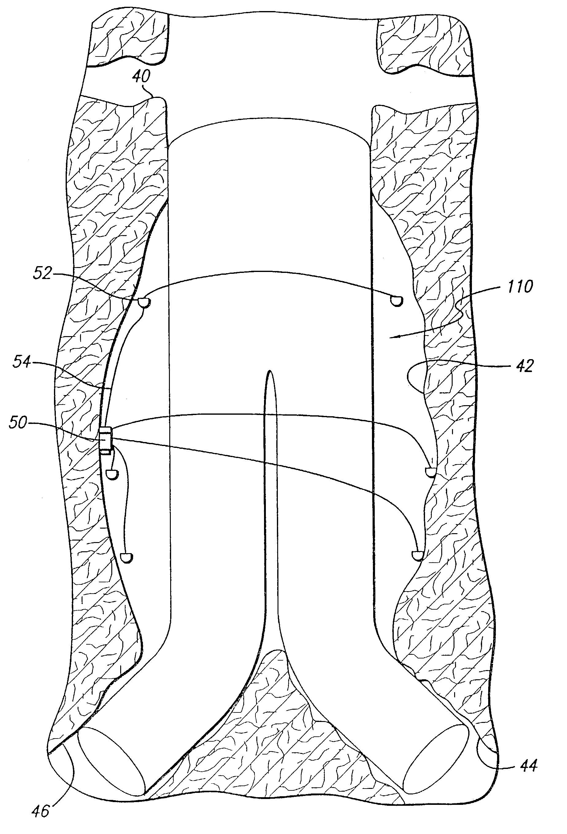
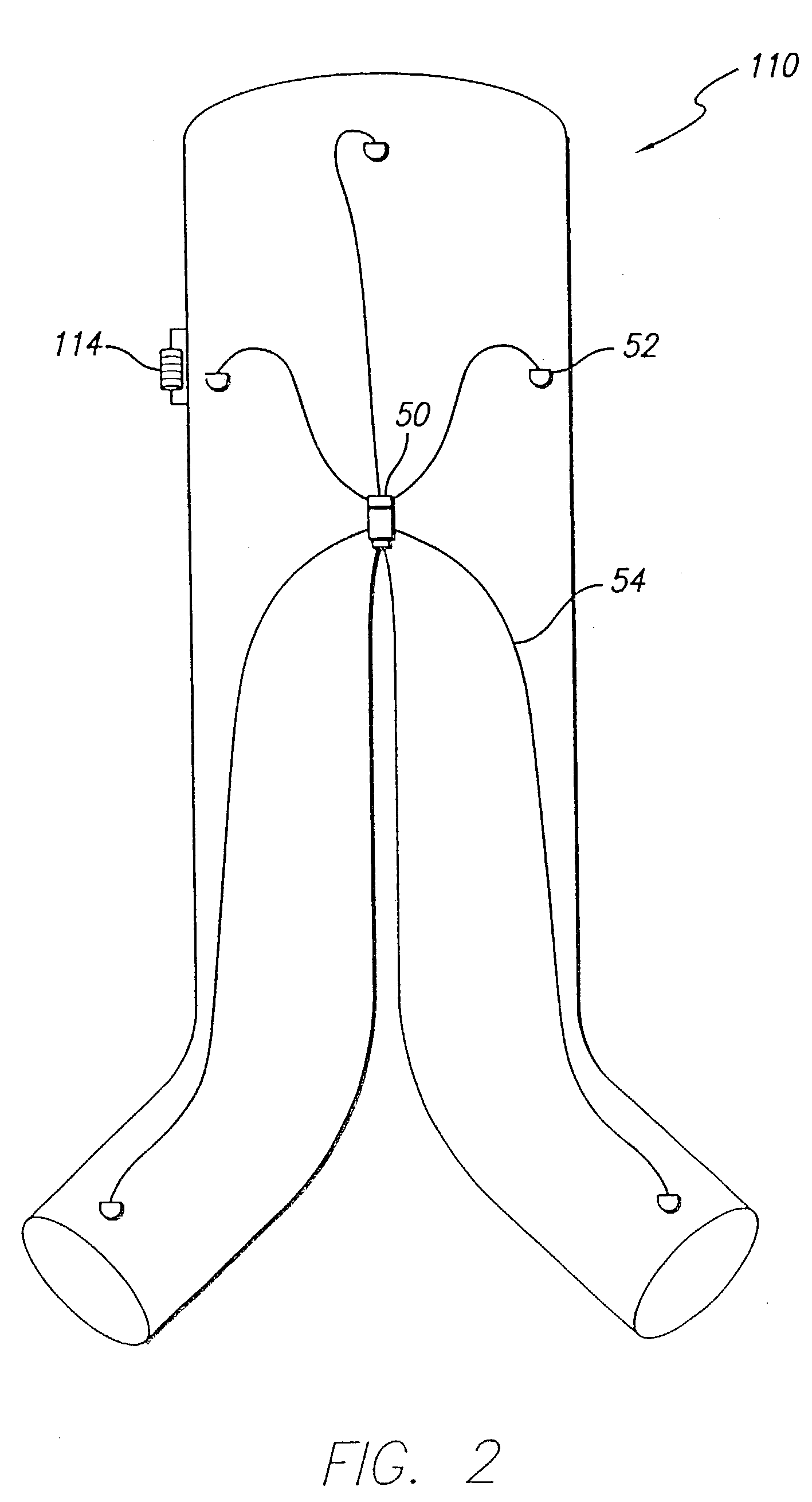
[0033]As shown in the exemplary drawings and for purposes of illustration, the invention is embodied in a prosthetic endovascular graft having the ability to measure pertinent parameters inside the lumen into which it is implanted and transmit the measurements to a receiver located external to the patient.

[0034]Referring to FIG. 1, an embodiment of the invention is shown in which a modular bifurcated endovascular graft 10 of the type known within the art is implanted in a body vessel 40 across an aneurysm sac 42 in the area of the contra-lateral 44 and ipsi-lateral 46 iliac arteries using methods known within the art (only the contra-lateral limb is shown). The bifurcated endovascular graft 10 may be assembled in-vivo from a tubular trunk component 20 and two limb components 30. The trunk component 20 has a superior end 22 adapted to be secured above the aneurysm and an inferior end 24 adapted to accept the limb components 30. The limb component 30 has a transmitter 12, power source...


PUM


Abstract

A endovascular graft having sensing devices attached thereto to facilitate measurement of pertinent parameters within the vasculature into which the graft is implanted. Power sources and transmitters may be attached to the graft to facilitate transmission of measurements to a receiving device outside the patient's body. The sensing devices, may be electrically passive or integrated devices with measurement and transmission capability. The sensing devices may be attached to specific locations on the graft material or attached to the lumen, thereby providing pertinent parameters from critical points inside the vasculature, or may be dispersed over the surface of the graft material or within the lumen to provide a profile of pertinent parameters. The sensing devices may be attached to the graft material with one suture using a running stitch to minimize graft bulk and may be coated with a material to inhibit or control tissue growth. A bio-reabsorbable or hard wire tether or suture may be employed to attach devices to one or more graft components.

Description

[0001]This application is a continuation-in-part of U.S. application Ser. No. 10 / 165,763, filed Jun. 7, 2002.BACKGROUND OF THE INVENTION[0002]This invention relates to the treatment of body lumens and, more particularly, to the endovascular placement of a prosthetic graft within vasculature for the purpose of repairing the same.[0003]Ruptured abdominal aortic aneurysms (AAA) are a leading cause of death in the United States. Treatment options to repair AAA include conventional open surgery and implantation of an endovascular graft. Conventional open surgical repair of AAA involves major abdominal surgery with associated high rates of morbidity. Endovascular grafts have been developed to endoluminally bypass abdominal aortic aneurysms through minimally invasive surgery. Many patients that are unacceptable surgical risks for open repairs are eligible for endovascular graft implantation. Deployment of transfemoral, endovascular grafts to treat AAA is appealing for many reasons: avoidan...

Claims


Application Information

Patent Timeline
Patent Type & Authority Patents(United States)
IPC IPC(8): A61F2/06A61B5/00A61B5/0215A61B5/026A61B5/05A61FA61F2/02
CPCA61B5/0031A61B5/02014A61B5/0215A61F2/07A61B5/01A61B5/026A61F2250/0002A61B5/145A61B5/14532A61F2002/065A61F2002/067A61B5/05
Inventor BROWN, PETER S.KOVAC, TIMWEI, MICHAELECKERT, ROBIN W.
Owner LIFESHIELD SCI
Why Eureka
Try Eureka