Detecting and profiling molecular complexes

a molecular complex and complex technology, applied in the field of molecular complex detection and profiling, can solve the problems of difficult application of techniques, difficult study of phosphorylation states of signaling proteins, difficult study of signaling pathways, etc., and achieves convenient multiplexing capability, reduced background, and improved sensitivity

Inactive Publication Date: 2008-07-22
MONOGRAM BIOSCIENCES
View PDF113 Cites 46 Cited by
  • Summary
  • Abstract
  • Description
  • Claims
  • Application Information

AI Technical Summary

Benefits of technology

[0009]The present invention provides a method of detecting or measuring the formation of one or more molecular complexes that has several advantages over current techniques including, but not limited to, (1) the detection and / or measurement of molecular tags that are separated from an assay mixture provide greatly reduced background and a significant gain in sensitivity; and (2) the use of molecular tags that are specially designed for ease of separation and detection thereby providing convenient multiplexing capability. The present invention also provides a alternative measure of the state of signal transduction whenever phosphorylation is associated with the formation of complexes.

Problems solved by technology

Unfortunately, such signaling pathways have been difficult to study not only because of their complexity and interconnectedness, but also because of the disruptive procedures required for analysis of intracellular complexes, e.g. Weng et al, Science, 284: 92-96 (1999); Machida et al, Molecular & Cellular Proteomics, 2.4: 215-233 (2003); Price et al, Methods in Molecular Biology, 218: 255-267 (2003).
In particular, the study of phosphorylation states of signaling proteins has been difficult because during sample preparation the phosphorylation state is rapidly altered by the actions of enzymes, such as phosphatases.
Unfortunately, such techniques are difficult to apply, and generally lack sufficient sensitivity to provide an accurate picture of the state of a signaling pathway.

Method used


Image

  • Detecting and profiling molecular complexes
  • Detecting and profiling molecular complexes
  • Detecting and profiling molecular complexes

Examples


example 1

PI3K / Her-3 Receptor Activation Complex

[0170]In this example, assays were designed as shown in FIGS. 7A and 7C to measure a receptor complex comprising Her2, Her3, and PI3K in breast cancer cell line, MCF-7. Binding compound (1106) having a first molecular tag (“mT1” in the figure and “eTag1” below) is specific for the extracellular domain of Her3 receptor (1102), binding compound (1110) having a second molecular tag (“mT2” in the figure and “eTag2” below) is specific for the p85 component (1111) of PI3K protein (1100), and cleaving probe (1108) having a photosensitizer attached (is specific for the intracellular domain of Her3 receptor (1102) where “H2” indicates a Her2 receptor (1104), “H3” indicates a Her3 receptor (1102), “p85” and “p110” are components of PI3 kinase (1100), which binds to a phosphorylation site of H3 (denoted by “P”) through its p85 moiety. The two assay designs are similar, except that in the design of FIG. 7A the cleaving probe is specific for the Her3 recepto...

example 2

Shc / Her-3 Receptor-Adaptor Interaction

[0198]In this example, an assays were designed as shown in FIGS. 8A and 8C. In FIG. 8A, Her2 receptor (1200) and Her3 receptor (1202) form a dimer in cell surface membrane (1204) and each receptor is represented as having phosphorylated sites (1209 and 1210). Shc proteins (1206 and 1208) bind to phosphylation sites (1210) and (1209), respectively. A first binding compound (1214) and cleaving probe (1216) are specific for different antigenic determinants of the extracellular domain of Her2 receptor (1200). A second binding compound (1212) is specific for Shc proteins (1206 and 1208). The assay designs of FIGS. 8A and 8C are similar, except that in the design of FIG. 8A the cleaving probe is specific for the Her2 receptor, and in the design of FIG. 8C, the cleaving probe is specific for the Her3 receptor. Thus, in the former case, total Her2 receptor is measured, whereas in the latter case total Her3 receptor is measured. The assays were carried o...

example 3

SHC / / Grb2 Protein-Protein Interaction

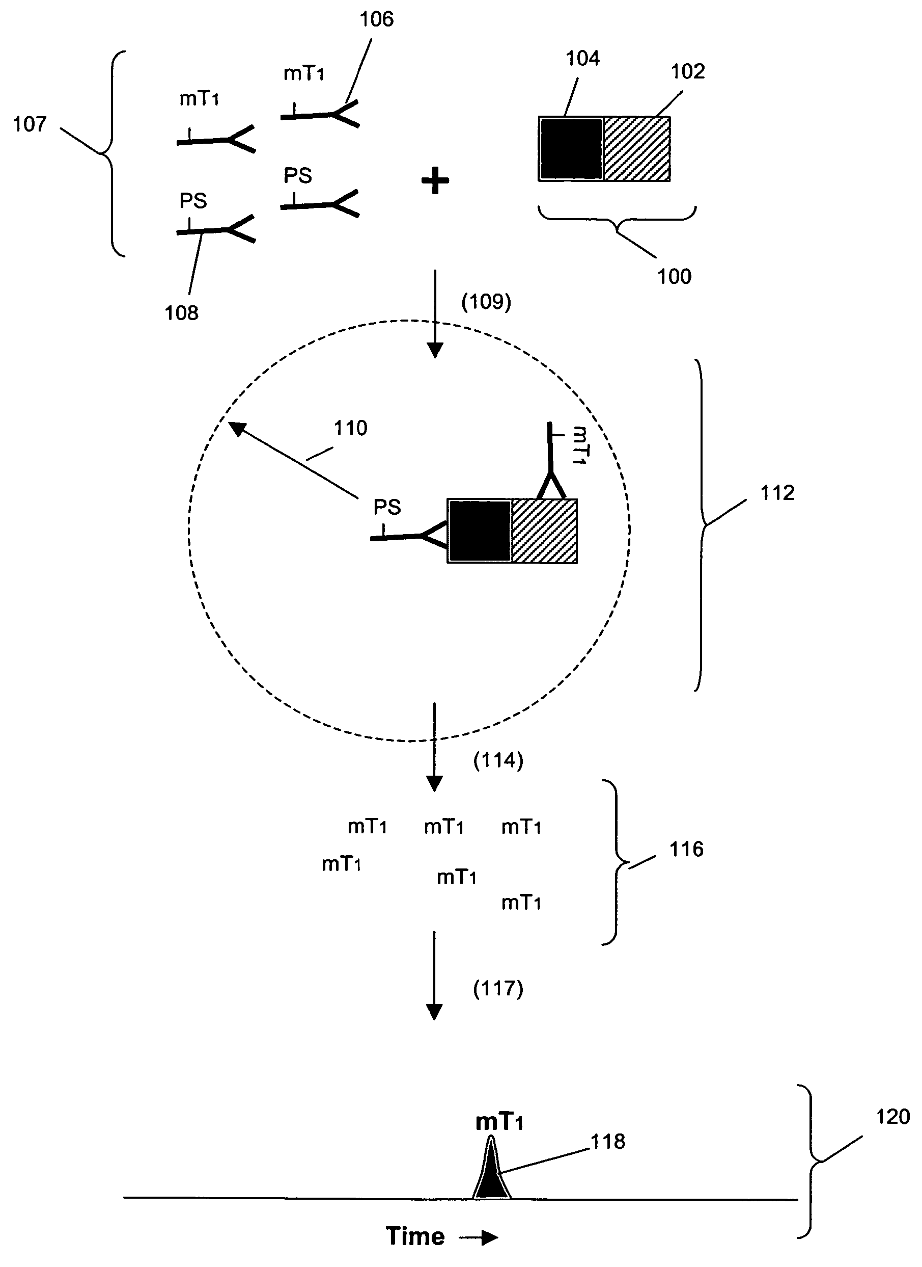
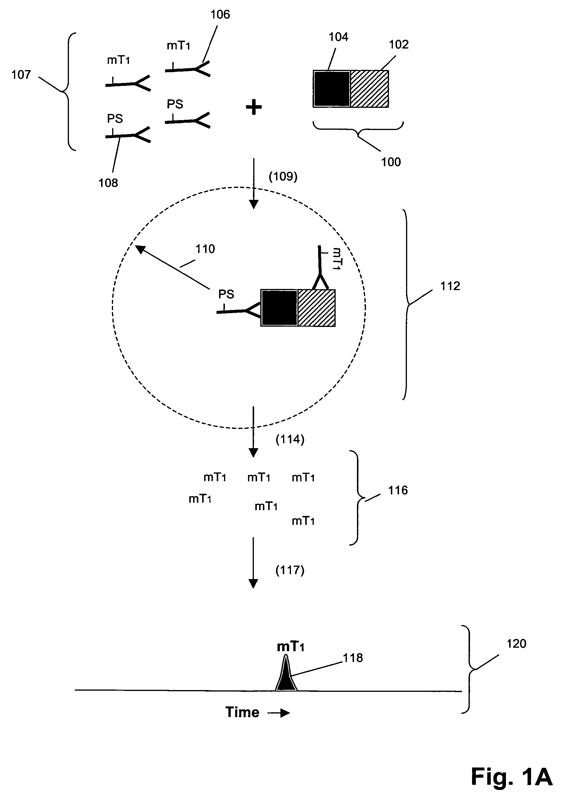
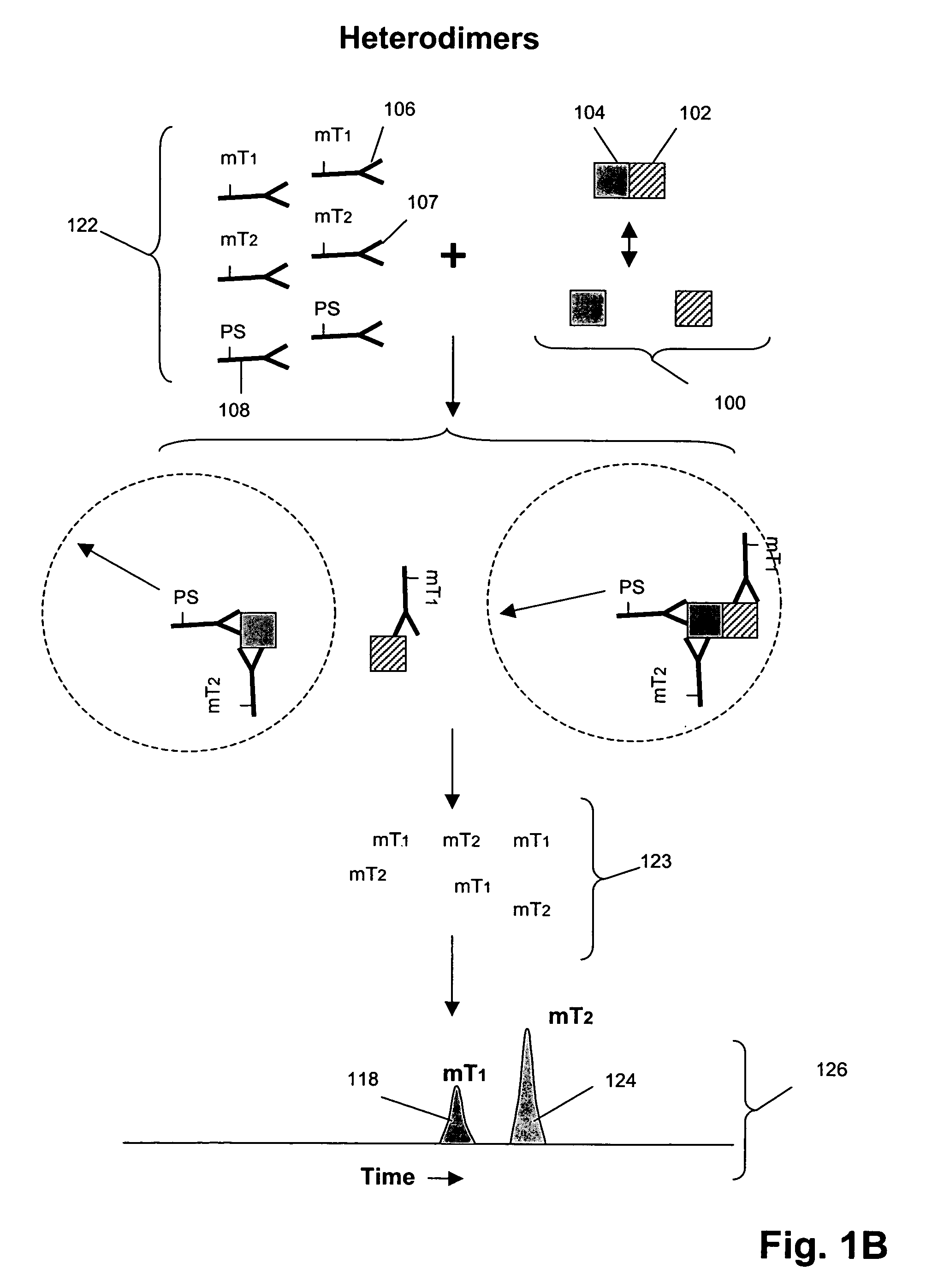
[0222]In this example, an assay was designed as shown in FIG. 1B to detect Grb2 / / SHC protein-protein complexes in cell lysates. A first binding compound having a first molecular tag was specific for Grb2 and a cleaving probe was provided that was specific for a separate antigenic determinant of Grb2 than that of the first binding compound. A second binding compound specific for SHC having a second molecular tag was also provided. The assays were carried out as follows. Sample preparation was carried out as above.

[0223]The total assay volume is 40 ul. The lysate volume is adjusted to 10 ul with lysis buffer.

The antibodies are diluted in lysis buffer up to 20 ul. Typically about 5000 to 500,000 cell-equivalent of lysates is used per reaction.

[0224]Procedure: Working concentrations of pre-mixed antibodies prior to adding into reaction:[0225]For Grb2 / / SHC complex with cleaving probe at Grb2:[0226]eTag1_anti-Grb2 at 10 nM (eTag1 was Pro14 in this assa...


PUM

PropertyMeasurementUnit
molecular weightaaaaaaaaaa
diameteraaaaaaaaaa
pressureaaaaaaaaaa

Abstract

Methods are provided for detecting the formation of complexes of molecules, especially proteins, in a sample, such as a cell or tissue lysate. In one aspect, a cleaving probe specific for a first protein in a complex and one or more binding compounds specific for one or more second proteins in a complex are provided. Upon binding, the cleaving probe is induced to generate an active species, such as singlet oxygen, that cleaves molecular tags attached to the binding compounds only in the local region of the cleaving probe. The released molecular tags are separated from the assay mixture and from one another to provide a readout that is related to the number and types of proteins present in the complex.

Description

CROSS-REFERENCE TO RELATED APPLICATIONS AND PATENTS[0001]This is a continuation-in-part of U.S. patent applications Ser. No. 10 / 623,057 filed 17 Jul. 2003 now U.S. Pat. No. 7,105,308 and Ser. No. 10 / 154,042 filed 21 May 2002, now U.S. Pat. No. 7,255,999, and further claims priority from U.S. provisional applications Ser. No. 60 / 494,482 filed 11 Aug. 2003, Ser. No. 60 / 508,034 filed 1 Oct. 2003, and Ser. No. 60 / 512,941 filed 20 Oct. 2003, each of the foregoing applications being incorporated by reference in its entirety.FIELD OF THE INVENTION[0002]The present invention relates generally to methods for detecting and measuring molecular complexes, and more particularly, to methods for simultaneously measuring complexes among multiple proteins, especially within biological cells.BACKGROUND OF THE INVENTION[0003]The formation and disassociation of molecular complexes are crucial to regulatory processes in living organisms. In particular, signaling pathways between the surface and nucleus ...

Claims


Application Information

Patent Timeline
Patent Type & Authority Patents(United States)
IPC IPC(8): G01N33/53C07K1/10G01N33/532G01N33/554C07K1/14C12Q1/68G01N33/537G01N33/542G01N33/543G01N33/566
CPCG01N33/542G01N33/566G01N2333/726G01N2333/71Y10S436/824
Inventor CHAN-HUI, PO-YINGSINGH, SHARATSALIMI-MOOSAVI, HOSSEINTAHIR, HASANWALLWEBER, GERALD J.KIRAKOSSSIAN, HRAIRMATRAY, TRACY J.
Owner MONOGRAM BIOSCIENCES
Why Eureka
Try Eureka