
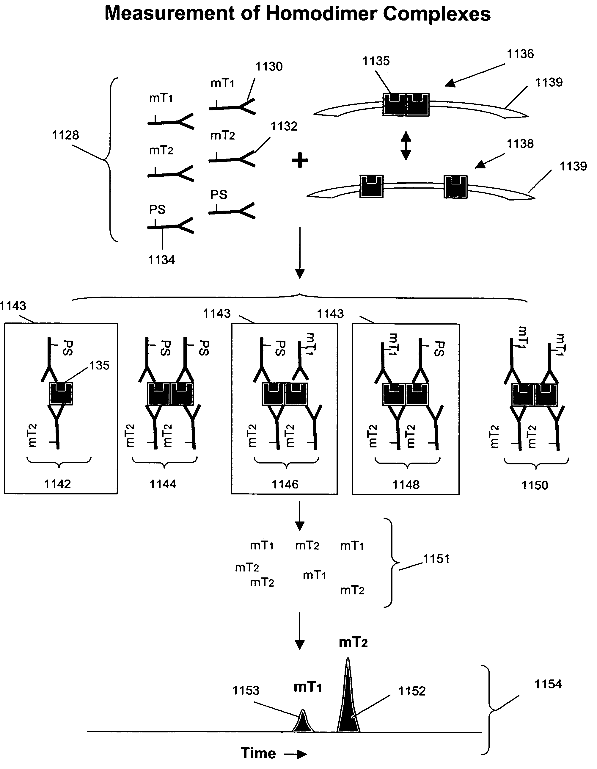
Measuring receptor homodimerization

a cell surface membrane and receptor technology, applied in the field of cell surface membrane receptor homodimerization, can solve the problems of lack of flexibility, lack of convenient and sensitive techniques, and difficult application of techniques for measuring such interactions, so as to reduce background, facilitate multiplexing, and improve the effect of sensitivity

Inactive Publication Date: 2008-07-22
MONOGRAM BIOSCIENCES
View PDF40 Cites 30 Cited by
  • Summary
  • Abstract
  • Description
  • Claims
  • Application Information

AI Technical Summary

Benefits of technology

"The invention provides methods for detecting and measuring the formation of dimers or oligomers of membrane-associated analytes, particularly receptor tyrosine kinases, using cleaving probes and binding compounds that are specific for different members of the dimer or oligomer. The methods involve inducing a cleavage-inducing moiety in the cleaving probe and the binding compound, which causes susceptible bonds to be cleaved, and detecting the molecular tags released from the cleavage. The methods can be used to measure the presence or absence of homodimers or homo-oligomers in cell membranes. The invention also provides kits for carrying out the methods. The technical effect of the invention is to provide a reliable and sensitive method for detecting and measuring the formation of dimers or oligomers of membrane-associated analytes."

Problems solved by technology

Unfortunately, despite the importance of receptor dimerization and oligomerization in signal transduction processes, the techniques for measuring such interactions are difficult to apply, lack flexibility, and lack sensitivity.
The lack of a convenient and sensitive technique for analyzing the oligomerization of cell surface molecules has greatly increased the difficulty of developing new therapeutics or diagnostic methods based on such phenomena.

Method used


Image

  • Measuring receptor homodimerization
  • Measuring receptor homodimerization
  • Measuring receptor homodimerization

Examples


example 1

Simultaneous Measurement of Her2-Her3 Heterodimerization and Erk1 Phosphorylation

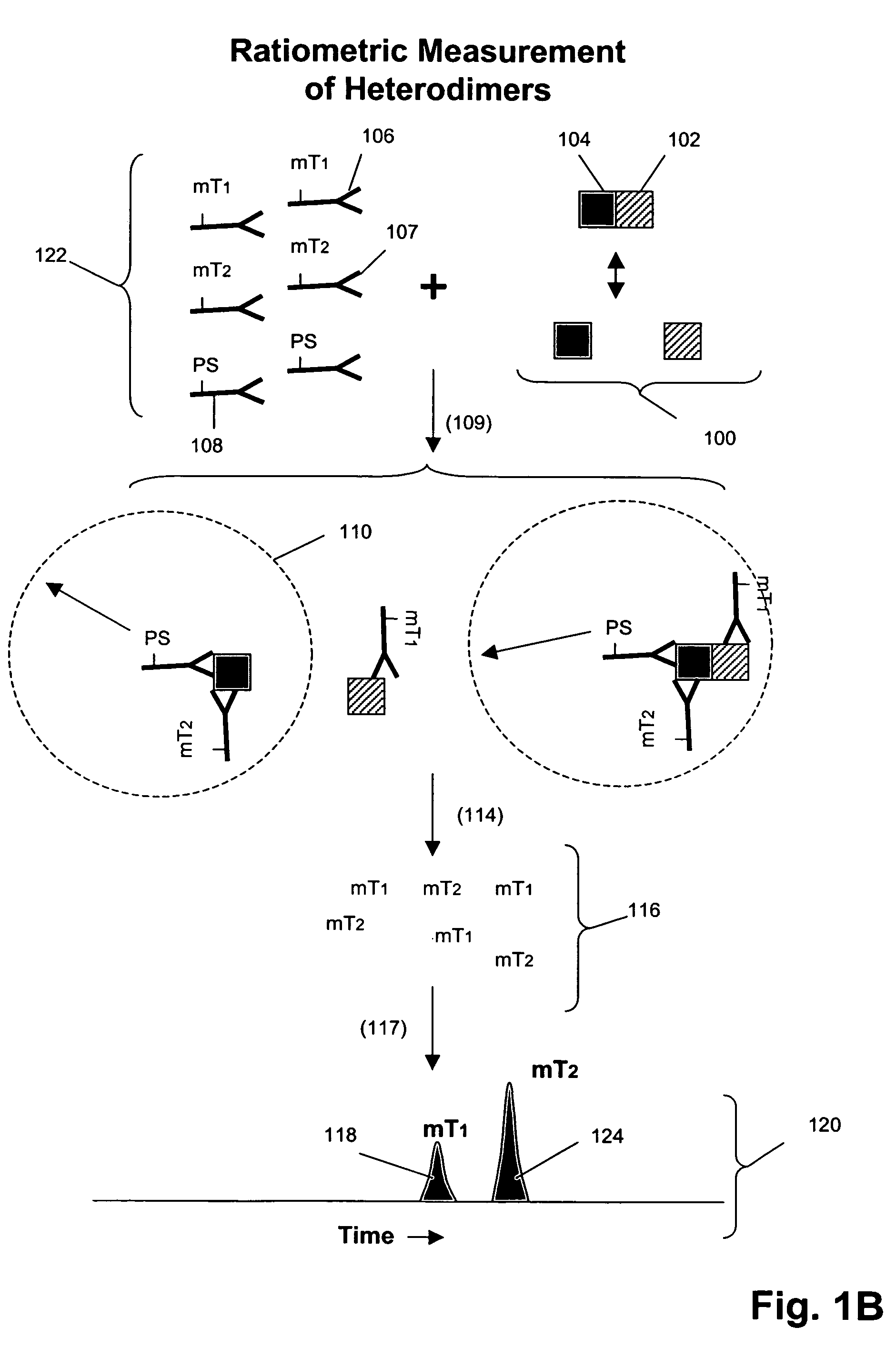
[0102]In this example, an assay is described for providing a ratiometric measure of phosphorylated Erk1 and Her2-Her3 heterodimerization. The assays are carried out as follows.

Sample Preparation:

[0103]1. Serum-starve breast cancer cell line culture (MCF-7) overnight before use.[0104]2. Stimulate cell lines with HRG in culture media for 10 minutes at 37° C. Exemplary doses of HRG are 0, 0.032, 0.16, 0.8, 4, 20, 100 nM for MCF-7 cells.[0105]3. Aspirate culture media, transfer onto ice, and add lysis buffer (described below) to lyse cells in situ.[0106]4. Scrape and transfer lysate to microfuge tube. Incubate on ice for 30 min. Microfuge at 14,000 rpm, 4° C., for 10 min.[0107]5. Collect supernatants as lysates and aliquot for storage at −80° C. until use.

Lysis Buffer (made fresh and stored on ice):

[0108]

FinalulStock1% Triton X-100100010% 20 mM Tris-HCl (pH 7.5)200  1 M100 mM NaCl200  5 M 50 mM NaF500  1 M ...

example 2

Analysis of Cell Lysates for Her-2 Heterodimerization and Receptor Phosphorylation

[0126]In this example, Her1-Her2 and Her2-Her3 heterodimers and phosphorylation states are measured in cell lysates from several cell lines after treatment with various concentrations of epidermal growth factor (EGF) and heregulin (HRG). Measurements are made using three binding compounds and a cleaving probe as described below.

Sample Preparation:

[0127]1. Serum-starve breast cancer cell line culture overnight before use.[0128]2. Stimulate cell lines with EGF and / or HRG in culture media for 10 minutes at 37° C. Exemplary doses of EGF / HRG are 0, 0.032, 0.16, 0.8, 4, 20, 100 nM for all cell lines (e.g. MCF-7, T47D, SKBR-3) except BT20 for which the maximal dose is increased to 500 nM because saturation is not achieved with 100 nM EGF.[0129]3. Aspirate culture media, transfer onto ice, and add lysis buffer to lyse cells in situ.[0130]4. Scrape and transfer lysate to microfuge tube. Incubate on ice for 30 m...

example 3

Analysis of Tissue Lysates for Her2 Heterodimerization and Receptor Phosphorylation

[0152]In this example, Her1-Her2 and Her2-Her3 heterodimers and phosphorylation states are measured in tissue lysates from human breast cancer specimens.

Sample Preparation:

[0153]1. Snap frozen tissues are mechanically disrupted at the frozen state by cutting.[0154]2. Transfer tissues to microfuge tube and add 3× tissue volumes of lysis buffer (from appendix I) followed by vortexing to disperse tissues in buffer.[0155]3. Incubate on ice for 30 min with intermittent vortexing to mix.[0156]4. Centrifuge at 14,000 rpm, 4° C., for 20 min.[0157]5. Collect supernatants as lysates and determine total protein concentration with BCA assay (Pierce) using a small aliquot.[0158]6. Aliquot the rest for storage at −80° C. until use.

Assay design:[0159]1. The total assay volume is 40 ul.[0160]2. The lysates are tested in serial titration series of 40, 20, 10, 5, 2.5, 1.25, 0.63, 0.31 ug total-equivalents and the volum...


PUM

PropertyMeasurementUnit
molecular weightaaaaaaaaaa
diameteraaaaaaaaaa
pressureaaaaaaaaaa

Abstract

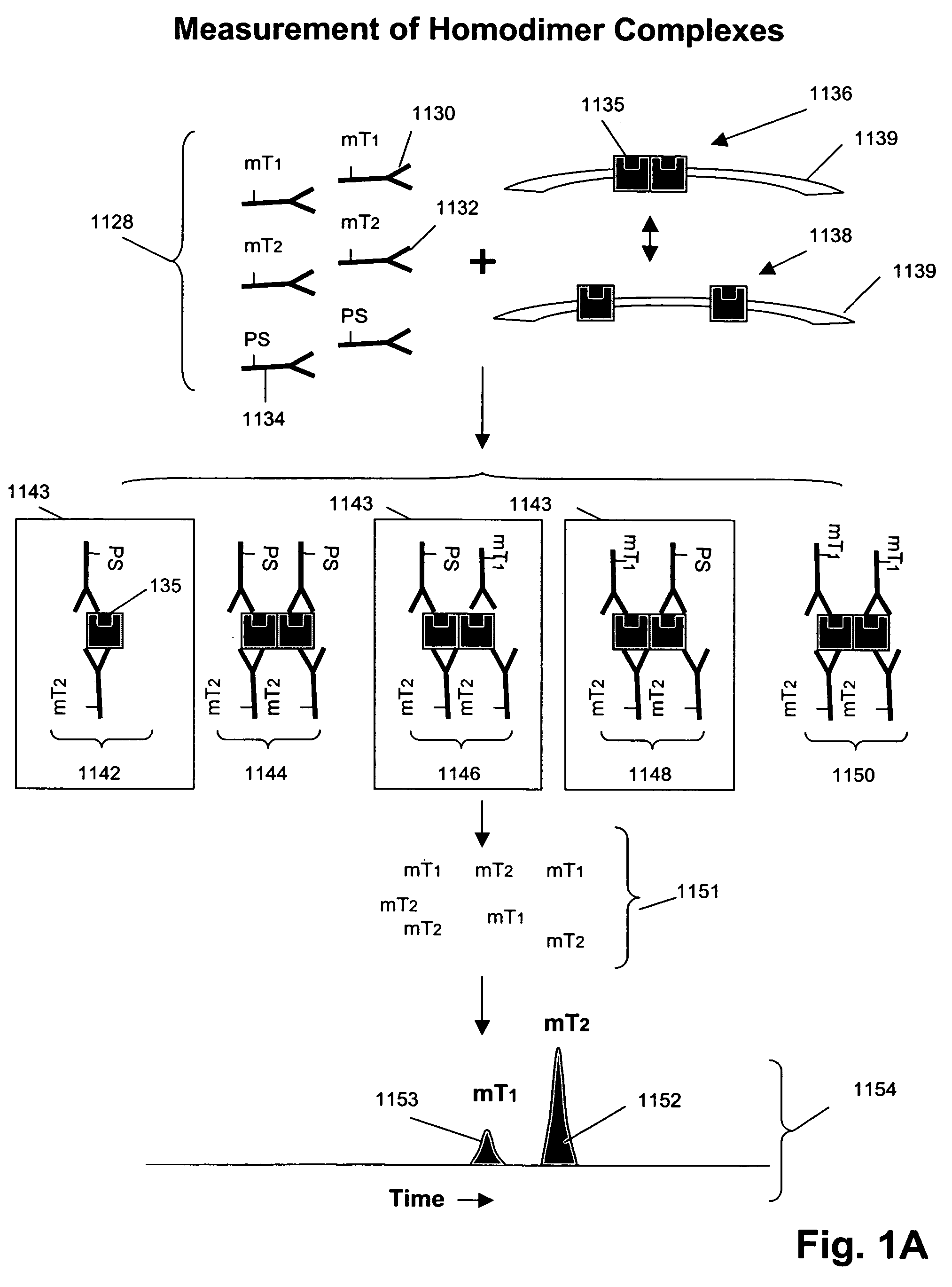
The invention provides methods and kits for detecting and / or measuring receptor homodimers on a cell surface membrane. In one aspect, the methods employ pairs of probes comprising binding compounds and a cleaving probe, such that at least one binding compound binds specifically to the same epitope of a membrane-bound analyte as the cleaving probe. The binding compound includes one or more molecular tags attached through a cleavable linkage, and the cleaving probe includes a cleavage-inducing moiety that can cleave the linkage when within a defined proximity thereto. Binding of the two probes to a homodimer of a cell surface molecules results in release of molecular tags from the binding compounds, providing a measure of formation of the homodimeric complex.

Description

[0001]This patent application is a continuation-in-part of U.S. patent application Ser. No. 10 / 623,057 filed 17 Jul. 2003 now U.S. Pat. No. 7,105,308 and claims priority from U.S. Provisional Applications Ser. No. 60 / 508,034 filed 1 Oct. 2003; Ser. No. 60 / 566,352 filed 28 Apr. 2004; and Ser. No. 60 / 577,256 filed 3 Jun. 2004, all of which are incorporated herein by reference in their entireties.FIELD OF THE INVENTION[0002]The present invention relates to methods for measuring oligomerization of cell surface molecules, particularly homodimers of cell surface membrane receptors.BACKGROUND OF THE INVENTION[0003]The interactions of cell surface membrane components play crucial roles in transmitting extracellular signals to a cell in normal physiology, and in disease conditions. In particular, many types of cell surface receptors undergo dimerization or oligomerization in connection with the transduction of an extracellular event or signal, e.g. ligand-receptor binding, into a cellular re...

Claims


Application Information

Patent Timeline
Patent Type & Authority Patents(United States)
IPC IPC(8): G01N33/53C07K1/10G01N33/532C07K1/14G01N33/554C07K16/00G01N33/533C12Q1/68G01N33/567
CPCG01N33/542G01N33/566G01N33/74G01N2333/726G01N2333/71Y10S436/824
Inventor PIDAPARTHI, SAILAJASHI, YININGDUA, RAJIVCHAN-HUI, PO-YINGSINGH, SHARAT
Owner MONOGRAM BIOSCIENCES
Learn More