Distributed architecture for mammographic image acquisition and processing

a distribution architecture and mammography technology, applied in the field of mammography, can solve the problems of slowed digital acceptance process, current digital imaging system does not provide a full field of view for a patient's breast, digital imaging equipment, etc., and achieves the effect of improving patient throughput, facilitating acquisition, and improving patient comfor

Inactive Publication Date: 2008-07-29
HOLOGIC INC
View PDF21 Cites 30 Cited by
  • Summary
  • Abstract
  • Description
  • Claims
  • Application Information

AI Technical Summary

Benefits of technology

[0011]Among other things, the processor may assist in transmitting the imaging information to or storing the imaging information in the image repository. The image repository may be located at the image acquisition site, the remote image review site, or at another location. In one implementation, the image repository is associated with a server separate from the acquisition and review sites. Such a client-server architecture allows for more efficient and/or convenient utilization of the acquisition and review equipment and certain processing advantages as discussed below.
[0012]The remote image review station includes a user interface such as a keyboard and/or mouse for receiving user inputs, a processor for assisting in image review workflow management based on the user inputs and one or more displays for displaying selected mammographic images. This station is located separate from the image acquisition site and, preferably, can be operated independent of the image acquisition equipment. In one implementation, the image acquisition equipment and image review equipment can be operated concurrently such that images for one patient can be acquired while images for another patient are being reviewed. This distributed architecture thereby allows for more efficient use of imaging equipment, higher patient throughput and more convenient image review.
[0013]According to another aspect of the present invention, at least one image-processing tool is provided in connection with a mammographic image server. The associated mammographic imaging system includes image acquisition equipment, image review equipment and the server that is located separate from the image acquisition equipment and image review equipment. The server may be located at a separate location on the medical facility premises or off-site. The image acquisition equipment and image review equipment may be at the same location or at separate locations. The server is interconnected to the image acquisition equipment and the image review equipment for communication of imaging information therebetween and provides access to at least one tool for processing imaging information. In this regard, the tool may run on the server hardware or on a separate platform in communication with the server. Some examples of tools that may be provided in connection with the server include image repository database management tools, CAD tools, other diagnostic aids or medical information privacy and security tools. Providing such tools in connection with a server allows for beneficial resource sharing, improved access to image information and improved access to updated versions of processing logic.
[0014]According to a further aspect of the invention, a mammographic imaging system includes multiple image acquisition stations and/or multiple image review stations associated with a central server subsystem (i.e., a single server subsystem associated with one or more machines for serving the multiple stations). The number of image acquisition stations may be the same as or different than the number of image review stations. In the context of a large medical facility, one on-site...

Problems solved by technology

As a result, mammography equipment is available in many locations and such equipment, as well as clinicians and physicians experienced in using such equipment and analyzing the mammographic images, are typically kept busy meeting the demand.
Moreover, it is sometimes possible to obtain improved diagnostic information by digital processing.
Despite these advantages, certain perceived disadvantages have slowed the process of full digital acceptance.
Some of the perceived disadvantages are specific to particular digital imaging equipment.
In this regard, some current digital imaging systems do not provide a full field of view for a patient's breast.
In addition, some current digital imagi...

Method used


Image

  • Distributed architecture for mammographic image acquisition and processing
  • Distributed architecture for mammographic image acquisition and processing
  • Distributed architecture for mammographic image acquisition and processing

Examples


Embodiment Construction

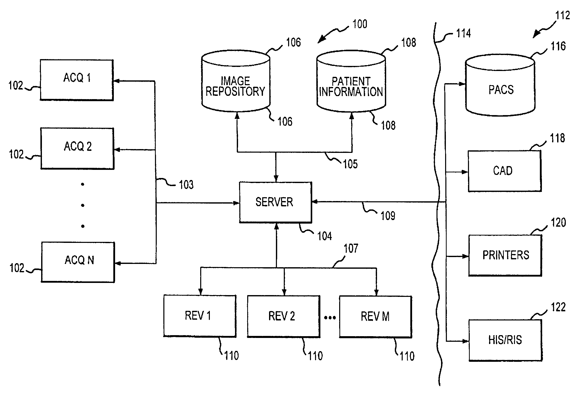
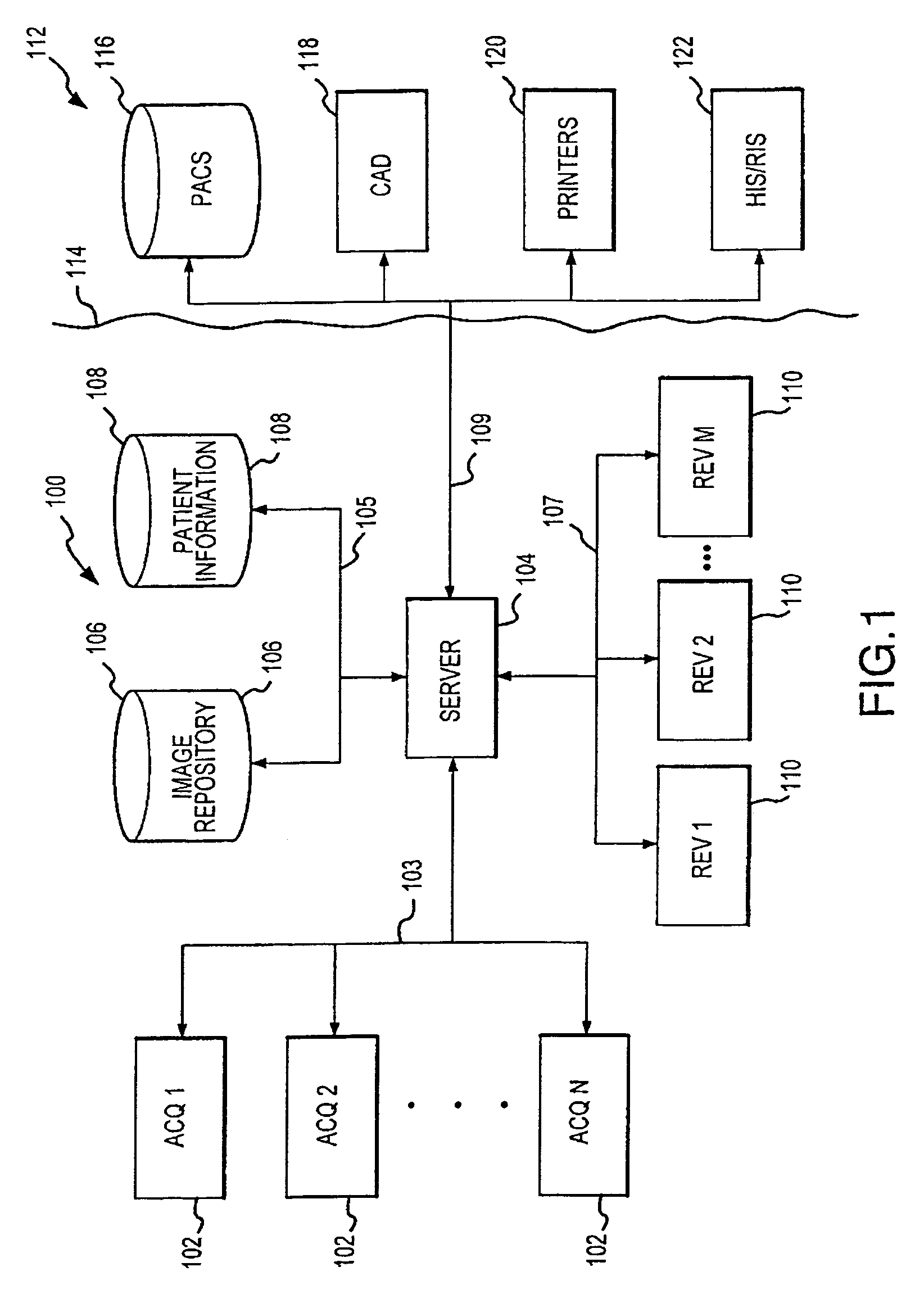
[0023]In the following description, the invention is set forth in the context of a mammographic image system employing a distributed architecture based on a client server model. In particular, the invention is described below in connection with an implementation at a large medical facility that includes multiple mammographic image acquisition sites and multiple image review sites all associated with a central server and central image repository. While this implementation effectively illustrates the operation and advantages of the present invention, it will be appreciated that the invention is not limited to this implementation or similar contexts. For example, various aspects of the present invention are applicable to environments having a single image acquisition site and / or a single image review site. Additionally, it is not necessary that the acquisition equipment, review equipment and server be located at one site. In this regard, particular elements of the system or combination...


PUM


Abstract

A distributed architecture allows for decoupling of mammographic image acquisition and review, thereby enabling more efficient use of resources and enhanced processing. In one embodiment, the system (100) includes a number of image acquisition stations (102) and a number of image review stations (110) all associated with a central server (104). The server (104) is operative to access an image repository (106), a patient information data base (108) and a number of DICOM tools (112). The invention allows for more efficient and/or more convenient use of the image acquisition equipment and image processing stations. Moreover, the distributed architecture including the central image repository provides certain processing and analysis advantages. The invention also provides certain processing and workflow enhancements that allow for a more full realization of potential digital mammography advantages.

Description

RELATED APPLICATION INFORMATION[0001]This patent application is a continuation-in-part patent application and claims priority from U.S. patent application Ser. No. 10 / 307,214, which was filed on Nov. 29, 2002 now abandoned and is titled “Distributed Architecture for Mammographic Image Acquisition Processing”.FIELD OF THE INVENTION[0002]The present invention relates generally to mammography and, in particular, to a distributed architecture that allows for decoupling of mammographic image acquisition and review, thereby enabling more efficient use of resources and enhanced processing.BACKGROUND OF THE INVENTION[0003]Mammography, including x-ray imaging and other imaging modalities, is widely used in detection and analysis of cysts, lesions, microcalcifications and other areas of interest within a patient's breast. Because of its proved effectiveness in early detection and analysis, including detection of nonpalpable lesions, mammography continues to be recommended for many women. As a...

Claims


Application Information

Patent Timeline
IPC IPC(8): H05G1/00A61B5/00A61B6/00A61B6/04A61B8/08
CPCA61B5/0002A61B5/7435A61B5/7475A61B6/463A61B8/565A61B6/502A61B6/563A61B6/468A61B8/0825
Inventor MINYARD, THOMAS J.NIELDS, MORGAN W.KREMER, TED V.
Owner HOLOGIC INC
Why Eureka
Try Eureka