Method and apparatus of CT cardiac diagnostic imaging using motion a priori information from 3D ultrasound and ECG gating

a technology of motion a priori information and ct cardiac diagnostic imaging, applied in the field of diagnostic imaging, can solve the problems of significant time required to collect a complete set of projection data, motion correction or compensation can add significant complexity in post processing of images, and conventional ecg gating described above does not provide mechanical motion detection

Active Publication Date: 2008-08-19
GENERAL ELECTRIC CO
View PDF17 Cites 28 Cited by
  • Summary
  • Abstract
  • Description
  • Claims
  • Application Information

AI Technical Summary

Problems solved by technology

In known studies, this motion correction or compensation can add significant complexity in post processing of the images.
Because of various limitations in conventional CT scanners, the time necessary to collect a complete set of projection data is significant relative to object motion.
The conventional ECG gating described above does not provide mechanical motion detection.
That is, while an ECG signal can indicate that motion is occurring, has occurred, or is about to occur, it is a boundary measurement (electrical signals within the heart are measured on the surface of a patient) and does not provide accurate real-time displacement data of the heart.
Since actual mechanical motion, or displacement, of the heart contributes to sub-optimal image quality, cardiac images that depend solely on ECG signals often require significant post processing to correct for motion artifacts or often require a very high acquisition rate to minimize the extent of cardiac motion during acquisition.
Conventionally, half-scan imaging techniques (requiring a fraction of the time for one complete rotation of the gantry about the subject) are used to reduce the impact of motion; however, their effectiveness is less than optimal since half-scan weighting techniques used for image reconstruction combine CT projection data acquired at both extents the of angular range of data acquisition covering 180 degree plus the fan angle of the X-ray beam.
Further, even with a gantry speed of 0.3 s / rotation, the central region of the projection range constitutes a slightly larger than 150 ms temporal window, which is prohibitively slow to sufficiently “freeze” cardiac motion.

Method used


Image

  • Method and apparatus of CT cardiac diagnostic imaging using motion a priori information from 3D ultrasound and ECG gating
  • Method and apparatus of CT cardiac diagnostic imaging using motion a priori information from 3D ultrasound and ECG gating
  • Method and apparatus of CT cardiac diagnostic imaging using motion a priori information from 3D ultrasound and ECG gating

Examples


Embodiment Construction

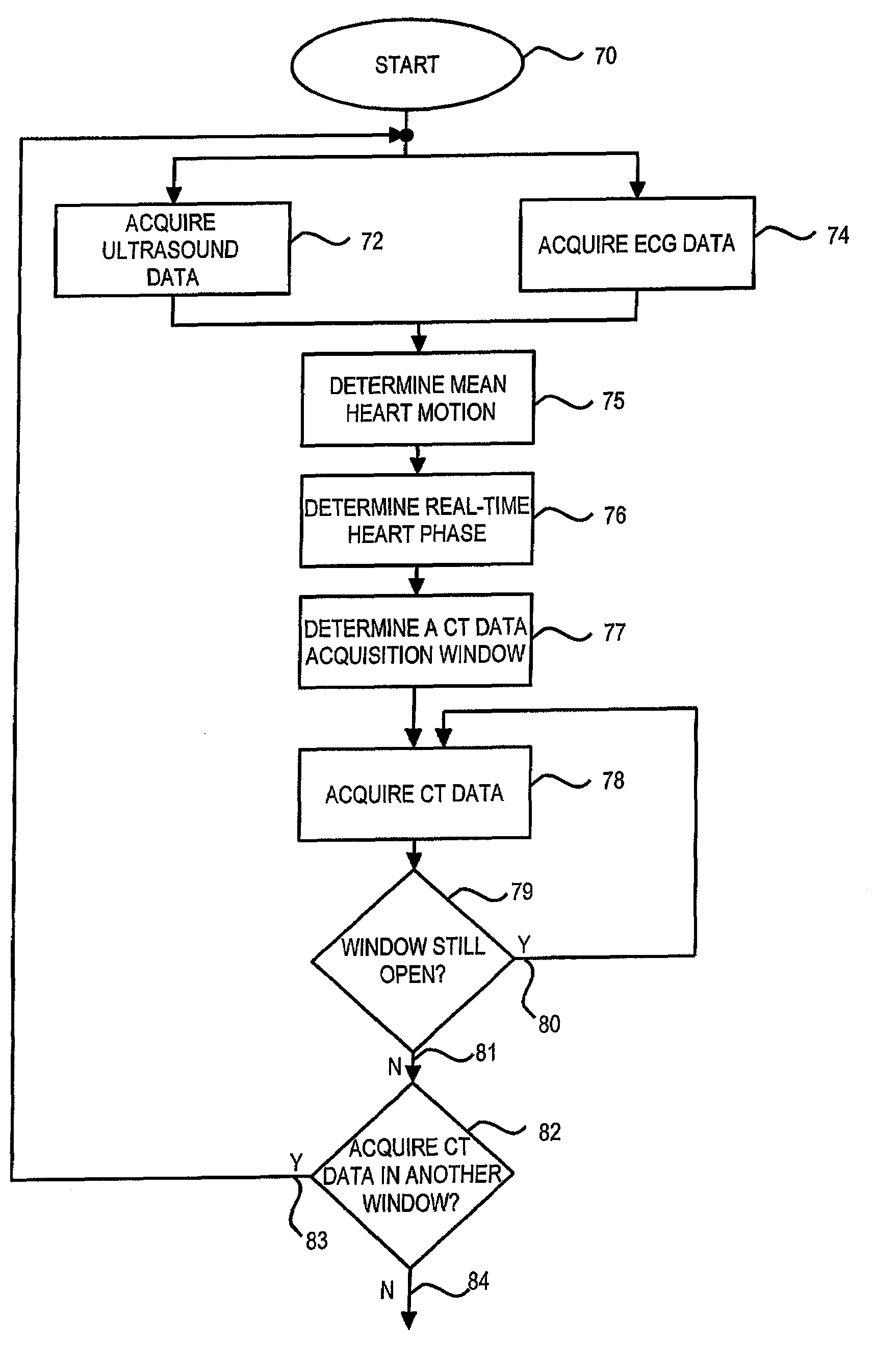
[0018]The present invention is directed to a method and apparatus for acquiring heart motion and heart phase data, and using this data to prospectively acquire CT imaging data of the heart. The heart motion data and heart phase data are also used for motion correction or compensation of the imaging data and for facilitating reconstruction of a CT image.

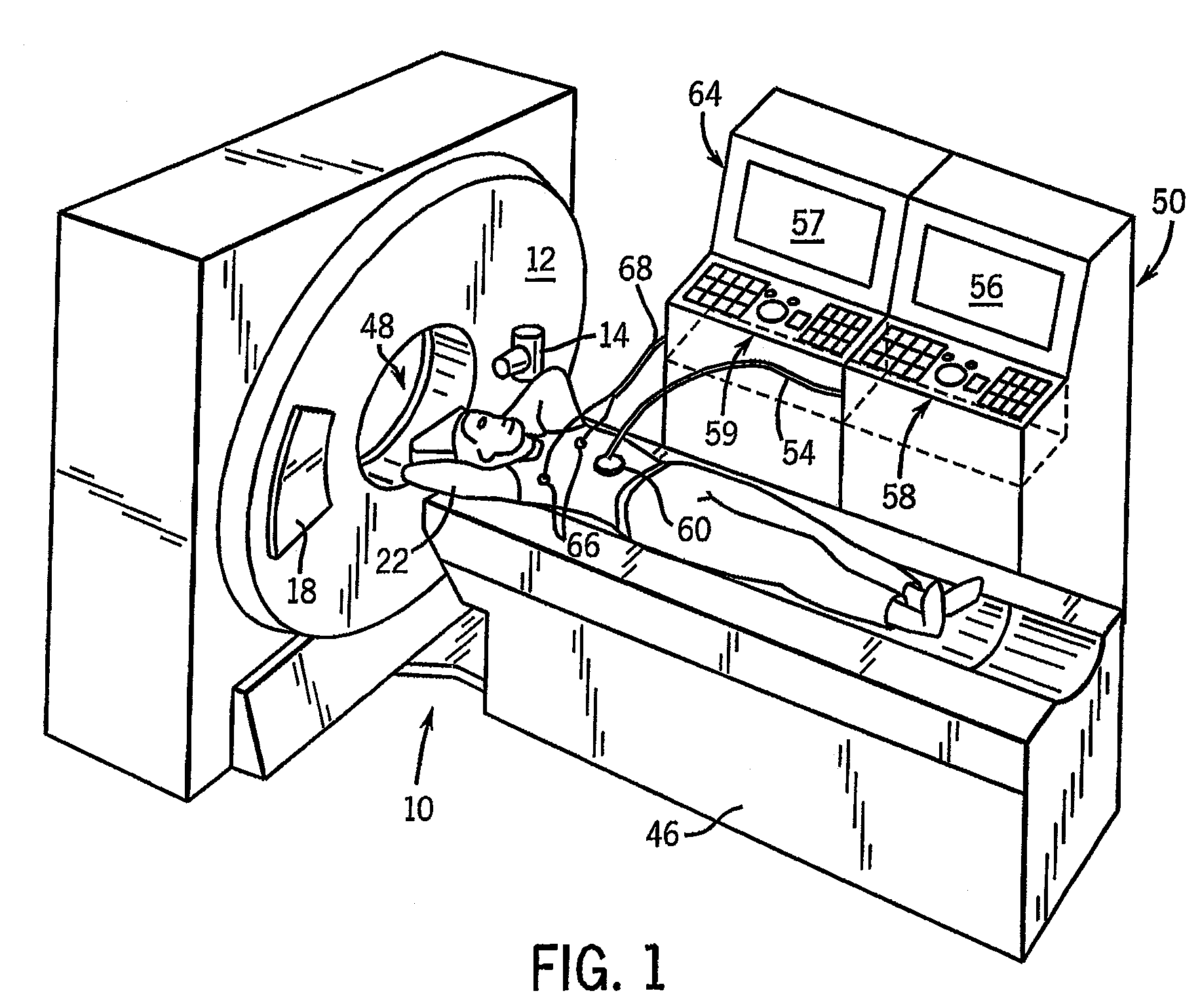
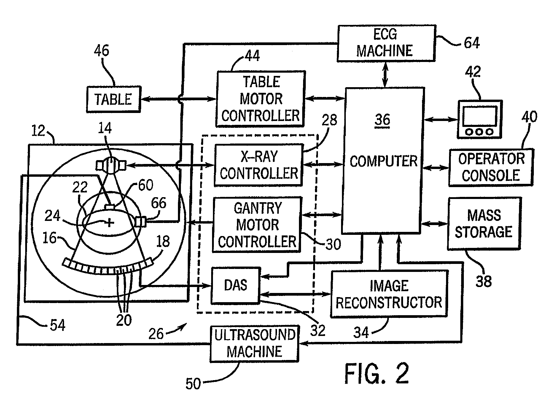
[0019]The present invention will be described with respect to a “third generation” CT scanner, but is equally applicable to other generations of CT system, as well as with other imaging modalities. Moreover, the present invention will be described with respect to an imaging system that includes a CT scanner that acquires image data, an ECG machine that acquires cardiac electrical data, and an ultrasound machine that acquires cardiac motion data from a patient. The CT scanner, ECG machine, and ultrasound machine are stand-alone devices that can be used independently from one another, but, as will be described, are configured to operate...


PUM


Abstract

ECG and ultrasound data of the heart are acquired in real-time during a scan. A data acquisition module is controlled during the scan to prospectively gate acquisition of CT data as a function of the real-time ECG data and the real-time ultrasound data. An image is reconstructed from the acquired CT data.

Description

BACKGROUND OF THE INVENTION[0001]The present invention relates generally to diagnostic imaging and, more particularly, to correcting motion errors in imaging data acquired from an object prone to motion.[0002]Various imaging modalities are useful to image objects in or prone to motion, such as the heart in cardiac studies. For example, in computed tomography (CT), magnetic resonance imaging (MRI) and other imaging modalities directed to the acquisition of data from an object prone to motion, one or more motion correction techniques are generally used to reduce motion-induced artifacts in the reconstructed images. In known studies, this motion correction or compensation can add significant complexity in post processing of the images.[0003]In one specific example, CT imaging requires measurement of more than 180 degrees of projection data to formulate an image. Because of various limitations in conventional CT scanners, the time necessary to collect a complete set of projection data i...

Claims


Application Information

Patent Timeline
Patent Type & Authority Patents(United States)
IPC IPC(8): A61B6/00
CPCA61B6/541A61B8/08
Inventor TKACZYK, JOHN ERICEDIC, PETER MICHAELLEBLANC, JAMES W.
Owner GENERAL ELECTRIC CO
Why Eureka
Try Eureka