Antibodies which bind B7RP1

a technology of b7rp1 and antibodies, applied in the field of polypeptides, can solve the problems of dramatically high t-cell levels in the ctla-4 gene of the mime, and achieve the effect of inhibiting t-cell proliferation

Inactive Publication Date: 2008-10-14
AMGEN INC
View PDF63 Cites 42 Cited by
  • Summary
  • Abstract
  • Description
  • Claims
  • Application Information

AI Technical Summary

Benefits of technology

[0063]FIG. 15. T-cell co-stimulation by B7RP-1-Fc fusion protein. A) T-cell proliferation induced by different quantities of B7RP-1-Fc (closed squares), B7.2-Fc (closed circles), or OPG-Fc fusion protein control (open squares) in conjunction with anti-CD3 antibody. Fusion proteins were used at various concentrations to coat 96 well plates pre-coated with anti-human Fc FAb2 (12.5 μg/ml) and anti-CD3 antibody (0.9 μg/ml). B7RP-1-Fc and B7.2-Fc co-stimulate T-cells in a dose-dependent fashion up to 0.3 μg/ml, at which the maximal effect is achieved. B) T-cell proliferation induced by B7RP-1-Fc (closed squares), B7.2-Fc (closed circles), non-fused Fc (open squares), or no Fc (open circles) in conjunction an anti-CD3 antibody (0.85 μg/ml) and in the presence of various concentrations of a rabbit anti-B7RP-1-Fc polyclonal antibody. Fc fusion proteins were

Problems solved by technology

Mice without the CTLA-4 gene exhibit dramatically high levels of T-cells, since the s

Method used


Image

  • Antibodies which bind B7RP1
  • Antibodies which bind B7RP1
  • Antibodies which bind B7RP1

Examples


example 1

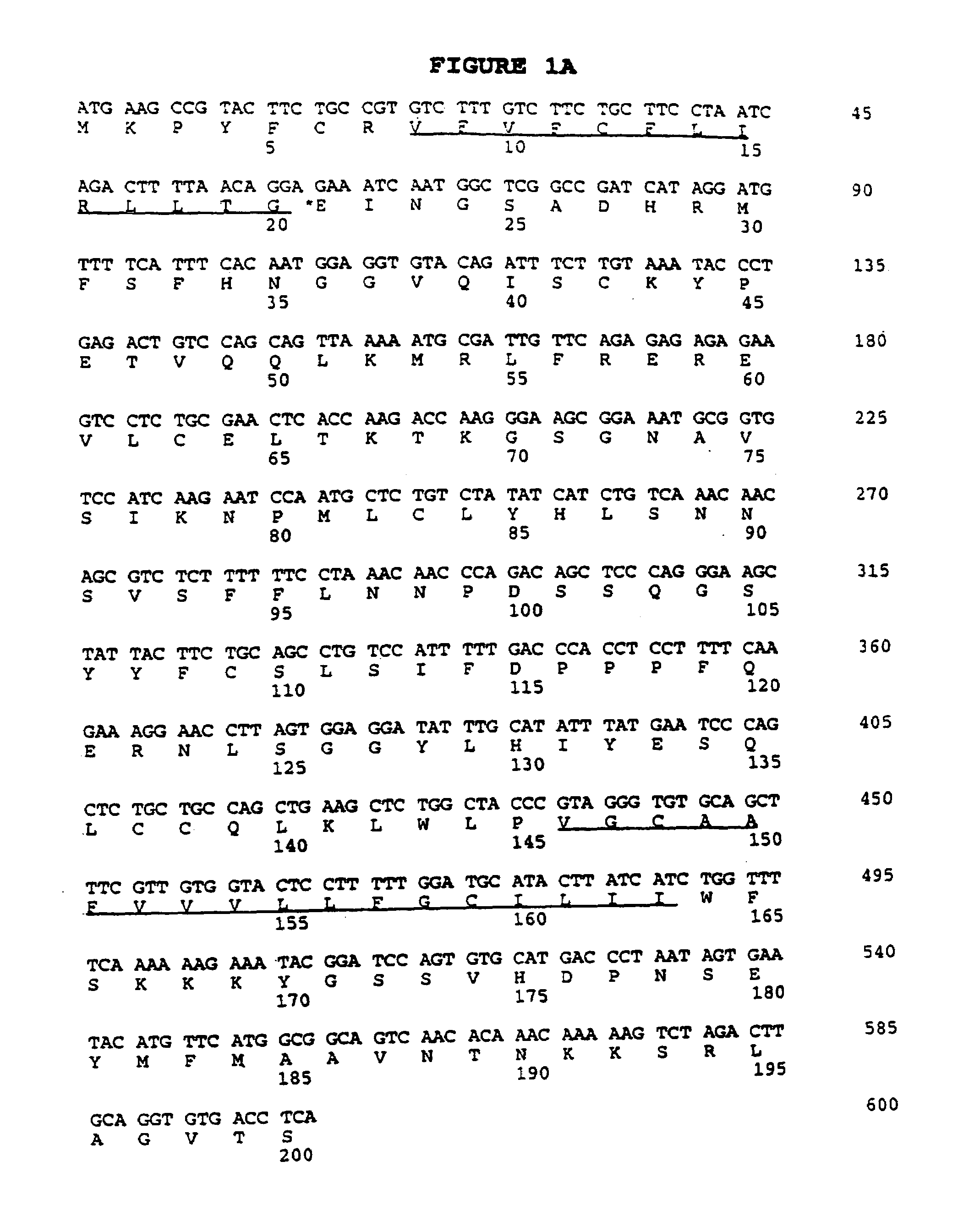
CRP1 cDNA and Amino Acid Sequence

[0204]Female C57 / Black 6 mice were sacrificed, and the small intestines were excised, and the Peyer's patches were removed. The small intestine tissue was sliced open and washed to remove mucus and other debris. The epithelial layer, which contains the intestinal intraepithelial cells (iIELs), was released by gentle agitation in RPMI-1640 supplemented with 1 mM dithiothreitol (DTT), for 20 minutes at 37° C. Disassociated cells were passed through a 100μ filter, washed in 50 ml of RPMI-1640, mixed to further break up clumps of cells, and then passed through a 40μ strainer to obtain single cell populations. These cells were then washed again in a 50 ml volume of RPMI-1640 to ensure the removal of the residual DTT. The tissue was then agitated and washed as before to gather the remaining iIELs. The iIELs were separated from the adipose cells and most epithelial cells on a 3-step Percol gradient, with the iIELs banding at the 40% to 80% interface. These ...

example 2

Cloning of Human CRP1 cDNA

[0206]The nucleic acid sequence encoding human CRP1 protein is identified by the following procedures. A human cDNA library was prepared from enriched lymphocytes from peripheral human blood from normal human volunteers. The lymphocytes were purified and red blood cells were removed by Lymphocyte Separation Media (ICN Pharmaceuticals, Inc., Costa Mesa, Calif.). The cells were then activated overnight in media containing 10 ng / ml PMA, 500 ng / ml ionomycin, and plate-bound activating antibodies to CD3. Total RNA was prepared from the activated cells by the Trizol method (Gibco / BRL) and poly A RNA was isolated by Dynal bead purification. cDNA was made from the isolated poly A RNA and size selected for largest cDNA fragments. The size selected cDNA was then ligated into the plasmid pSPORT (Gibco / BRL). DNA encoding human CRP1 protein is obtained by screening the activated lymphocyte cDNA library by either recombinant bacteriophage plaque, or transformed bacteria ...

example 3

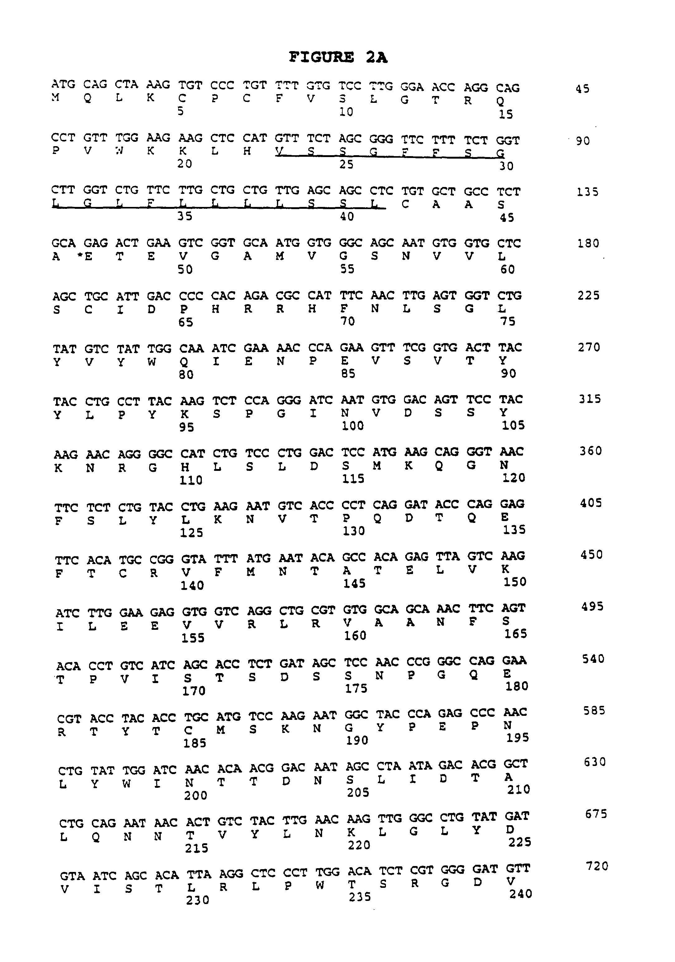
B7RP1 DNA and Amino Acid Sequence

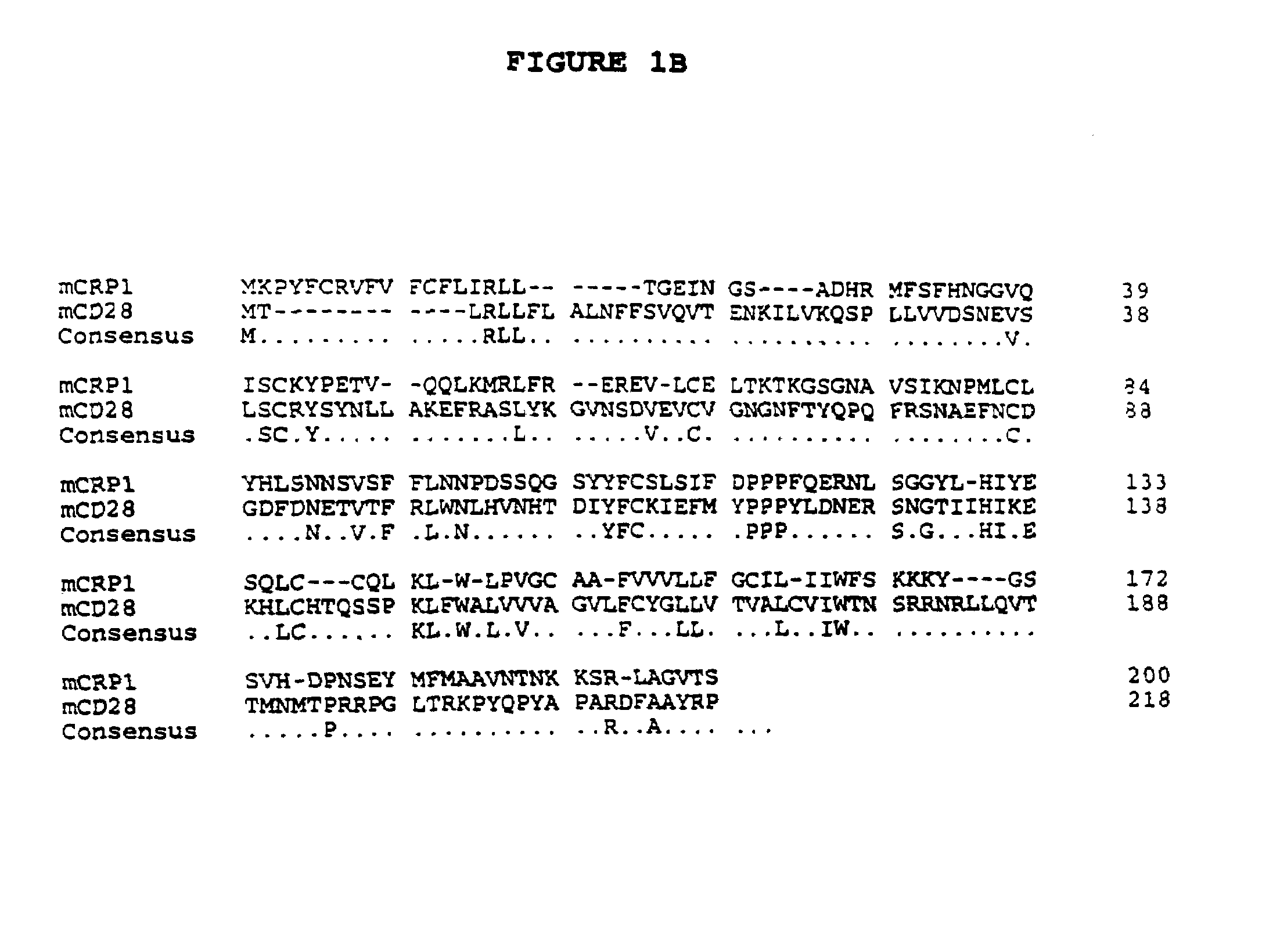
[0207]A cDNA clone, designated smil1-00003-g5, contained nucleotide sequence homology to B7.1 (CD80) and B7.2 (CD86). Translation of the sequence (FIG. 2A) and subsequent comparison to known proteins in a public database revealed 20% amino acid identity with murine B7.1 (FIG. 2B). This low homology was significant because murine B7.1 shares only 24% amino acid identity with murine B7.2. Despite this low homology, critical cysteine residues are conserved between the open reading frame of this clone and murine B7.1 and B7.2 at residues 62, 138, 185, and 242 (relative to the initiating methionine, FIG. 2B). The approximate mature protein length and the location of the transmembrane region relative to the carboxy terminus are also similar in the putative ORF of this clone, as compared to B7.1 and B7.2. We named the gene B7RP1, for B7-Related Protein-1.


PUM

PropertyMeasurementUnit
Currentaaaaaaaaaa
Currentaaaaaaaaaa
Currentaaaaaaaaaa

Abstract

Novel polypeptides which comprise a receptor-ligand pair involved in T-cell activation are disclosed. Nucleic acid molecules encoding said polypeptides, and vectors and host cells for expressing same are also disclosed. The polypeptides, or agonists and antagonists thereof, are used to treat T-cell mediated disorders.

Description

[0001]This application is a Section 371 filing of PCT / US00 / 01871 filed Jan. 27, 2000, now published as WO00 / 46240, which is a continuation-in-part of U.S. Ser. No. 09 / 264,527, filed Mar. 8, 1999 now abandoned, which is a continuation-in-part of U.S. Ser. No. 09 / 244,448, filed Feb. 3, 1999 now abandoned.FIELD OF THE INVENTION[0002]The present invention relates to polypeptides that are involved in T-lymphocyte activation. Specifically, the invention relates to T-lymphocyte costimulatory polypeptides, the nucleic acids encoding the polypeptides, expression vectors and host cells for production of the polypeptides, and compositions and methods for the treatment of diseases related to immunosuppression and immune activation.BACKGROUND OF THE INVENTION[0003]For the generation of a proper T-lymphocyte (T-cell) immune response, two signals must be provided to the T-cell by antigen presenting cells (APC). First, antigen must be presented to the T-cell receptor (TCR) via a major histocompatib...

Claims


Application Information

Patent Timeline
IPC IPC(8): C07K16/00A61K39/395C07K16/28
CPCC07K16/2827C07K2316/96C07K2319/30C07K2317/73C07K2317/76
Inventor YOSHINAGA, STEVEN KIYOSHI
Owner AMGEN INC
Why Eureka
Try Eureka