Early detection of sepsis

a sepsis and early detection technology, applied in the field of early detection of sepsis, can solve the problems of complex process, high cost, and high complexity of early detection of disease symptoms, and achieve the effect of reducing the number and heterogeneity of factors, and accelerating the process of sepsis detection

Active Publication Date: 2008-12-16
BECTON DICKINSON & CO
View PDF14 Cites 22 Cited by
  • Summary
  • Abstract
  • Description
  • Claims
  • Application Information

AI Technical Summary

Benefits of technology

[0032]As discussed above, the course by which a patient progresses to death from sepsis is well-known and has been described as a continuum from a state termed systemic inflammatory response syndrome (SIRS) to successive states of sepsis, severe sepsis, septic shock, multiple end-organ failure (MODS), and death. The present methods

Problems solved by technology

Unfortunately, the early detection of disease symptoms in many instances is problematic, and a disease may be relatively advanced before clinical suspicion of the disease occurs.
Characterizing sepsis in a host, however, is made very complex by the number and heterogeneity of factors that play into the final outcome.
The process by which this occurs is often remarkably rapid, leaving the clinician with little time to make a considered clinical judgment.
More significant yet is the high morbidity and mortality associated with sepsis.
Diagnosing sepsis at a point in time when the clinician can intervene with preventive measures has been and continues to be a very challenging task.
Investigators point out that disease definitions that are too broad limit the ability of clinicians to direct appropriate therapies at patients who are at high risk for developing sepsis.
In addition, these definitions do not permit the clinician to differentiate between an at risk patient who may derive a net benefit from a new, expensive therapy and a patient who will either not benefit, given his/her underlying disease co-morbidities, or who may be placed at higher risk from the therapy's inherent safety profile.
Because many microorganisms require specialized conditions to promote their rapid growth, and may be more commonly present in certain body microenvironments, and because microbe numbers may initially be low at the site of infection, cultures are not always positive, even when a microorganism may be present.
Moreover, cultures can also

Method used


Image

  • Early detection of sepsis

Examples


example 1

Preferred Protocols

Measurement of Cell-Surface Markers by Flow Cytometry

[0089]Cell-surface markers, HLA-DR, CD64, and CD11b, are assayed by flow cytometry. Flow cytometry is carried out using a BD FACSCalibur™ flow cytometry system and BD FACStation™ data management system with BD FACSComp™ software, version 4.0, and BD CellQuest™ software, version 3.1.

[0090]In flow cytometric assays for a cell-surface marker, peripheral blood cells first are stained using fluorescently labeled monoclonal antibodies specific for the marker. Blood is well mixed prior to staining, and staining is carried out within four hours of the blood draw. Stained cells are kept cold and in the dark prior to data acquisition, and data acquisition is carried out within two hours of the addition of BD FACS™ Lysing Solution to the samples.

[0091]Both HLA-DR and CD64 are measured from blood collected in K3EDTA collection tubes, whereas CD11b is measured in blood collected in a Na Heparin collection tube.

[0092]The resu...

example 2

Clinical Trials

Selection of SIRS Patients and Controls

[0104]Three separate patient groups and one group of normal donors were involved in the following examples. All patients were 18 years of age or older, and appropriate informed consent was obtained in all cases.

[0105]A total of 50 patients at risk for sepsis were selected from patients admitted to the Surgical Intensive Care Unit (SICU) of the Robert Wood Johnson Medical School Hospital in New Brunswick, N.J. Patients were classified as at risk for sepsis if they met two or more SIRS criteria (i.e., were SIRS positive) within a 24 hr period during their stay in the SICU.

[0106]An additional 15 patients were selected who were scheduled for clean, elective surgeries, and who were SIRS-negative at the start of monitoring, which was prior to surgery. This grou served as hospitalized controls (also referred to herein as the “clean surgical control group”).

[0107]An additional group of 12 normal donor volunteers were selected. This group...

example 3

Analysis of Clinical Trial Data

[0121]The data obtained from the clinical trial described in Example 2 were analyzed as described below.

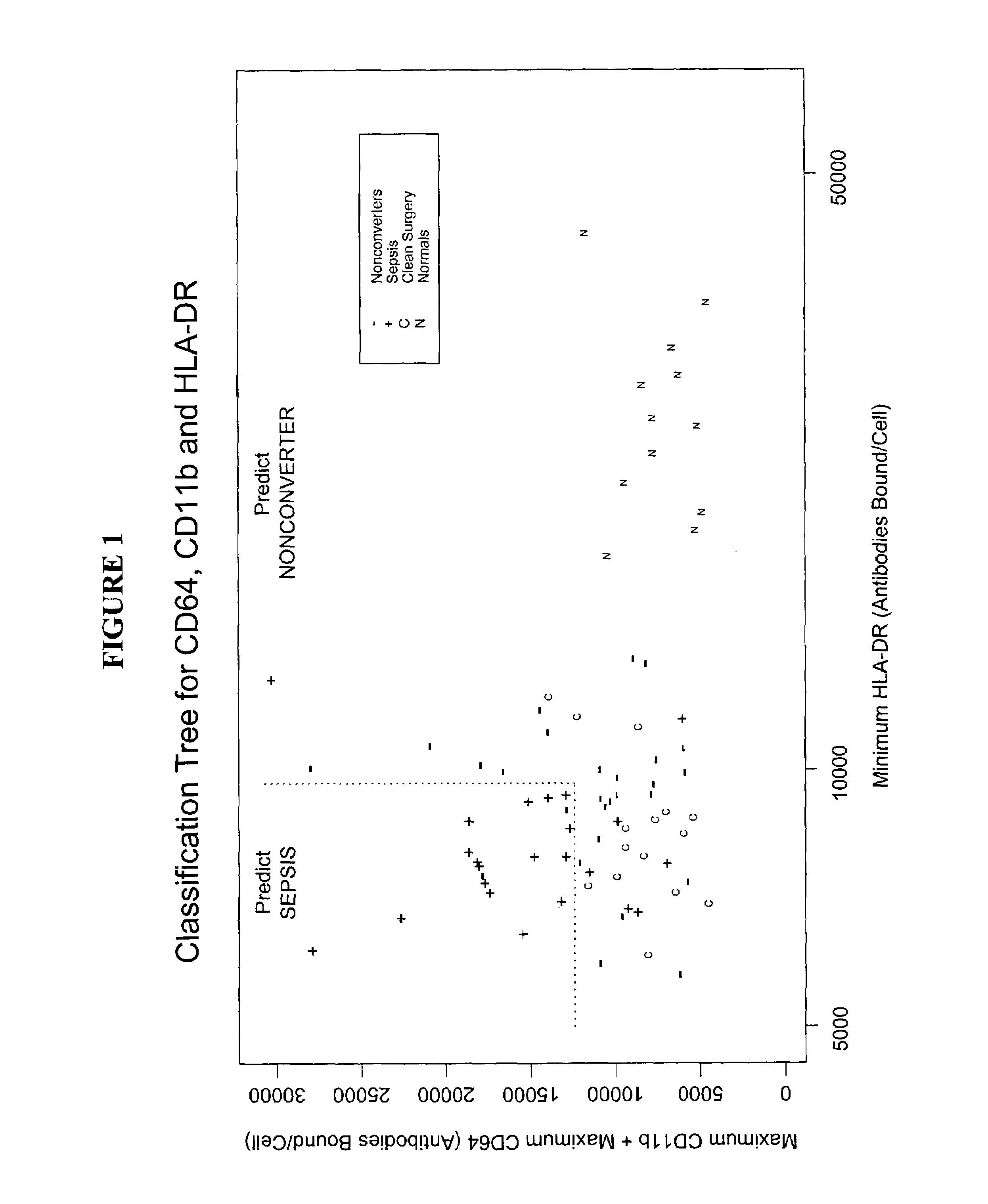
[0122]The following definitions are used throughout the examples to refer to subsets of patients based on their clinical status:[0123]Group 1: SIRS patients who never developed clinical suspicion of sepsis while in the SICU. These patients are referred to as “non-converters.” Of the 50 SIRS patients in the SICU, 27 were non-converters.[0124]Group 2: SIRS patients who developed clinical suspicion of sepsis while in the SICU. These patients are referred to as “converters.” Of the 50 SIRS patients in the SICU, 23 were converters. Of the 23 converters, 20 were confirmed as septic by cultures (culture-positive sepsis) and 3 could not be confirmed by culture (culture-negative sepsis).[0125]Group 3: The 15 patients in the clean surgical control group.[0126]Group 4: The 12 normal donor volunteers, i.e., the non-hospitalized control group.

[0127]The number of ...


PUM


Abstract

Diagnostic methods, systems, and kits for identifying patients with systemic inflammatory response syndrome who are likely to progress to sepsis.

Description

[0001]This application claims priority under 35 U.S.C. § 119(e) of provisional application Ser. No. 60 / 370,115, filed Apr. 2, 2002, which is incorporated herein by reference.BACKGROUND OF THE INVENTION[0002]1. Field of the Invention[0003]The present invention relates to methods and reagents for identifying patients at risk for developing a systemic inflammatory condition, such as sepsis.[0004]2. Description of Related Art[0005]Early detection of the presence of disease conditions in patients typically allows more effective therapeutic treatments and results in more favorable clinical outcomes than occur when disease conditions are not recognized until an advanced stage. Unfortunately, the early detection of disease symptoms in many instances is problematic, and a disease may be relatively advanced before clinical suspicion of the disease occurs.[0006]Systemic inflammatory conditions represent one class of diseases for which early diagnosis is particularly desirable, with sepsis bein...

Claims


Application Information

Patent Timeline
IPC IPC(8): G01N33/00G01N33/53G01N33/532G01N33/566G01N33/567G01N33/49G01N33/68
CPCG01N33/6893G01N2333/70535G01N2333/70553G01N2333/70596G01N2800/26
Inventor ANDERSON, STEPHEN J.HANEY, DOUGLAS J.WATERS, CORY A.
Owner BECTON DICKINSON & CO
Why Eureka
Try Eureka