

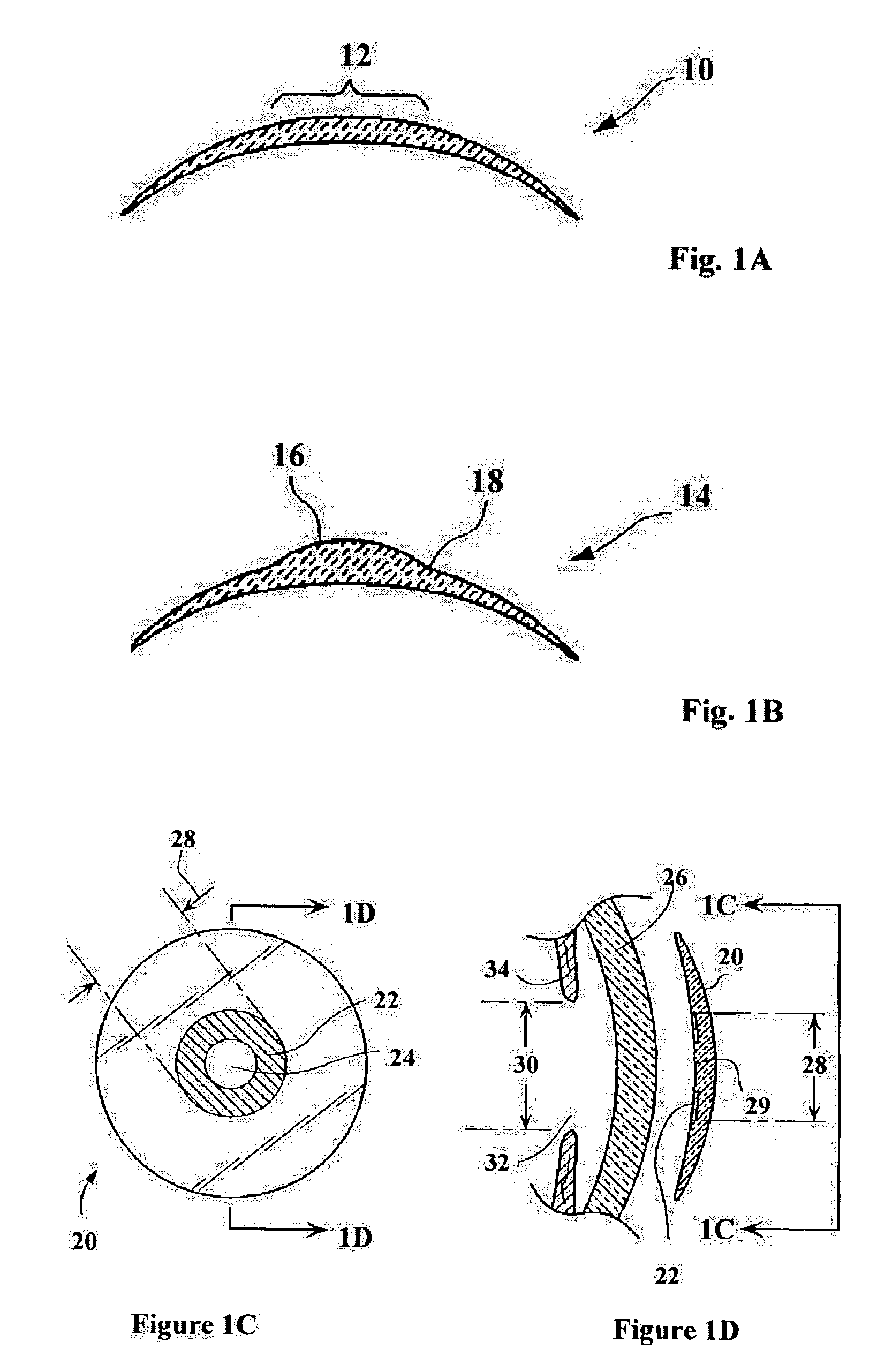
Methods for producing epithelial flaps on the cornea and for placement of ocular devices and lenses beneath an epithelial flap or membrane, epithelial delaminating devices, and structures of epithelium and ocular devices and lenses
a technology of epithelial flaps and corneas, applied in the field of ophthalmology, can solve the problems of limited use of epithelial flaps, setbacks of synthetic onlays, and insufficient healing of epithelial cells on these lenses, and achieve the effect of enhancing the delamination process
- Summary
- Abstract
- Description
- Claims
- Application Information
AI Technical Summary
Benefits of technology
Problems solved by technology
Method used
Image

Examples
example
[0156]A mechanical epithelial delamination was performed using a device similar to those shown in FIGS. 4A, 4B, 4C, 4D, and 4E, in that it had a yoke assembly with a yoke arms that drooped and included a dissecting wire having a diameter of 2 mil. (0.002 inch). The yoke was vibrated along the axis of the handle at about 250 Hz. A jig as shown in FIGS. 5A-5E was placed on the anterior surface of a freshly harvested pig eye (6 hours post-mortem) and vacuum was applied.
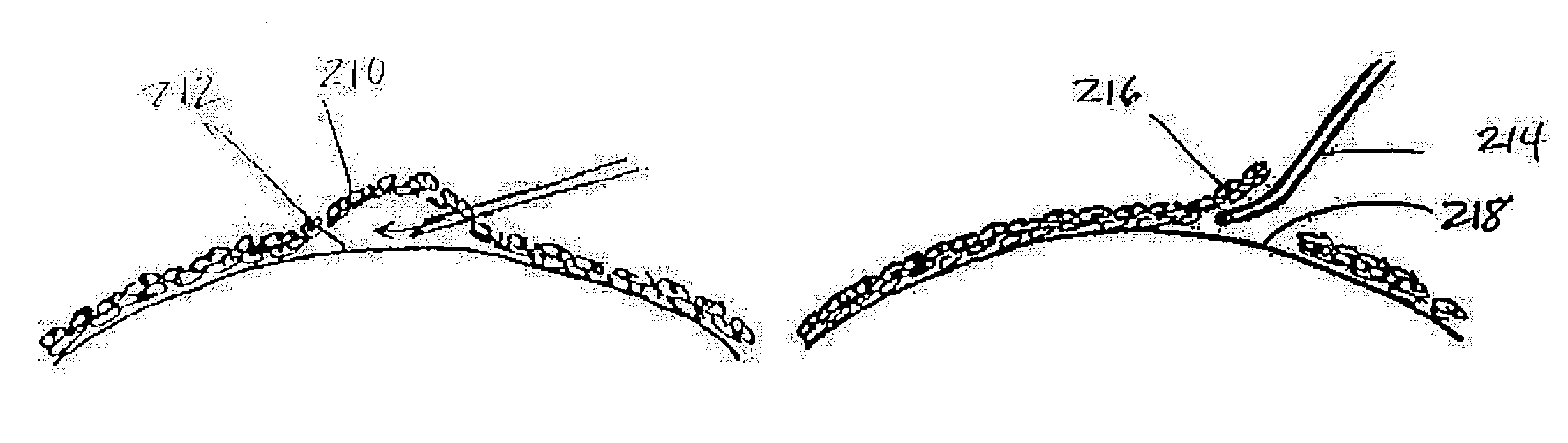
[0157]The wire was passed through the jig beneath the epithelium and a flap of the epithelium was raised. Histological samples of the remaining corneal surface, the raised epithelium, and the limn where the “hinge” of the flap met the cornea were taken. FIG. 8A shows the dissected and raised epithelium (600), the cornea (602), and the junction (604) where the dissection was terminated. FIG. 8B shows, in higher magnification, the junction (604) and further displays the precision of the blunt mechanical dissection: there i...
PUM
 Abstract
Description
Claims
Application Information