Devices and methods for percutaneous repair of the mitral valve via the coronary sinus

a technology of mitral valve and coronary sinus, which is applied in the field of endovascular devices and methods for interventional repair of the mitral valve via the coronary sinus, can solve the problems of incomplete coaptation, tethering of the mitral and tricuspid leaflets, etc., and achieve the effect of reducing the distance between the proximal and the distal anchor

Active Publication Date: 2009-03-10
EDWARDS LIFESCIENCES CORP
View PDF103 Cites 237 Cited by
  • Summary
  • Abstract
  • Description
  • Claims
  • Application Information

AI Technical Summary

Benefits of technology

[0012]In yet another alternate embodiment, the device includes a locking mechanism having transverse beam structures. A cinching thread having a series of conical elements is woven through the locking mechanism, allowing for the distance between a proximal and distal anchor to be shortened.

Problems solved by technology

These geometric alterations result in mitral and tricuspid leaflet tethering and incomplete coaptation during systole.

Method used


Image

  • Devices and methods for percutaneous repair of the mitral valve via the coronary sinus
  • Devices and methods for percutaneous repair of the mitral valve via the coronary sinus
  • Devices and methods for percutaneous repair of the mitral valve via the coronary sinus

Examples


Embodiment Construction

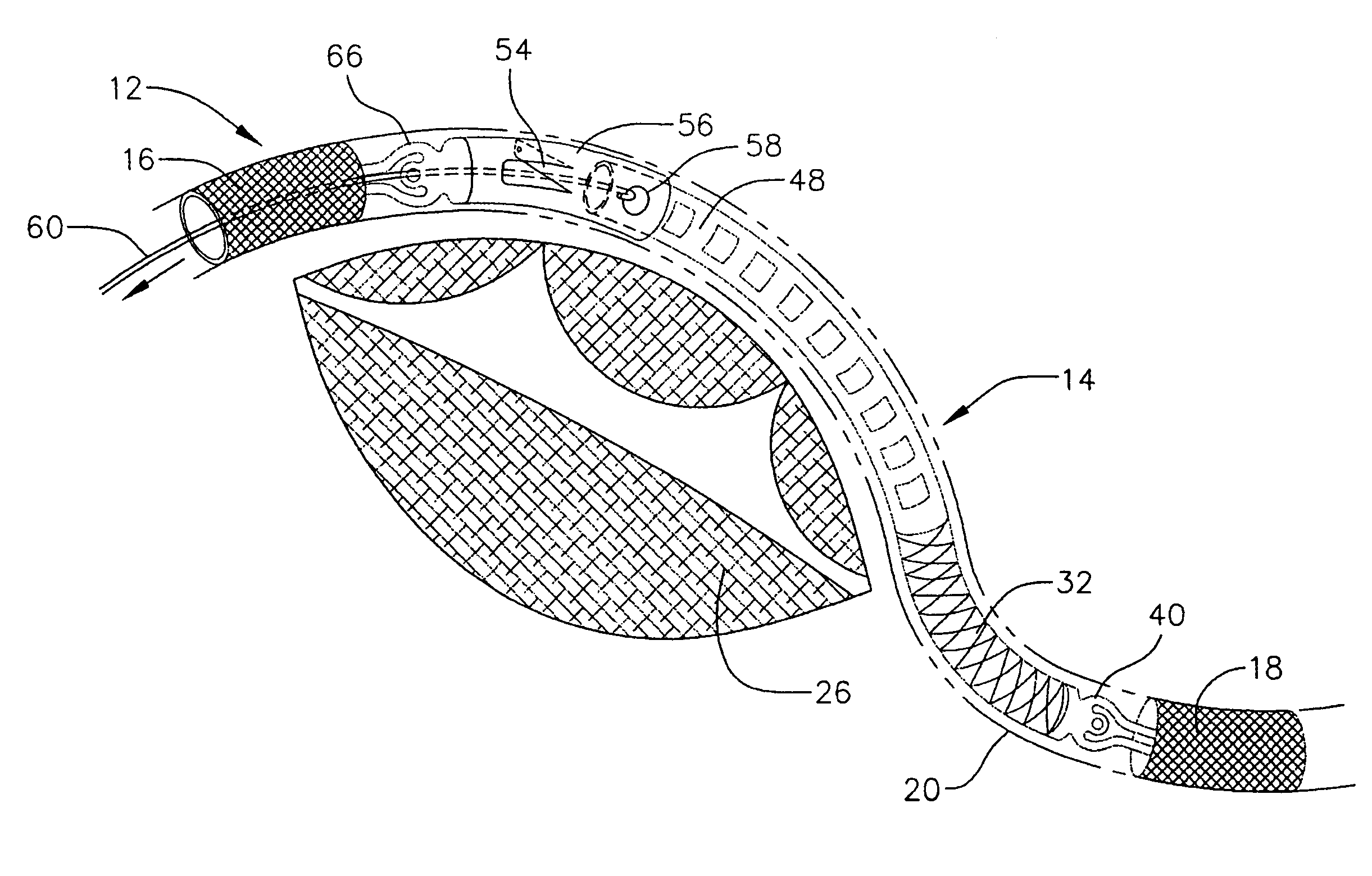
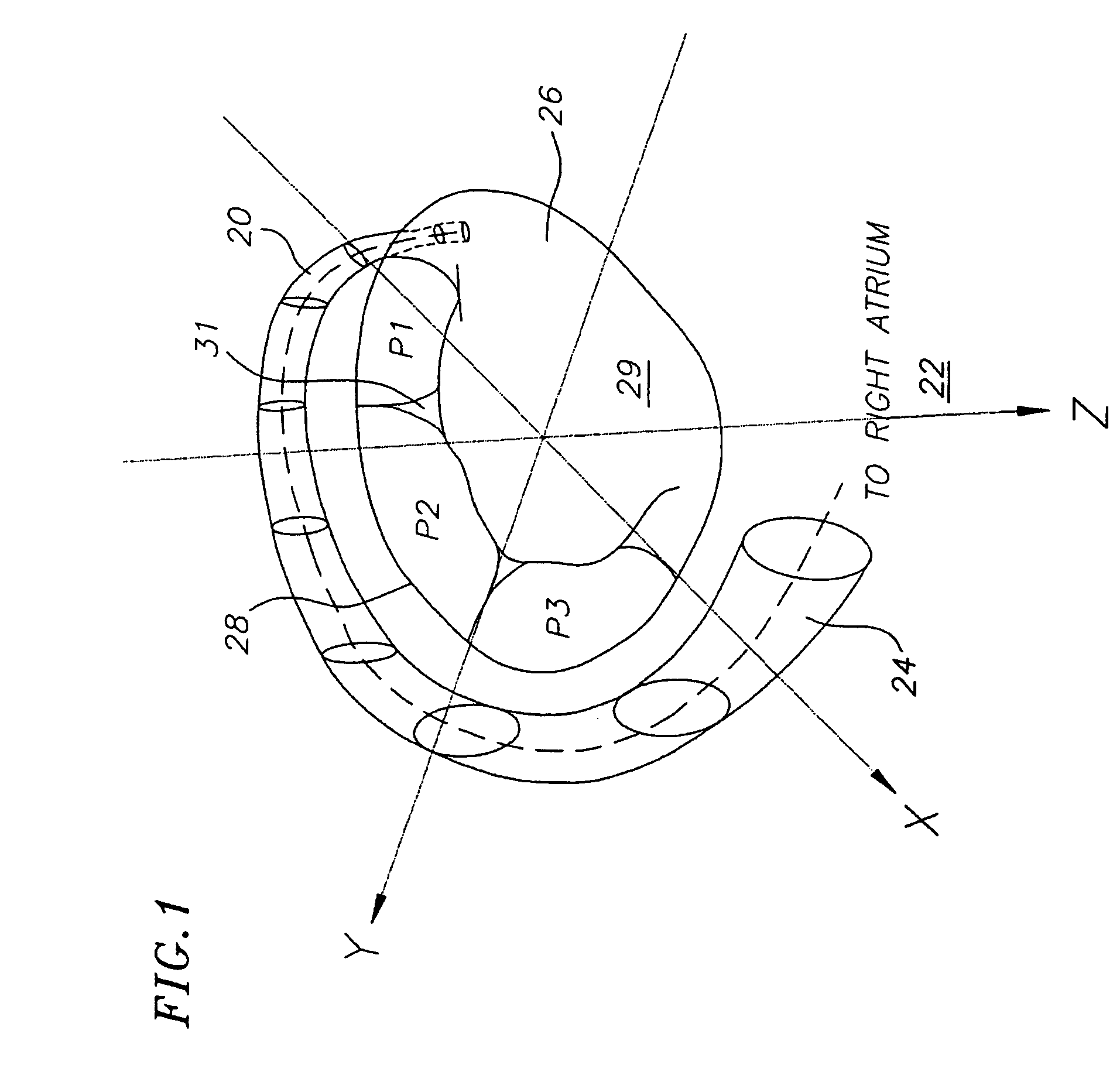
[0043]With reference now to FIG. 1, a coronary sinus 20 extends from a right atrium 22 and a coronary ostium 24 and wraps around a mitral valve 26. The term coronary sinus 20 is used herein as a generic term to describe a portion of the vena return system that is situated adjacent to the mitral valve 26 along the atrioventricular groove. The term coronary sinus 20 used herein generally includes the coronary sinus, the great cardiac vein and the anterior intraventricular vein. A mitral annulus 28 is a portion of tissue surrounding a mitral valve orifice to which several leaflets attach. The mitral valve 26 has two leaflets, namely an anterior leaflet 29 and a posterior leaflet 31. The posterior leaflet includes three scallops P1, P2 and P3.

[0044]The problem of mitral regurgitation typically results when a posterior aspect of the mitral annulus 28 dilates (i.e., enlarges), thereby displacing one or more of the posterior leaflet scallops P1, P2 or P3 away from the anterior leaflet 29. ...


PUM


Abstract

Devices and methods for treating mitral regurgitation by reshaping the mitral annulus in a heart. One preferred device for reshaping the mitral annulus is provided as an elongate body having dimensions as to be insertable into a coronary sinus. The elongate body includes a proximal frame having a proximal anchor and a distal frame having a distal anchor. A ratcheting strip is attached to the distal frame and an accepting member is attached to the proximal frame, wherein the accepting member is adapted for engagement with the ratcheting strip. An actuating member is provided for pulling the ratcheting strip relative to the proximal anchor after deployment in the coronary sinus. In one preferred embodiment, the ratcheting strip is pulled through the proximal anchor for pulling the proximal and distal anchors together, thereby reshaping the mitral annulus.

Description

BACKGROUND OF THE INVENTION[0001]1. Field of the Invention[0002]This invention generally relates to devices and methods for heart valve repair and, more particularly, to endovascular devices and methods for interventional repair of the mitral valve via the coronary sinus.[0003]2. Description of the Related Art[0004]Heart valve regurgitation occurs when a heart valve does not close tightly, thereby allowing blood to flow backward in the heart. Heart valve regurgitation typically occurs in the mitral valve, located between the left atrium and left ventricle, or in the tricuspid valve, located between the right atrium and right ventricle. Regurgitation in the mitral valve may result from changes in the geometric configurations of the left ventricle, papillary muscles and / or mitral annulus. Similarly, regurgitation in the tricuspid valve is caused by changes in the geometric configurations of the right ventricle, papillary muscles and / or tricuspid annulus. These geometric alterations re...

Claims


Application Information

Patent Timeline
Patent Type & Authority Patents(United States)
IPC IPC(8): A61F2/24
CPCA61F2/2451
Inventor SOLEM, JAN OTTOKIMBLAD, PER OLAFARIABI, SEPEHRSCHRECK, STEFANADZICH, VASOIANCEA, OCTAVIAN
Owner EDWARDS LIFESCIENCES CORP
Why Eureka
Try Eureka