Biological tissue elasticity measurement method and ultrasonographic device

a biochemical tissue and elasticity technology, applied in the field of biochemical tissue elasticity measurement method and ultrasonographic equipment, can solve the problems of not considering a measurement method for acquiring high-quality form and elasticity images, and achieve the effects of improving the quality of form images, elasticity ratio and elasticity distortion

Inactive Publication Date: 2009-04-21
HITACHI LTD
View PDF9 Cites 14 Cited by
  • Summary
  • Abstract
  • Description
  • Claims
  • Application Information

AI Technical Summary

Benefits of technology

[0007]In this case, an ultrasonic transmission signal is applied which has an amplitude, wave number or frequency preferable for each of the form image measurement and the elasticity image measurement as the ultrasonic transmission signal for each of the measurement. More specifically, there may be provided a first transmission signal generating unit for generating an ultrasonic transmission signal for the form image, and a second transmission signal generating unit for generating an ultrasonic transmission signal for the elasticity image having at least one ultrasonic wave among an ultrasonic wave having a larger amplitude, an ultrasonic wave having a larger wave number and an ultrasonic wave having a lower frequency than those of the ultrasonic transmission signal for the form image. As a result, the image quality of the form image such as a B-mode image and the quality of the elasticity image such as an elasticity ratio and an elasticity distortion can be improved, and images suitable for both of the diagnoses can be provided.

Problems solved by technology

However, the ultrasonographic apparatus disclosed in the publication does not consider a measurement method for acquiring both high quality form and elasticity images.

Method used


Image

  • Biological tissue elasticity measurement method and ultrasonographic device
  • Biological tissue elasticity measurement method and ultrasonographic device
  • Biological tissue elasticity measurement method and ultrasonographic device

Examples


first embodiment

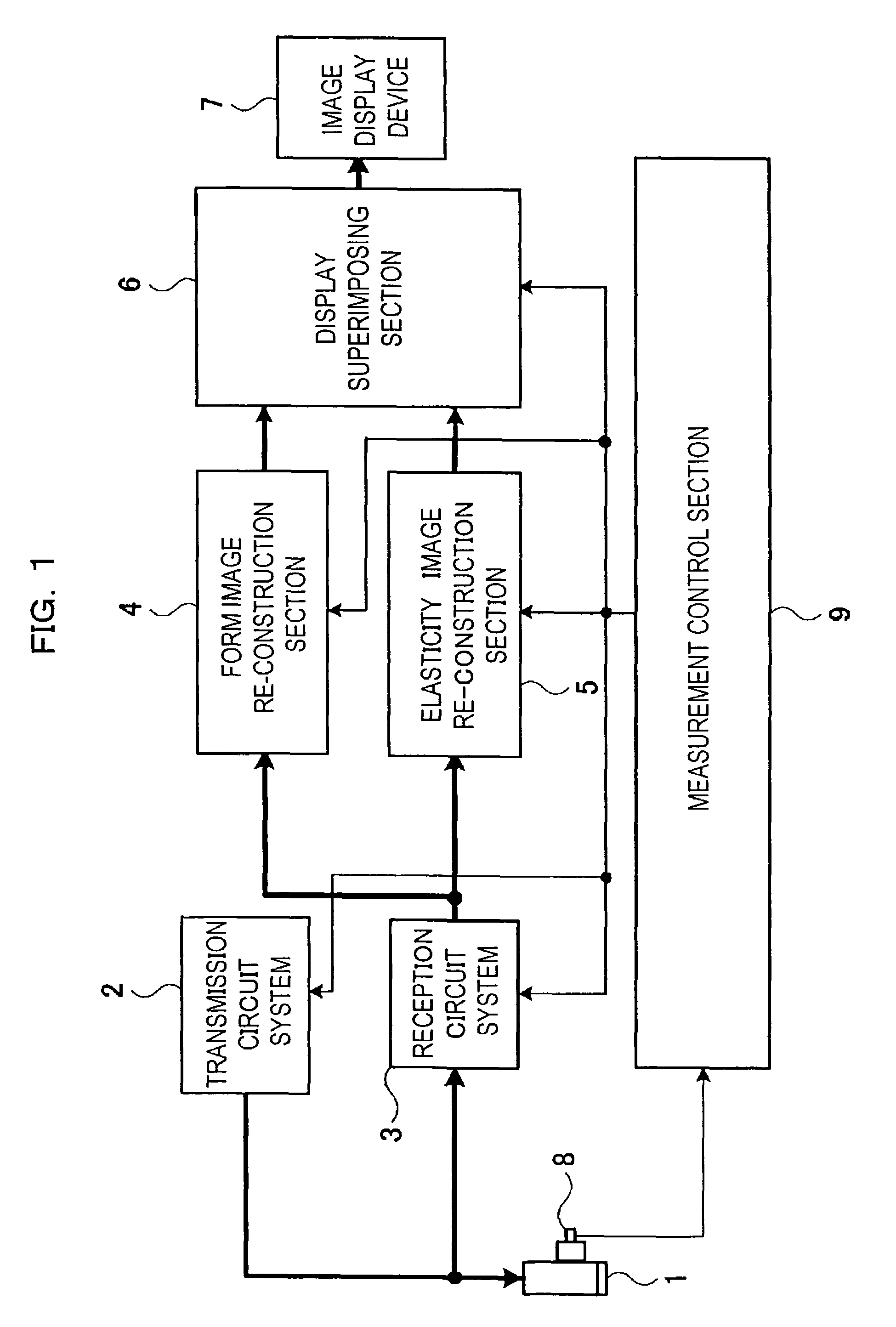
[0025]Here, the first embodiment relating to control of the elasticity diagnosis mode to be performed by the measurement control section 9 will be described with reference to FIGS. 3 to 5. When an operator presses and turns on the switch 8 associated with the ultrasonic probe 1 in order to start an elasticity diagnosis, this is input to the measurement control section 9 as an elasticity diagnosis mode start command. When the switch 8 is released and turned off, this is input to the measurement control section 9 as an elasticity diagnosis mode end command. Based on this, the measurement control section 9 controls the starting and ending of the elasticity diagnosis mode for performing a compound measurement of the elasticity image measurement and the form image measurement. On the other hand, in the elasticity diagnosis mode, the operator must perform an operation for pressing the ultrasonic probe 1 against the patient when the switch 8 associated with the ultrasonic probe 1 is turned...

second embodiment

[0037]Next, a second embodiment relating to control of an elasticity diagnosis mode to be performed by the measurement control section 9 will be described with reference to FIGS. 6 to 9. FIG. 6 is a timing chart of an operation. FIGS. 7 to 9 are flowcharts illustrating details of the operation. FIG. 6 shows, like FIG. 4, changes in states of “switch (8)”, “ultrasonic scanning position”, “transmission / reception sequence”, “ultrasonic transmission wave number(s)” and “reception of ultrasonic waves” in order from the top of the figure. The horizontal axis indicates the number of times of scanning, and DF and FF indicate a dynamic filter and a fixed filter, respectively. The meaning of the other symbols is identical to those of FIG. 4. This embodiment is different from FIG. 4 in that the form image measurement and elasticity image measurement are selectively switched not for each image frame but for each ultrasonic beam.

[0038]As shown in FIG. 6, during form image measurement B, an ultra...


PUM


Abstract

An ultrasonographic apparatus includes: an ultrasonic probe 1 for transmitting / receiving an ultrasonic wave to / from a patient, means 2 for generating an ultrasonic transmission signal and transmitting it to the ultrasonic probe, means 3 for receiving a reflection echo signal received by the ultrasonic probe; means 4 for re-constructing a form image according to the reception signal processed by the reception processing means; means 5 for re-constructing an elasticity image according to the reception signal processed by the reception processing means; means 7 for displaying the form image and the elasticity image; means 8 for switching between the form image mode and elasticity image mode; and means 9 for performing control so as to selectively acquire the form image and the elasticity image in the measurement period of the elasticity image mode switched by the mode switching means. Thus, it is possible to perform measurement associated with acquisition of both the form image and the elasticity image of the biological tissue and improve the image quality of the form image such as a B mode image and the image quality of the elasticity image such as elasticity ratio and elasticity distortion.

Description

TECHNICAL FIELD[0001]The present invention relates to a biological tissue elasticity measurement method and ultrasonographic apparatus, and, in particular, to a measurement method and ultrasonographic apparatus for acquiring a high quality elasticity image such as an elasticity ratio and an elasticity distortion of the biological tissue of a patient and a high quality form image of the biological tissue.BACKGROUND ART[0002]An ultrasonographic apparatus irradiates an ultrasonic wave to the patient by using an ultrasonic probe, measures the acoustic characteristic of a biological tissue within the patient by using the reflection echo signal, re-constructs a form image (such as a B-mode image and an M-mode image) of the biological tissue, for example, based on a difference or change in acoustic characteristic of an arbitrary area of the patient and displays the form image on a screen for a diagnosis.[0003]Recently, it has been proposed that the ultrasonographic apparatus is used to mea...

Claims


Application Information

Patent Timeline
Patent Type & Authority Patents(United States)
IPC IPC(8): A61B8/00A61B8/08G01S7/52G01S15/89
CPCA61B8/08A61B8/485G01S7/52042G01S7/52074G01S7/52085G01S15/8952G01S7/52071
Inventor TAMANO, SATOSHIOSAKA, TAKASHIOSHIKI, MITSUHIROMATSUMURA, TAKESHISHIINA, TSUYOSHI
Owner HITACHI LTD
Why Eureka
Try Eureka