
Collaborative medical imaging portal system

Inactive Publication Date: 2016-08-25
THANGARAJ VENKATESAN
View PDF1 Cites 18 Cited by
  • Summary
  • Abstract
  • Description
  • Claims
  • Application Information

AI Technical Summary

Benefits of technology

The invention is a system and method for processing patient clinical image data and meta data in a clinical trial setting. The system includes a processor, a trial-specific definition, configuration, and implementation of the system, an acquisition interface, a middle-tier server for verifying content, a conversion unit, a comparator, a data storage unit, a master indexing engine, a user interface module, a processing interface, a workflow engine, and a clinical efficacy data capture module. The system allows for seamless processing of image data and meta data according to trial-specific configurations, provides efficient data management, analysis, and reporting. It also reduces the time spent preparing data for biomarker development, improves decision making, and enhances data standardization for secondary analysis. Overall, the invention aims to improve clinical trial efficiency and outcomes by automating and streamlining the image data processing workflow.

Problems solved by technology

Existing systems employ largely manual processes in performing the described tasks and these processes offer limited efficiency and data comparability.
As a result no consistent strategy for collecting or storing the data exists.
Known systems typically manually perform clinical trial image information management steps that are not comprehensively standardized.
This, in conjunction with the lack of interface technology and processes results in an increased burden in performing image-based clinical trials that is compounded by incompatible system results.
Further, existing solutions are not readily adapted for use in new trials and environments.

Method used


Image

  • Collaborative medical imaging portal system
  • Collaborative medical imaging portal system
  • Collaborative medical imaging portal system

Examples


Embodiment Construction

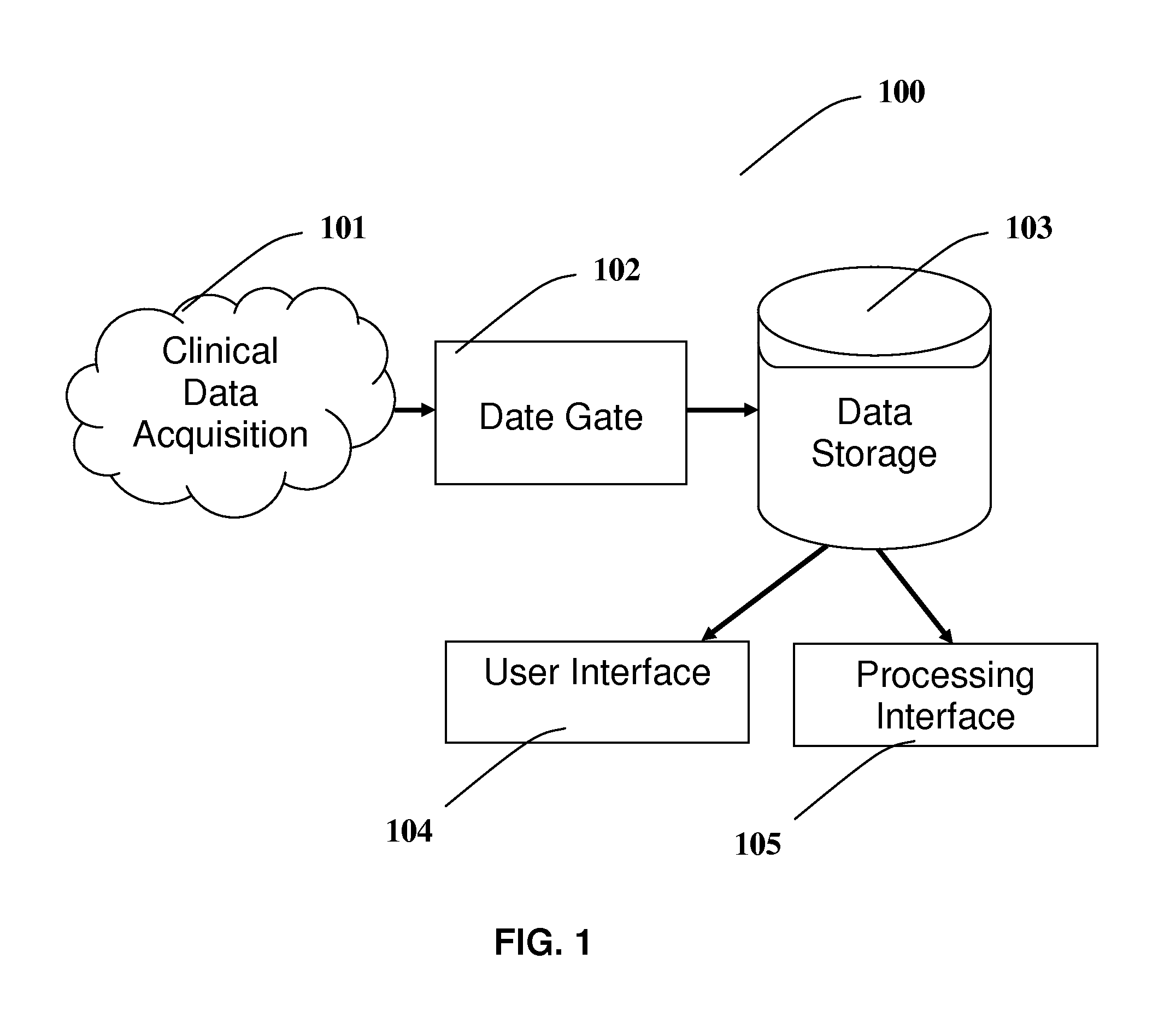
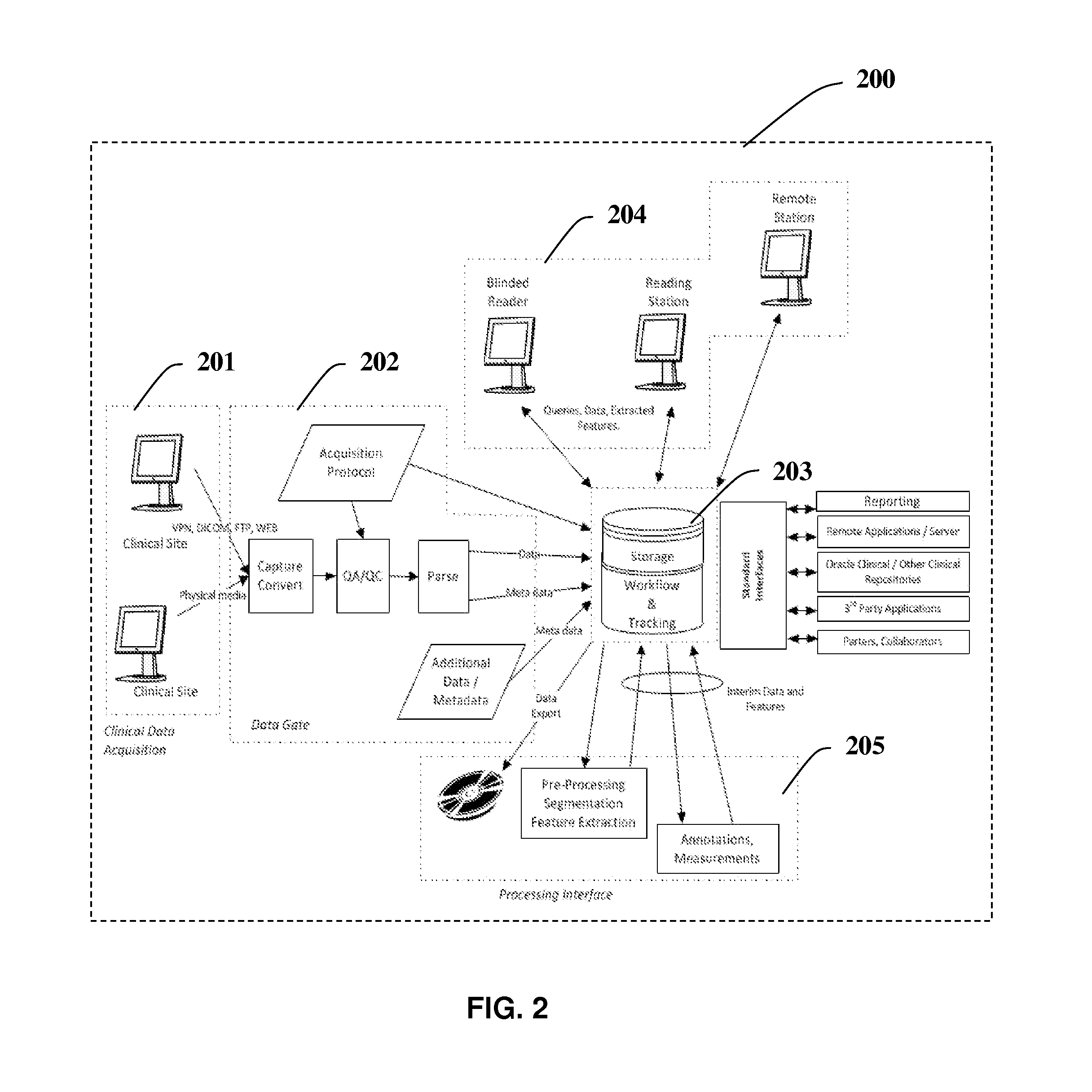
[0019]FIG. 1 a high-level architecture of the Image collaborative portal system. The FIG. 1 system comprises a continuous infrastructure comprising the basic system 100 components including Acquisition 101, Data Gate 102, Storage 103, User Interface 104 and Processing Interface 105. The Data Acquisition component 101 is external to the system and represents the instrument that generates the original data. For imaging data this would be the scanner (i.e. MRI, PET, or CT scanner). The Data Gate 102 component is the gatekeeper to the system and serves two primary functions, wherein 1) to convert the data to a suitable and consistent format for storage. 2) to perform a data QA / QC to ensure that any data stored in the system was acquired according to protocol and is of sufficient quality for meeting its intended purpose. The Storage Component 103 or the backend of the system is the data repository. This component includes the security, backup and retrieval mechanisms. Security includes a...


PUM


Abstract

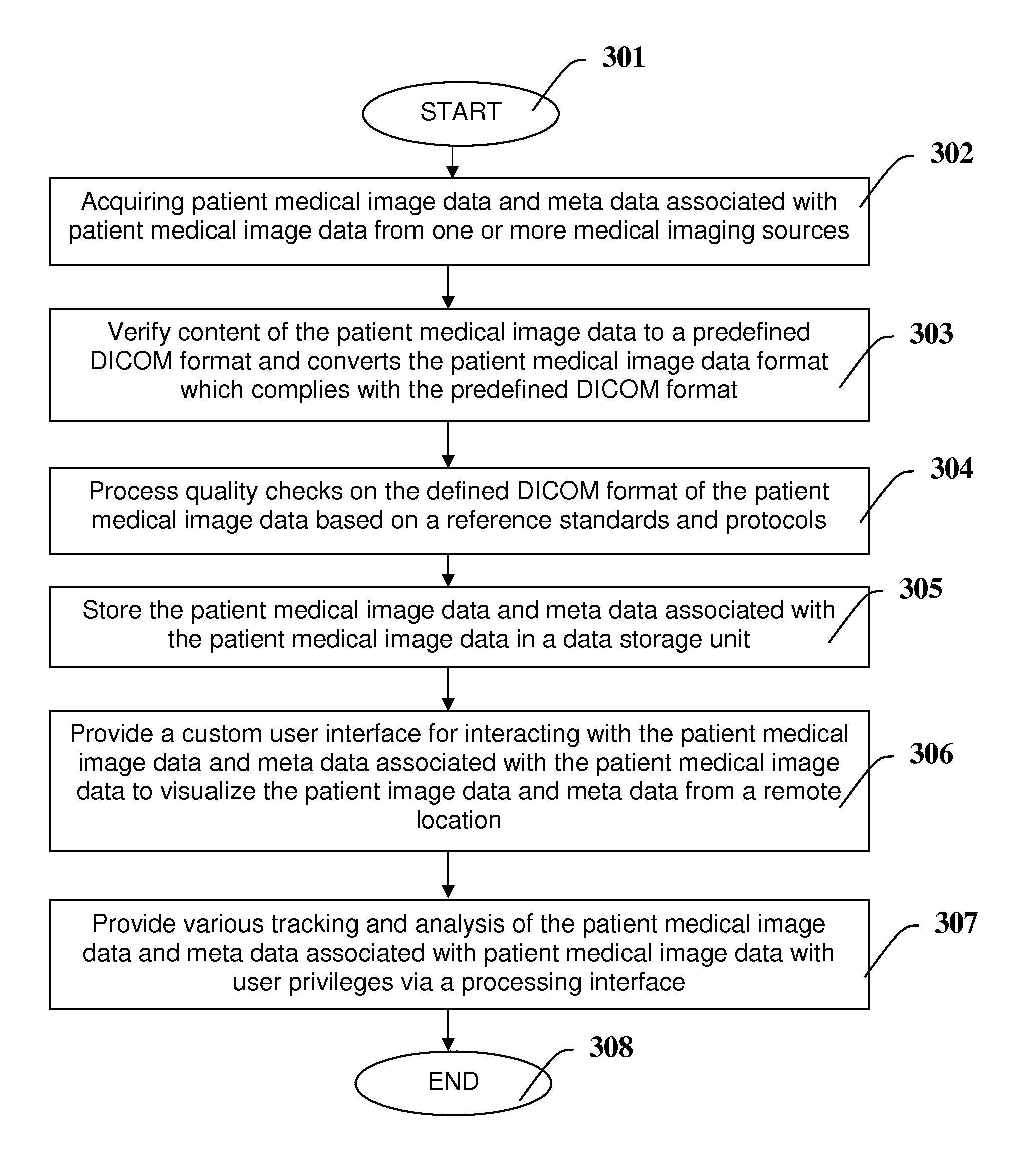
A patient clinical image data processing system, comprising a processor, an acquisition interface for acquiring patient medical image data and meta data from one or more medical imaging sources; and a middle-tier server to verify content of the patient medical image data to a predefined DICOM format; and a conversion unit which converts the patient medical image data format which complies with the predefined DICOM format; a comparator configured to process quality checks on the defined DICOM format of the patient medical image based on a reference standards and protocols; a data storage unit, operable for storing the patient medical image data and meta data; a user interface module for providing a custom user interface for interacting with the patient medical image data and meta data to visualize the patient medical image data and meta data from a remote location; a processing interface for providing various tracking and analysis of the patient medical image data and meta data with user privileges.

Description

FIELD OF THE INVENTION[0001]The invention relates to collaborative medical imaging portal system more particularly to the utilization of collaborative medical imaging portal system user interface supporting clinical trials remotely.BACKGROUND AND DESCRIPTION OF PRIOR ART[0002]Existing systems typically acquire image information in clinical trial processes manually. A site with an imaging modality for e.g. CT, MR, X-ray, ultra-sound etc. uses imaging procedures and parameters that seem to produce images in a more or less reproducible way according to each user's expertise with the imaging modality. Existing systems employ largely manual processes in performing the described tasks and these processes offer limited efficiency and data comparability.[0003]Large datasets are routinely collected during clinical trials. Traditionally the endpoints, such as tumor size or QT intervals, were extracted from these datasets and little concern was paid to the original data. As a result no consist...

Claims


Application Information

Patent Timeline
IPC IPC(8): G06F17/30G06T1/60G06F3/0484G06T1/00
CPCG06F17/3028G06T1/0007G06T2210/32G06F3/04842G06T1/60G16H10/20G16H10/60G16H15/00G16H30/20G16H30/40G16H40/63G16H40/67G16H80/00G06F16/51
Inventor THANGARAJ, VENKATESAN
Owner THANGARAJ VENKATESAN
Learn More