Method for extracting timing parameters using a cardio-mechanical sensor

a timing parameter and sensor technology, applied in the field of conditioning signals, can solve the problems of not always available noninvasive echocardiography measurement equipment, difficult to predict the trend of the time, and difficult to predict the time of the tim

Inactive Publication Date: 2009-08-04
ST JUDE MEDICAL
View PDF29 Cites 6 Cited by
  • Summary
  • Abstract
  • Description
  • Claims
  • Application Information

AI Technical Summary

Benefits of technology

[0017]An object of the present invention is to provide a system and method for measuring and trending EF and providing the trend information to a clinician non-invasively. A further object is to provide such a system and method that allow trend values to be determined based on short term values.

Problems solved by technology

The assessment of cardiac function is a common problem in the evaluation of patients.
Noninvasive measurement of ejection fraction by echocardiography requires expertise and (expensive) equipment that is not always available.
None of the techniques described above allows for trending when the patient is at home.

Method used


Image

  • Method for extracting timing parameters using a cardio-mechanical sensor
  • Method for extracting timing parameters using a cardio-mechanical sensor
  • Method for extracting timing parameters using a cardio-mechanical sensor

Examples


Embodiment Construction

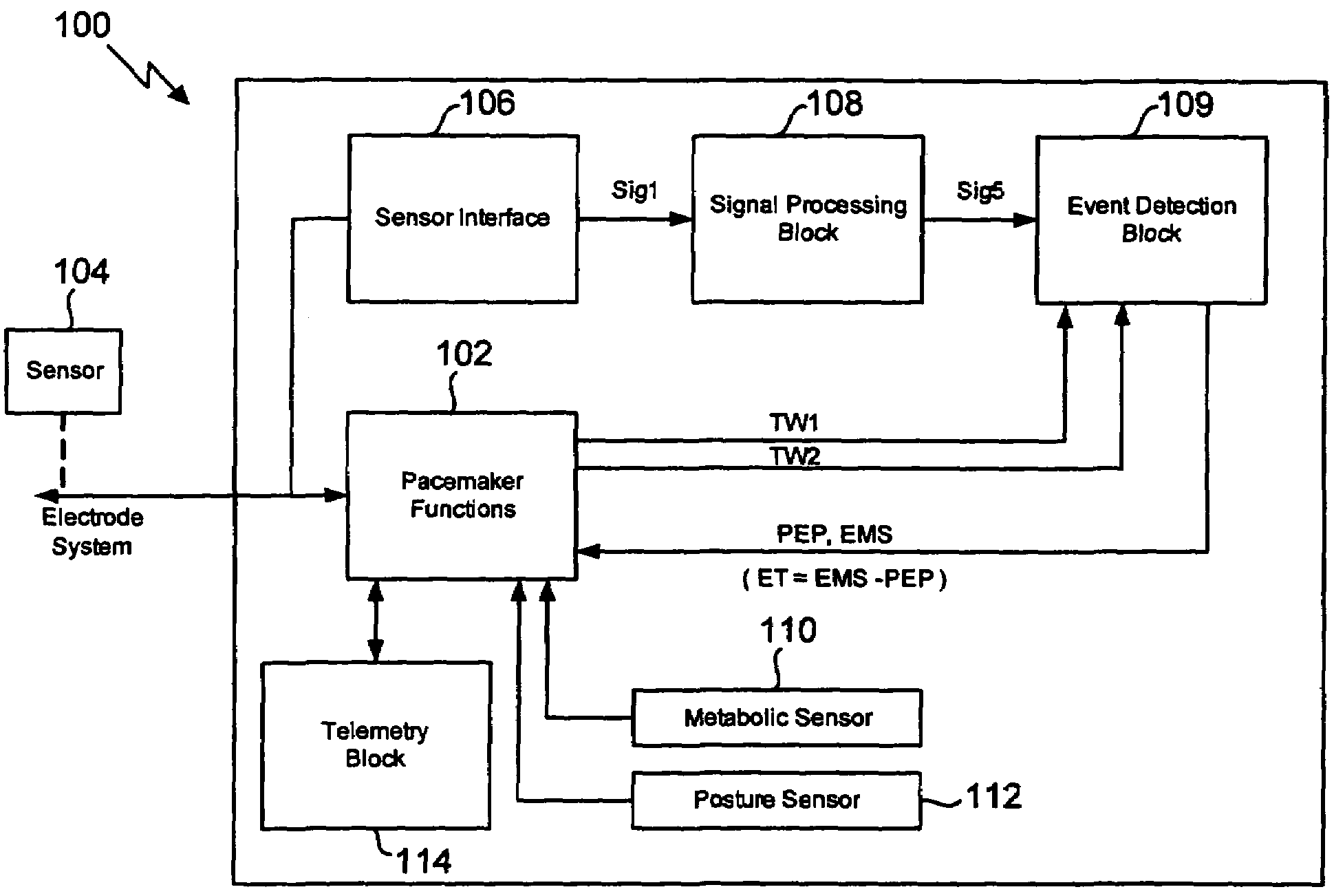
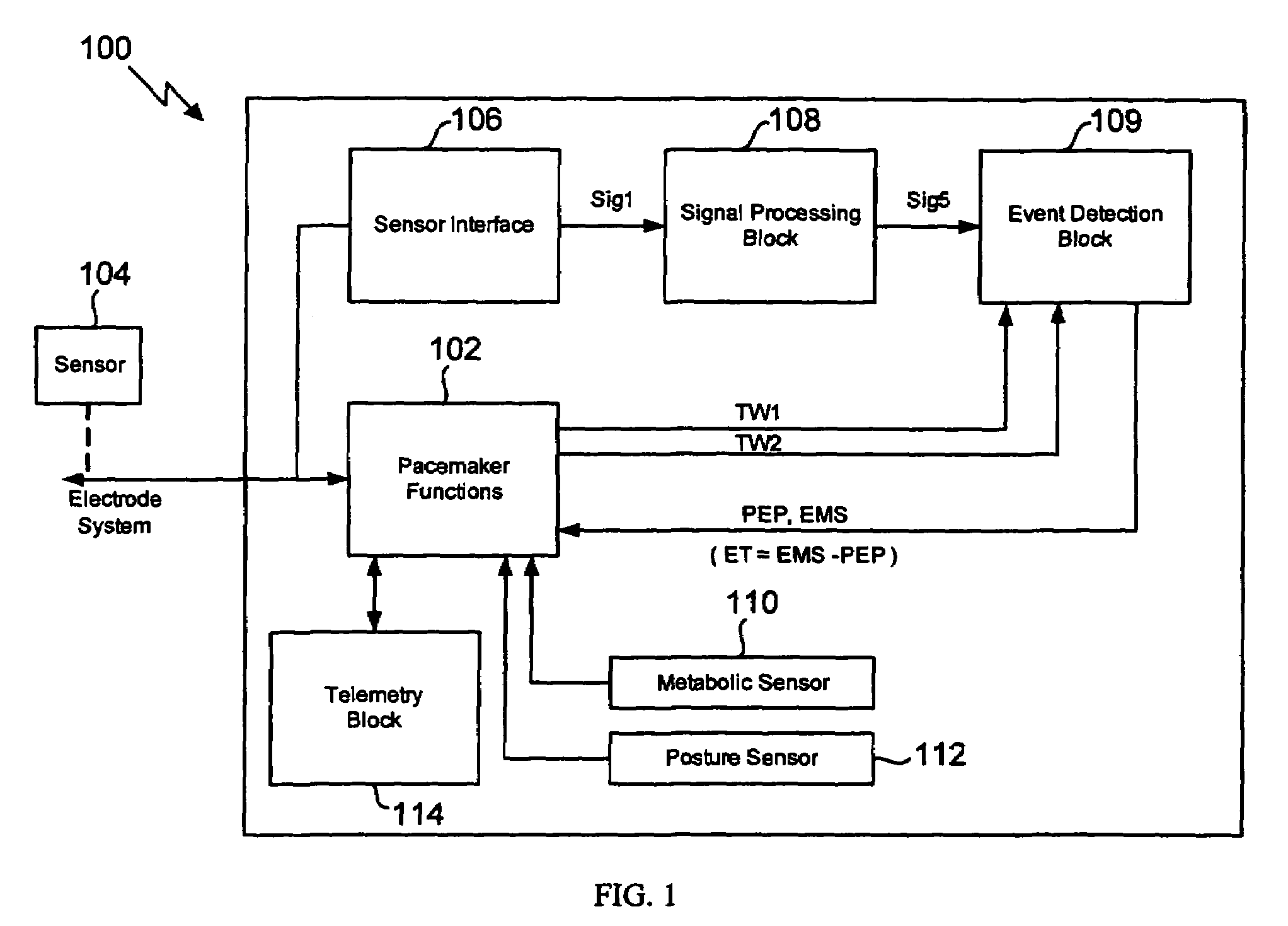
[0026]The invention involves measuring pre-ejection period (PEP) and left ventricular ejection time (LVET) to determine the ejection fraction (EF) and to trend the data over time. This provides an advantageous monitoring of a patient's cardiac function in a minimally invasive manner with a minimum of disruption to the patient.

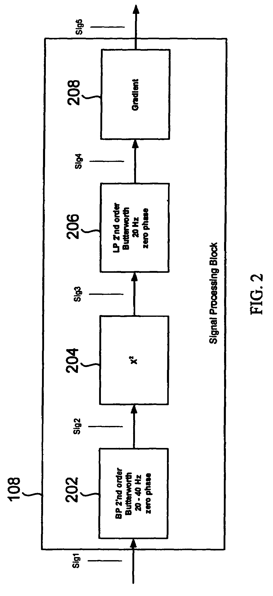
[0027]The invention achieves a robust detection of the opening and closing of the aortic valve of a heart chamber by processing the measured raw signal from an implanted sensor. It was found that a robust detection of the opening and closing of the aortic valve can be achieved by analyzing the signal energy (power) in a frequency range much higher than the heart rate. The signal processing of the invention is insensitive to circadian signal morphology changes for a patient. In one embodiment of the invention, the time derivative of the raw signal rather than the raw signal itself is processed as the high frequency parts are enhanced in the time derivative.

[0028...


PUM


Abstract

In a method and apparatus for measuring the ejection fraction in a mammalian heart, the opening and closing of a heart valve is sensed with an implanted sensor, and a pre-ejection period is measured as a function of the sensed opening and closing of the heart valve. The ventricular ejection time also is measured, and the ejection fraction is determined as a function of the measured pre-ejection period and the ejection time.

Description

BACKGROUND OF THE INVENTION[0001]1. Field of the Invention[0002]The present invention relates to systems and methods for conditioning signals obtained from a sensor preferably located in a coronary vessel.[0003]2. Description of the Prior Art[0004]The assessment of cardiac function is a common problem in the evaluation of patients. The gold standard is the measurement of the ejection fraction (EF or LVEF) by echocardiography. The ejection fraction is the amount of blood pumped out of a ventricle during each heart beat. The EF evaluates how well the heart is pumping. The normal value is 65 percent (with a typical range being 50-70 percent). Lower values indicate ventricular dysfunction. The normal left ventricle has an average volume of 100 ml. Each heart beat ejects approximately 70 ml, which results in an EF≈70%. Noninvasive measurement of ejection fraction by echocardiography requires expertise and (expensive) equipment that is not always available. Further the patient must travel...

Claims


Application Information

Patent Timeline
Patent Type & Authority Patents(United States)
IPC IPC(8): A61B5/026
CPCA61B5/02416A61B5/0452A61N1/36514A61B5/7239A61B5/1116A61B5/0031A61B5/083A61B5/686A61N1/36578A61B5/349
Inventor NOREN, KJELLHOLMSTROM, NILS
Owner ST JUDE MEDICAL
Why Eureka
Try Eureka