System and method for tree-model visualization for pulmonary embolism detection

a pulmonary embolism and tree-model technology, applied in the field of visualization and computer-aided diagnosis and detection of pulmonary embolism, can solve the problems of time-consuming analysis, potentially life-threatening short-term complications, and critical correct diagnosis

Active Publication Date: 2009-10-06
SIEMENS HEALTHCARE GMBH
View PDF4 Cites 65 Cited by
  • Summary
  • Abstract
  • Description
  • Claims
  • Application Information

AI Technical Summary

Benefits of technology

[0015]The step of fitting the tree-model comprises: defining a root site for the anatomical tree structure; computing a skeleton of the segmented anatomical tree structure; forming a raw tree-structure using the skeleton; eliminating false branches in the raw tree-structure; smoothing and centering the skeleton; and assigning direction vectors to points in the segmented anatomical tree structure.
[0019]The step of fitting the tree-model comprises: defining a root site for the anatomical tree structure; computing a skeleton of the segmented anatomical tree structure; forming a raw tree-structure using the skeleton; eliminating false branches in the raw tree-structure; smoothing and centering the skeleton; and assigning direction vectors to points in the segmented anatomical tree structure.
[0023]The processor is operative with the program code, when fitting, to: define a root site for the anatomical tree structure; compute a skeleton of the segmented anatomical tree structure; form a raw tree-structure using the skeleton; eliminate false branches in the raw tree-structure; smooth and center the skeleton; and assign direction vectors to points in the segmented anatomical tree structure.

Problems solved by technology

This short-term complication is potentially life threatening and occurs in about ten percent of patients with acute DVT events.
However, such treatment carries risks, thus making correct diagnosis critical.
This analysis, however, can be time consuming, especially for peripheral arteries.
However, because the radiologist can only track a limited number of vessels at one time, the entire tracking process must be repeated.
Since PE CAD candidates are automatically identified, some PE locations may be missed and false positives may occur.
In addition, the radiologist is typically given no information as to why a particular PE location was chosen or not.
Lung areas showing lower than average density are typically suggestive of an upstream clot; however, other conditions such as emphysema may also result in below average intensity.
However, the visualized tree must be navigated in 3D, and because branches tend to occlude other branches, navigation around the tree requires a significant amount of interaction.
In addition, the entire surface of each branch may need to be examined, thus requiring a full rotational view of each branch resulting in a time-consuming process.

Method used


Image

  • System and method for tree-model visualization for pulmonary embolism detection
  • System and method for tree-model visualization for pulmonary embolism detection
  • System and method for tree-model visualization for pulmonary embolism detection

Examples


Embodiment Construction

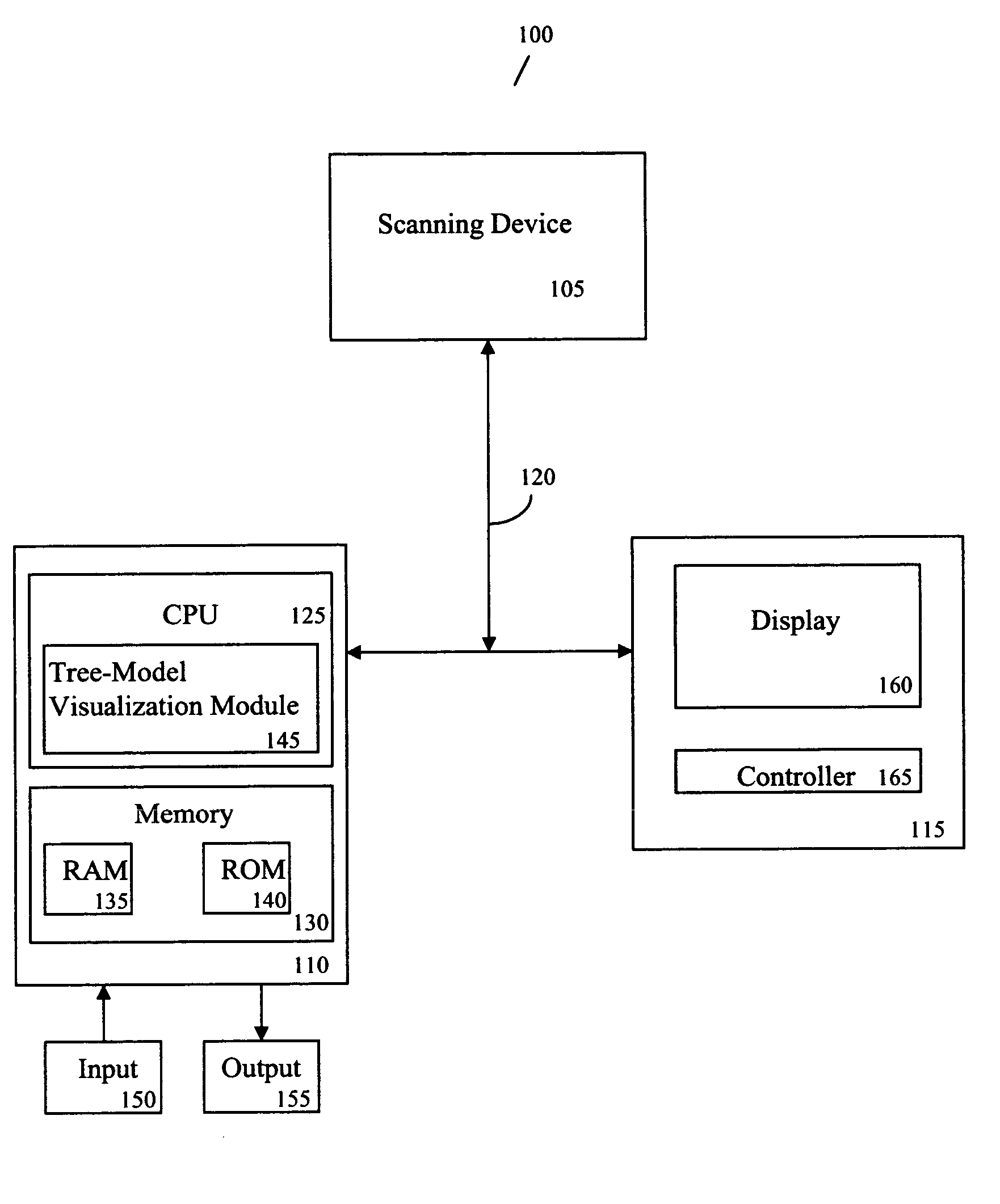
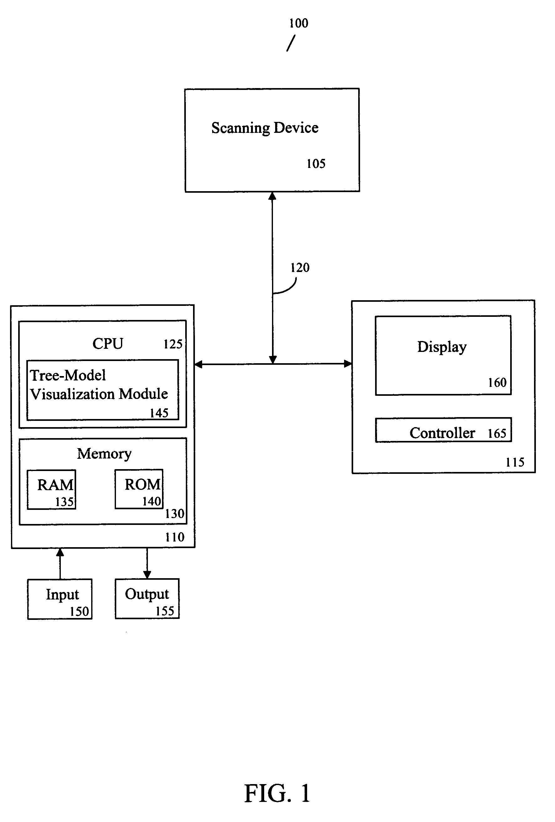
[0034]FIG. 1 is a block diagram of a system 100 for tree-model visualization for pulmonary embolism detection according to an exemplary embodiment of the present invention.

[0035]As shown in FIG. 1, the system 100 includes, inter alia, a scanning device 105, a personal computer (PC) 110 and an operator's console 115 connected over, for example, an Ethernet network 120. The scanning device 105 may be a magnetic resonance (MR) imaging device, a CT imaging device, a helical CT device, a positron emission tomography (PET) device, a 2D or 3D fluoroscopic imaging device, a 2D, 3D, or four-dimensional (4D) ultrasound imaging device, or an x-ray device. The scanning device 105 may also be a hybrid-imaging device capable of CT, MR, PET or other imaging techniques.

[0036]The PC 110, which may be a workstation, portable or laptop computer, a personal digital assistant (PDA), etc., includes a central processing unit (CPU) 125 and a memory 130, which are connected to an input 150 and an output 155...


PUM


Abstract

A system and method for tree-model visualization for detecting an abnormality in an anatomical tree structure are provided. The method comprises: fitting a tree-model to an anatomical tree structure; converting branches of the tree-model into first two-dimensional branch images; and arranging the first two-dimensional branch images in a hierarchical order to form a second two-dimensional image.

Description

CROSS-REFERENCE TO RELATED APPLICATION[0001]This application claims the benefit of U.S. Provisional Application No. 60 / 598,098, filed Aug. 2, 2004, a copy of which is herein incorporated by reference.BACKGROUND OF THE INVENTION[0002]1. Technical Field[0003]The present invention relates to visualization and computer aided diagnosis and detection of pulmonary embolism, and more particularly, to a system and method for tree-model visualization for pulmonary embolism detection.[0004]2. Discussion of the Related Art[0005]A pulmonary embolism (PE) occurs when a piece of a blood clot from a deep vein thrombosis (DVT) breaks off and travels to an artery in a lung where it blocks the artery, damages the lung and puts a strain on the heart. This short-term complication is potentially life threatening and occurs in about ten percent of patients with acute DVT events. It may be even more common than generally realized because the majority of embolisms occur without symptoms.[0006]Although PE is...

Claims


Application Information

Patent Timeline
Patent Type & Authority Patents(United States)
IPC IPC(8): G06K9/00G06T19/00
CPCG06T7/0081G06T7/0083G06T7/0091G06T7/60G06T19/00G06T2219/021G06T2207/10088G06T2207/20044G06T2207/30061G06T2207/30101G06T2207/10081G06T7/11G06T7/12G06T7/155
Inventor KIRALY, ATILLA PETERNOVAK, CAROL L.
Owner SIEMENS HEALTHCARE GMBH
Why Eureka
Try Eureka