Method of examining colon cancer and colon adenoma

a colon cancer and colon adenoma technology, applied in the field of colon cancer and colon adenoma detection, can solve the problems of insufficient positive rate of methods, inability to find suitable methods of detection, and inability to establish the actual system for colorectal cancer diagnosis

Inactive Publication Date: 2009-10-13
JAPAN SCI & TECH CORP +2
View PDF4 Cites 5 Cited by
  • Summary
  • Abstract
  • Description
  • Claims
  • Application Information

AI Technical Summary

Problems solved by technology

Although, immunological fecal occult blood test and various tumor markers are used at present for the examination of colorectal cancers, these methods do not have satisfactory positive rates.
As to the laboratory diagnosis of colorectal adenomas, which is regarded as the birthplace of colorectal cancers, suitable methods of detecting are not available and people rely inevitably on endoscopy at the present time.
However, an actual system for diagnosis of colorectal cancers has not been established.

Method used


Image

  • Method of examining colon cancer and colon adenoma
  • Method of examining colon cancer and colon adenoma
  • Method of examining colon cancer and colon adenoma

Examples


example 1

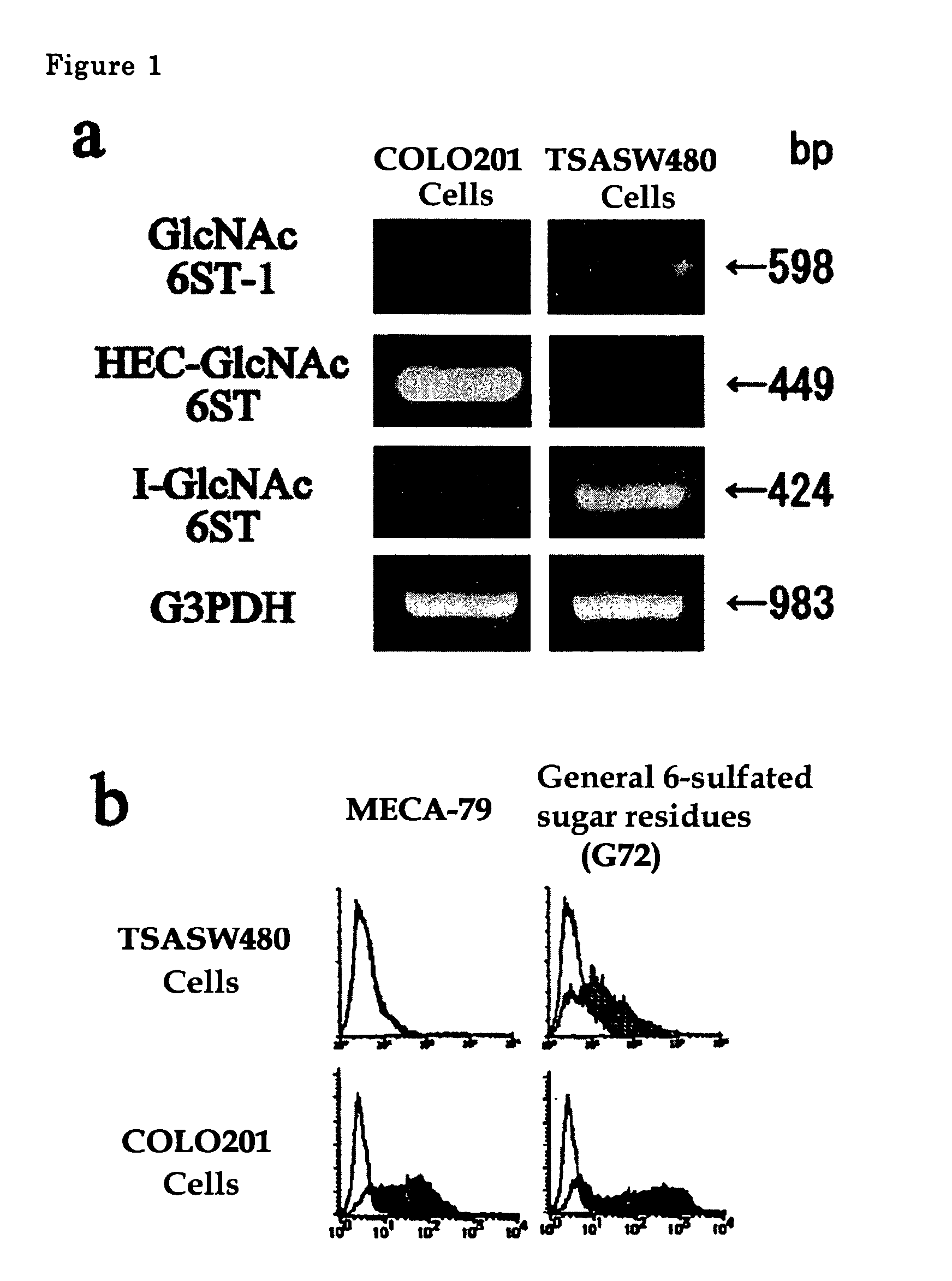
[0038]Gene expression of GlcNAc-6-sulfotransferase isozymes was examined by RT-PCR on human-derived colorectal cancer cells and on normal colorectal epithelial cells. In the RT-PCR analysis, PCR primers for detection of the expression of HEC-GlcNAc6ST gene (Genebank, AF131235) are synthetic oligonucleotides of SEQ ID NO. 1 for upper strand side and those of SEQ ID NO. 2 for lower strand side (Tm=59° C.), those for GlcNAc6ST-1 gene (Genebank, AB011451) are synthetic oligonucleotides of SEQ ID NO. 3 for upper strand side and those of SEQ ID NO. 4 for lower strand side (Tm=62° C.) and those for I-GlcNAc6ST gene (Genebank, AF176838) are synthetic oligonucleotides of SEQ ID NO. 5 for upper strand side and those of SEQ ID NO. 6 for lower strand side (Tm=60° C.).

[0039]The results are shown in FIG. 1a. COLO201 cells are typical cells showing colorectal cancer pattern, which expresses strongly HEC-GlcNAc6ST gene and little GlcNAc6ST-1 and I-GlcNAc6ST genes. TSA-SW480 cells treated SW480 cell...

example 2

[0041]In this example, cells transduced with HEC-GlcNAc6ST gene, GlcNAc6ST-1 gene or I-GlcNAc6ST gene were prepared. Then flowcytometric analysis for these cells was performed using MECA-79 antibody.

[0042]For the preparation of cells transduced with HEC-GlcNAc6ST gene, the gene (Genebank, AF131235) inserted into pcDNA3.1 vector was used. For those with GlcNAc6ST-1 gene, the gene (Genebank, AB011451) inserted into pIRES1hygro vector was used. For those with I-GlcNAc6ST gene, the gene (Genebank, AF176838) inserted into pCDNA3.1 vector was used. The flowcytometric analysis was performed as in Example 1.

[0043]The results are shown in FIG. 2. MECA-79 antibody reacted strongly with those cells transduced with HEC-GlcNAc6ST, but reacted with slightly those cells transduced with GlcNAc6ST gene or I-GlcNAc6ST gene.

example 3

[0044]Colorectal cancer tissues derived from patients (31 cases) were stained with immunohistological staining using MECA-79 antibody. For immunohistological staining, frozen sections with 10 μm thick were used, 1.0 μg / ml MECA-79 antibody was used as the first antibody, and a reagent kit (Vectastain) of Vecta Co. containing anti-rat IgM antibody was used as the second antibody according to the protocol of the company.

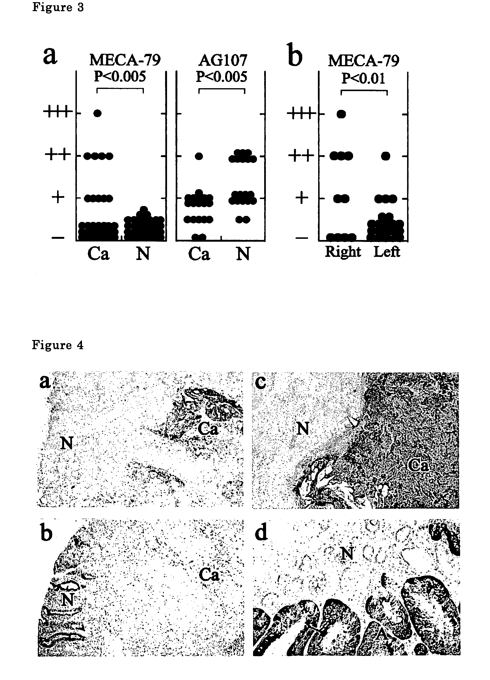
[0045]The results are shown in FIG. 3. The antibody does not react with non-cancer colorectal mucosa (N) (0 case / 31 ca31 cases, 32%). Positive reaction rate is higher for cancers in right-half colorectum (60%) and lower for those in left-half colon (19%).

[0046]A typical stained photographs are shown in FIG. 4.

[0047]FIG. 4a shows the stained photograph of a colorectal cancer tissue (ca) and a colorectal non-cancer tissue (N) of a patient. The colorectal cancer tissue is strongly stained but the non-cancer tissue is little stained.

[0048]FIG. 4b shows the photograph of the...


PUM

PropertyMeasurementUnit
thickaaaaaaaaaa
structureaaaaaaaaaa
reactivityaaaaaaaaaa

Abstract

The present invention provides a method for examining colorectal cancer and colorectal adenoma, which enables to detect colorectal cancer patients and patients at high risk of colorectal cancer at a high probability and is useful for diagnosis of colorectal cancer and colorectal adenoma, and provides the examination reagents thereof. The present inventors discovered that there are significant differences in the distribution of GlcNAc-6-sulfotransferase isozymes, sulfation enzymes of sugar residues, among non-cancer colorectal tissues, colorectal cancer tissues and colorectal adenoma tissues. Furthermore the inventors applied the discovery to diagnosis and found that colorectal cancers and adenomas are detected specifically by assaying a definite range of GlcNAc-6-sulfated sugar residues in tissues from patients or feces samples. MECA-79 antibody (Pharmingen, catalog No. 09961D, Distributor: Becton Dickinson), reacting with GlcNAc-6-sulfated sugar residues, which are produced specifically by the enzyme present in colorectal cancer and colorectal adenoma tissues could be used for the examination of colorectal cancers and colorectal adenomas.

Description

FIELD OF THE INVENTION[0001]The present invention relates to a method for examining human colorectal cancers and colorectal adenomas, and to antibodies and examination reagents thereof.PRIOR ART[0002]Since the number of colorectal cancer patients is increasing year by year, a method for early detection is necessary. Although, immunological fecal occult blood test and various tumor markers are used at present for the examination of colorectal cancers, these methods do not have satisfactory positive rates. Namely, the positive rate of immunological fecal occult blood test used for the examination of colorectal cancers is 50-60%. As to the tumor markers of colorectal cancers, carcino-embryonic antigen (CEA), CA19-9, STX, which are used for examining the therapeutic effect and for monitoring recurrence, are not satisfactory as tumor markers for early detection of colorectal cancers.[0003]The colon is divided into right-half and left-half drawing a line at the flexure coli and colorectal...

Claims


Application Information

Patent Timeline
Patent Type & Authority Patents(United States)
IPC IPC(8): A61K39/395C07K16/40C12N15/09G01N33/574
CPCG01N33/57419G01N2400/02G01N2333/91097
Inventor KANNAGI, REIJIIZAWA, MINEKOMURAMATSU, TAKASHIUCHIMURA, KENJIHOSOKAWA, HIDEAKI
Owner JAPAN SCI & TECH CORP
Why Eureka
Try Eureka