
Ultrasound probe support and stepping device

a technology of ultrasound probes and support devices, which is applied in the direction of catheters, applications, therapy, etc., can solve the problems of damage to surrounding tissues, preventing or at least inhibiting the wide application of these treatments, etc., to achieve quick and easy mounting and removal

Active Publication Date: 2009-10-20
CIVCO MEDICAL INSTR CO
View PDF25 Cites 34 Cited by
  • Summary
  • Abstract
  • Description
  • Claims
  • Application Information

AI Technical Summary

Benefits of technology

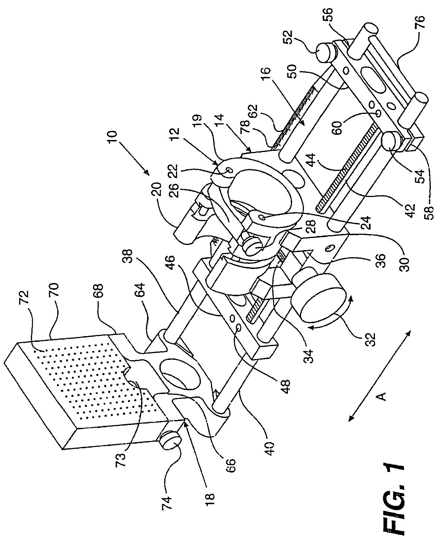
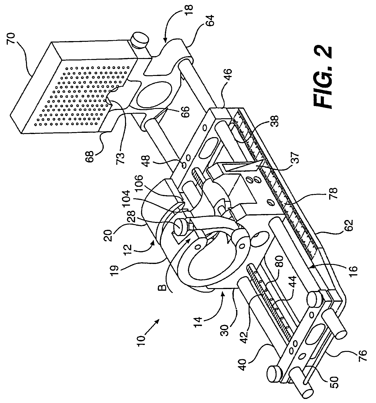
[0011]A medical instrument mount is rotatably mounted to the carriage assembly, wherein the medical instrument mount is configured to receive a medical instrument. Additionally, the medical instrument mount is mounted to the carriage assembly, such that the height of the medical instrument mount is adjustable on the carriage assembly. The rotatable mounting of the medical instrument mounts enables the medical instrument to be rotated about it longitudinal axis, without have to adjust the positions of the carriage assembly or the medical instrument mount.
[0012]A quick release member is operatively associated with the medical instrument mount and carriage assembly. The quick release member enables the medical instrument mount to be quickly and easily mounted to and removed from the carriage assembly when the carriage assembly is in the imaging position without losing position and orientation of the carriage assembly. The quick release member includes a grooved portion of the carriage assembly and a tongued portion of the medical instrument mount, the grooved portion configured and dimensioned to receive the tongued portion and the tongued portion releasable from the grooved portion by rotation of the medical instrument mount with respect to the carriage assembly.

Problems solved by technology

Although the effectiveness of these treatments has been established, the risks associated with these treatments prevent or at least inhibit the wide application that they might otherwise achieve.
The risks in each instance are related to the difficulties in achieving full control and accurate monitoring of the treatment.
The risks of damaging surrounding tissues are present in every case, potentially catastrophic and require great care and experience to control.
However, no vertical adjustments of the ultrasound probe with respect to template grid is possible.
As previously noted, the major concern and risk of morbidity from cryotherapy and thermotherapy is thermal damage to the rectal area.
This worry often limits effective treatment at the posterior margins of the prostate.
In the case of cryotherapy, freezing this area is required and if the freezing is too aggressive a postoperative fistula from the prostate to the rectum may result from also freezing the rectal wall.
However, all these current methods for improving safety require actively moving the ultrasound probe away from the anterior rectal wall using methods that are clumsy, time consuming and that significantly compromise good visibility using the ultrasound image.

Method used


Image

  • Ultrasound probe support and stepping device
  • Ultrasound probe support and stepping device
  • Ultrasound probe support and stepping device

Examples


Embodiment Construction

[0027]In the description which follows, any reference to either direction or orientation is intended primarily and solely for purposes of illustration and is not intended in any way as a limitation to the scope of the present invention. Also, the particular embodiments described herein, although being preferred, are not to be considered as limiting of the present invention.

[0028]Referring now to the drawing figures in which like reference designators refer to like elements, there is shown in FIG. 1 the device 10 according to the present invention. The device 10 of the present invention includes an ultrasound probe mount 12, a carriage 14, a base assembly 16, and a template grid mount 18. The probe mount 12 is adapted to receive and securely clamp around a central enlarged portion of an ultrasound probe. This probe mount 12 is held for rotation within carriage 14. The carriage 14 is, in turn, held for slidable longitudinal movement along the base assembly 16 and the template grid mou...


PUM


Abstract

The present invention provides an apparatus for guiding a medical instrument. The medical apparatus includes a bracket member having a cavity dimensioned and configured to receive at least a portion of a medical instrument. A carriage member is slidably connected to a base assembly adapted to move the carriage member into an imaging position. The carriage assembly is adapted to rotationally support the bracket member, as well as provide a vertically adjustable height of the bracket member. A quick release member is operatively associated with the bracket and carriage members for removably attaching and detaching the bracket member to and from the carriage member when the carriage member is in the imaging position without losing the position and orientation of the carriage member.

Description

CROSS-REFERENCE TO RELATED APPLICATION[0001]This application claims the benefit under 35 U.S.C § 119 (e) of U.S. Provisional Patent Application No. 60 / 401,830, filed Aug. 7, 2002, entitled ULTRASOUND PROBE SUPPORT AND STEPPING DEVICE, the entirety of which is incorporated herein by reference.FIELD OF THE INVENTION[0002]This invention relates generally to support apparatus for precision medical instruments, and more particularly to an adjustable support and stepping device for use with transrectal ultrasound imaging probes and a template grid or matrix.BACKGROUND OF THE INVENTION[0003]Brachytherapy (radioactive seeds), thermotherapy (heating), and cryotherapy (freezing) are proven therapies for tumors, both benign and malignant. Although the effectiveness of these treatments has been established, the risks associated with these treatments prevent or at least inhibit the wide application that they might otherwise achieve. The risks in each instance are related to the difficulties in a...

Claims


Application Information

Patent Timeline
Patent Type & Authority Patents(United States)
IPC IPC(8): A61B19/00A61B8/12A61N5/10
CPCA61B8/12A61N5/1007A61B8/4209A61N2005/1012
Inventor BARZELL, WINSTON E.WHITMORE, WILLET F.BRAUNER, STEPHEN E.WILSON, ROGER
Owner CIVCO MEDICAL INSTR CO
Learn More