
Plugged tip delivery for marker placement

a marker and tip technology, applied in the field of markers delivery devices and methods, can solve the problems of inaccuracy, misdirected follow-up treatment to an undesired portion of the patient's tissue, and inability to heal or otherwise change, so as to avoid errors in the placement of markers, prevent tissue ingress, and improve accuracy

Inactive Publication Date: 2010-01-26
SENORX
View PDF123 Cites 107 Cited by
  • Summary
  • Abstract
  • Description
  • Claims
  • Application Information

AI Technical Summary

Benefits of technology

[0012]Assemblies embodying features of the invention include marker delivery devices having a delivery tube with an orifice at its distal tip, an inner bore leading to an orifice, and at least one marker (preferably more than one) within the bore of the delivery tube. A plug is disposed at least in part within the bore and orifice to prevent markers from prematurely passing through the orifice and to prevent tissue from entering the bore when the delivery tube is advanced through tissue. The plug may itself serve as a marker, and may be the sole marker, although typically the delivery tube contains a plug and at least one other marker. The plug is releasably secured within or adjacent to the orifice in order to retain a marker within the delivery tube bore proximate thereto but to allow passage of the marker out of the orifice when the plug is ejected from the orifice. The plug may partially or completely occlude the orifice, and is configured to retain the marker within the delivery tube before the marker is to be placed at a desired location within a patient's body, and to allow the marker to pass out of the orifice when delivery of the marker is desired. A movable plunger may be slidably disposed within the tube from an initial position accommodating the marker or markers and the plug within the tube, to a delivery position to push a marker against the plug to push the plug out of the orifice and to then eject one or more markers through the orifice.
[0016]The invention provides the advantages of securely retaining markers within a marker delivery device, improving accuracy and avoiding errors in of placement of markers at desired locations within a patient's body, preventing ingress of tissue into the distal tip of the device when it is advanced through tissue, and guiding the device by use of an imaging device. These and other advantages of the invention will become more apparent from the following description when taken in conjunction with the accompanying drawings.

Problems solved by technology

However, radiographically visible tissue features, originally observed in a mammogram, may be removed, altered or obscured by the biopsy procedure, and may heal or otherwise become altered following the biopsy.
This, in turn, can result in follow-up treatments being misdirected to an undesired portion of the patient's tissue.
Such a marker enables a follow-up procedure to be performed without the need for traditional radiographic mammography imaging which, as discussed above, can be subject to inaccuracies as a result of shifting of the location wire as well as being tedious and uncomfortable for the patient.

Method used


Image

  • Plugged tip delivery for marker placement
  • Plugged tip delivery for marker placement
  • Plugged tip delivery for marker placement

Examples


Embodiment Construction

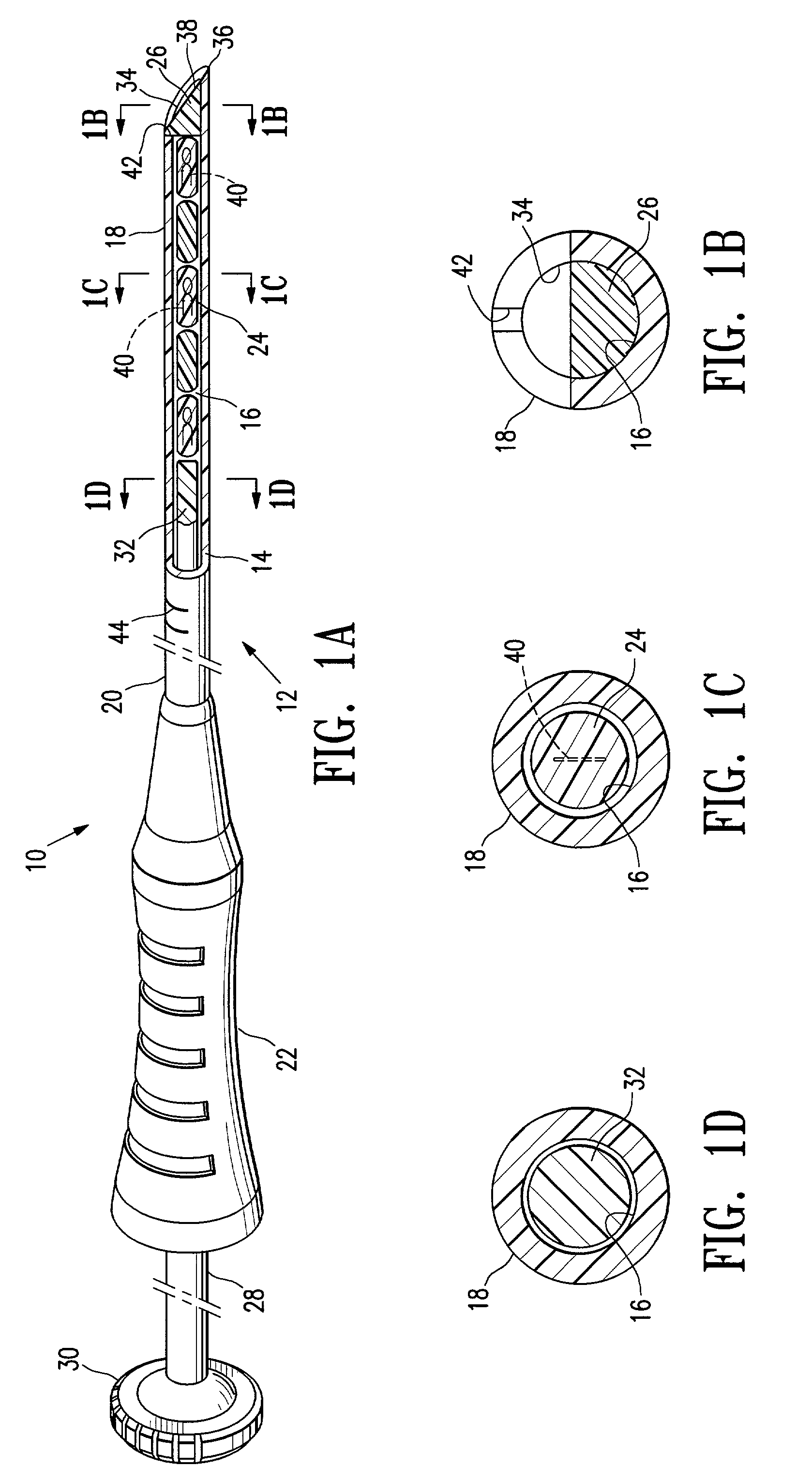
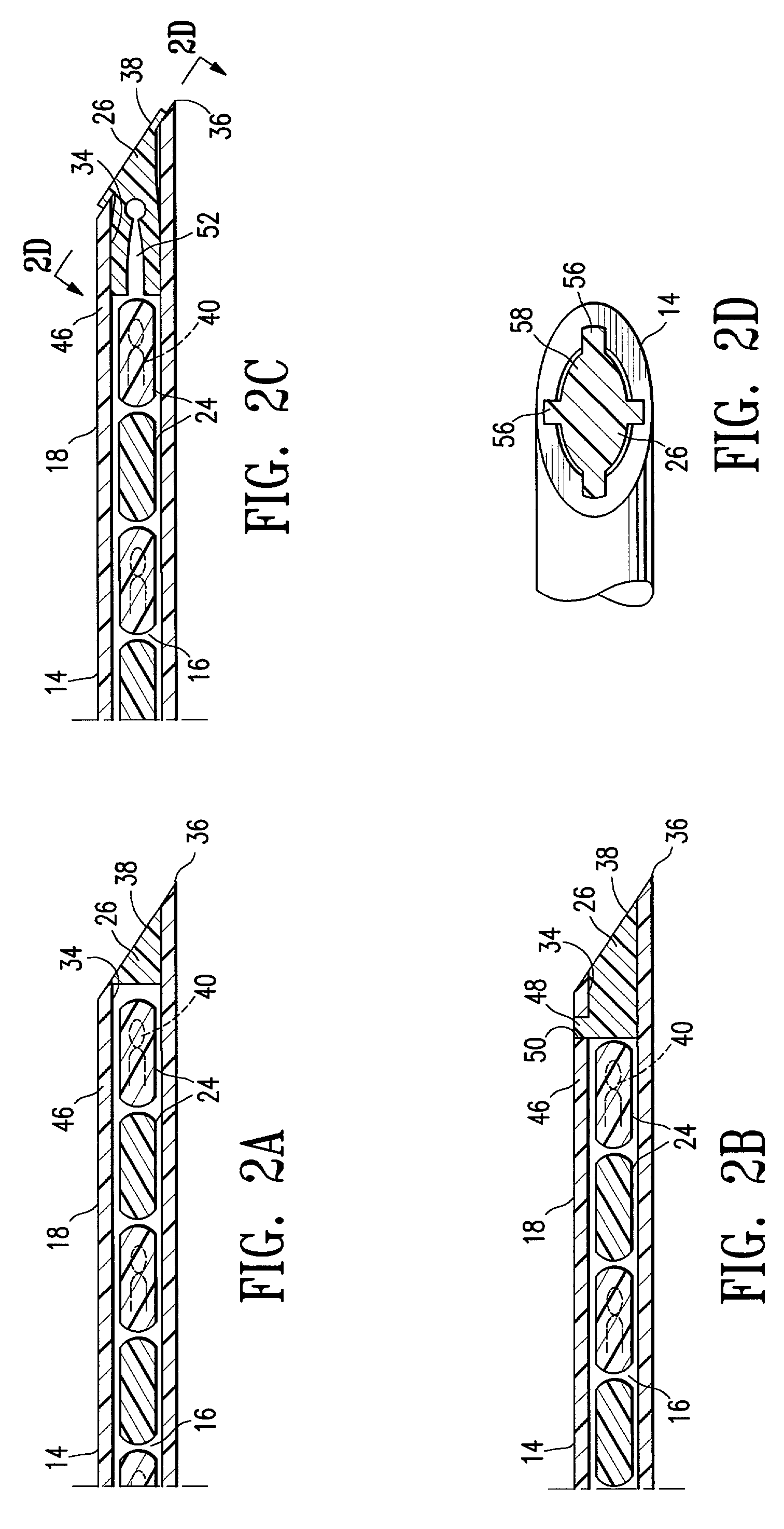
[0035]Marker delivery assemblies embodying features of the invention are illustrated in FIGS. 1A-1D. Such assemblies include marker delivery devices, markers, and a plug occluding a distal portion of the delivery device. The assembly 10 shown in FIG. 1A includes a delivery device 12, delivery tube 14 with a bore 16, a distal portion 18, and a proximal portion 20 with a handle 22. Several markers24, and a plug 26 are shown disposed within the bore 16. A plunger 28 with a plunger handle 30 and a plunger distal end 32 is movable within the tube bore 16, and is configured to push markers 24 and plug 26 out of orifice 34 at the distal tip 36 of delivery tube 14 when the distal end 32 of plunger 28 moves in a distal direction. Plunger handle 30 allows an operator to readily manipulate plunger 28. A device 12 may include a plunger locking mechanism to prevent inadvertent longitudinal movement of plunger 28; for example, a plunger 28 and a handle 22 may be configured so that plunger 28 must...


PUM


Abstract

The invention provides marker delivery devices, assemblies, and methods. Assemblies embodying features of the invention include marker delivery devices having a delivery tube with an orifice, markers, and a plug releasably disposed in the tube occluding the orifice. The plug prevents markers from passing through the orifice before the marker is to be placed at a desired location within a patient's body, prevents ingress of tissue into the bore of the tube, and allows markers to pass out of the orifice when marker delivery is desired. Guidance of a delivery tube to a desired location within a patient's body may include the use of an imaging device, with or without the use of a guide cannula. The plug may itself serve as a marker, and may be the sole marker. Markers may be detectable by ultrasound, X-ray, magnetic resonance imaging, and other imaging devices, and may include bioactive elements.

Description

FIELD OF THE INVENTION[0001]The invention is directed generally to devices and methods for delivering markers to a desired location within a patient's body. In particular, the invention is directed to devices, assemblies, and methods configured to retain a biological marker within a delivery device before delivery of the marker to a desired intracorporeal location.BACKGROUND OF THE INVENTION[0002]In diagnosing and treating certain medical conditions, it is often desirable to mark a suspicious body site for the subsequent taking of a biopsy, delivery of medicine, radiation, or other treatment, to mark a location from which a biopsy was taken, or at which some other procedure was performed. As is known, obtaining a tissue sample by biopsy and the subsequent examination are typically employed in the diagnosis of cancers and other malignant tumors, or to confirm that a suspected lesion or tumor is not malignant. The information obtained from these diagnostic tests and / or examinations is...

Claims


Application Information

Patent Timeline
Patent Type & Authority Patents(United States)
IPC IPC(8): A61B17/00A61B19/00A61M37/00
CPCA61M37/0069A61B90/39A61B2090/3987A61B2090/3925A61B2090/3908
Inventor LUBOCK, PAULSAFABASH, JASON H.SHABAZ, MARTINMERRITT, JOHN
Owner SENORX
Learn More