Assemblies, systems, and methods for vacuum assisted internal drainage during wound healing

a vacuum assisted and internal drainage technology, applied in the direction of wound drains, suction devices, intravenous devices, etc., can solve the problems of significant loss of patients' income, tissue damage, cellular death, etc., to reduce the “dead-space” or open area, improve wound healing benefits, and reduce the swelling of the overlying and underlying tissue.

Active Publication Date: 2010-04-20
IC SURGICAL INC
View PDF59 Cites 106 Cited by
  • Summary
  • Abstract
  • Description
  • Claims
  • Application Information

AI Technical Summary

Benefits of technology

[0024]The assembly, system, and / or method apply a vacuum of significant pressure internally and directly in a wound area or body cavity for enhanced wound healing benefits. By applying a vacuum of significant consistent pressure internally and directly in the wound area or body cavity, the assembly, system, and / or method reduce the “dead-space” or open area inside the wound or cavity, and thereby aid in decreasing tissue edema and swelling of the overlying and underlying tissue. The assembly, system, and / or method increase the nature and extent of wound drainage, promote tissue adherence and closure of wounds, and thus decrease seroma formation and promote primary wound healing. The assembly, system, and / or method thereby decrease the costly and increased patient morbidity caused by seroma formation and the resultant delay in primary wound healing or need for additional surgical procedures or drainage.

Problems solved by technology

Following surgery or due to trauma, there can also be resulting tissue damage, regardless of how careful the surgeon is.
This tissue damage results in cellular death, and the body's natural defense reaction is an inflammatory one.
Such complications result in a significant amount of lost income to patients, as well as expenses to insurers and physicians who have to care for these patients that require serial drainage.
Such complications also delay wound healing, may entail additional surgical procedures, and ultimately delay the patient's return to work and routine functional activity.
Seroma management can also be costly and, further, can place health care workers to additional needle exposure risks and related outcomes such as hepatitis, etc.
The problem with these devices is that, although they may drain fluid, fluid drainage is limited to fluid directly around the drain itself.
As a result, current drains may manage fluid collection, but they do not effectively clear all of the fluid in the space and, more importantly, they do not clear enough fluid to effectively seal down and close off the dead space.
Conventional VAC devices, however, are only approved and used for external wounds only.
Conventional VAC devices are not approved or used for internal wounds or operative sites.
Current wound drain devices assemblies at times do not remove a substantial amount of fluid from within a wound and have other performance issues.
For example, external VAC devices clear fluid directly around external wounds (as FIG. 3 shows), and they are limited to the application to external wounds only.
Furthermore, the clinical use of external VAC devices may not make wound drainage more cost-effective, clinician-friendly, and patient-friendly.
Removal of the foam structures in the presence of granulating tissue and the force of pressure on the wound bed that this removal can cause pain or discomfort.
Furthermore, the multiple steps of the conventional external VAC procedure—removing the adhesive membrane, then removing the old foam structures, then inserting the new foam structures, and then reapplying the adhesive member along the entire periphery of the wound—are exacting, tedious and time consuming.
They only prolong pain or discomfort, and cause further disruption to the patient, and also demand dedicated nursing time and resources.
Obtaining such a seal can be difficult, particularly in body regions where the surrounding skin is tortuous, and / or mucosal and / or moist.
Furthermore, prolonged wearing of wet dressings can cause further breakdown and maceration of the surrounding skin thereby increasing the wound size.
This can cause further discomfort to the patient, and the exudate can often be offensive in odor and color causing further embarrassment to the patient.
This may, in turn, require more numerous dressing changes and re-padding throughout the day, which is disruptive to the patient and costly both in terms of nursing time and resources.

Method used


Image

  • Assemblies, systems, and methods for vacuum assisted internal drainage during wound healing
  • Assemblies, systems, and methods for vacuum assisted internal drainage during wound healing
  • Assemblies, systems, and methods for vacuum assisted internal drainage during wound healing

Examples


Embodiment Construction

[0038]Although the disclosure hereof is detailed and exact to enable those skilled in the art to practice the invention, the physical embodiments herein disclosed merely exemplify the invention that may be embodied in other specific structure. While the preferred embodiment has been described, the details may be changed without departing from the invention, which is defined by the claims.

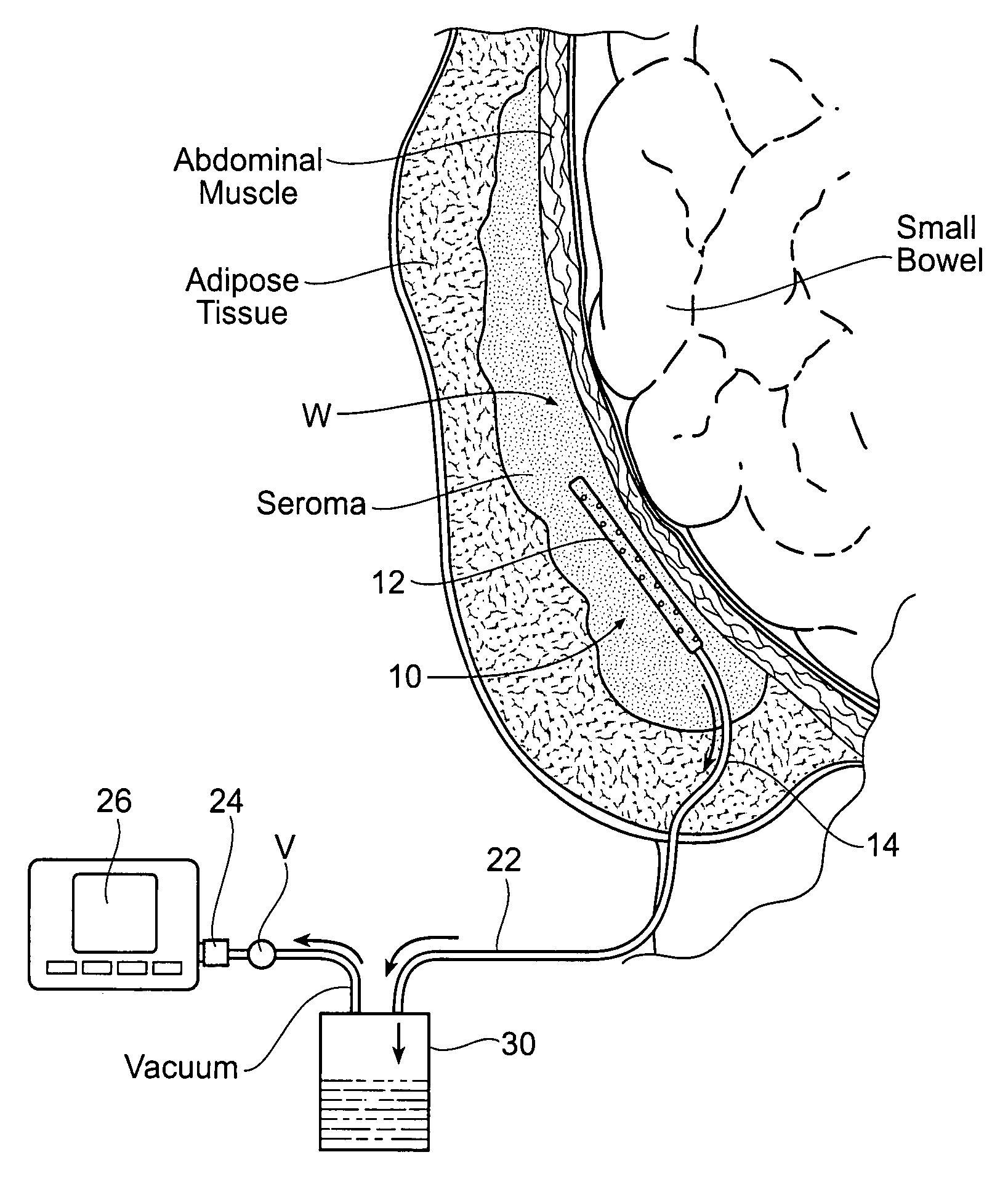
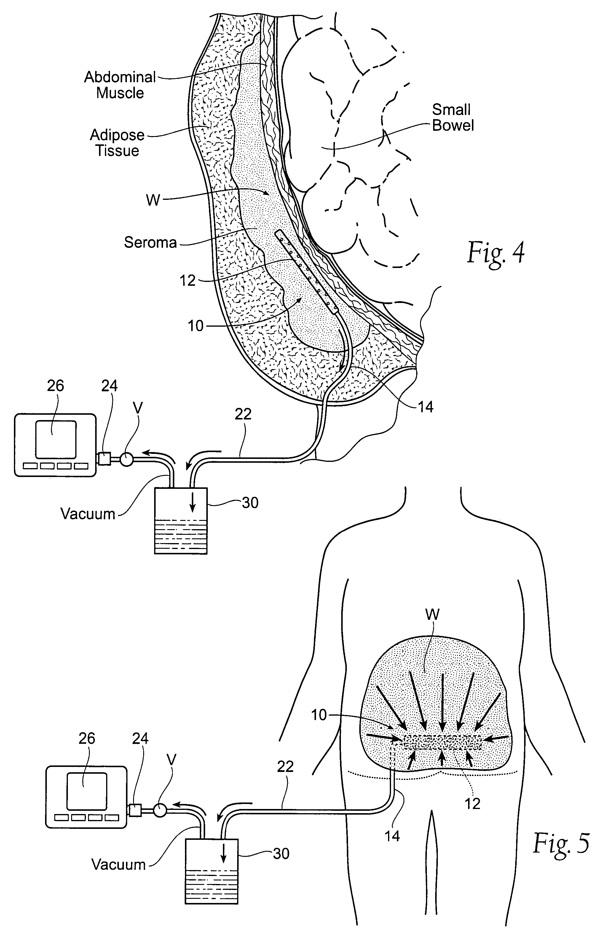
[0039]FIG. 4 shows a wound drainage system 10 comprising an internal drain assembly 12 that is sized and configured for surgical placement within a wound area W (or body cavity). The wound area W may be anywhere in a human or animal, e.g., within a body cavity, or beneath the skin, or in muscle, or within the soft tissues. As will be described in greater detail later (see FIG. 6), the internal drain assembly 12 includes a housing 18 that encloses a foam sponge component 16. The foam sponge component 16 communicates with the wound area W through one or more apertures 20 formed in the housing 18.

[0040...


PUM


Abstract

Assemblies, systems, and methods convey fluid from an internal wound site or body cavity by applying negative pressure from a source outside the internal wound site or body cavity through a wound drain assembly that is placed directly inside the internal wound site or body cavity.

Description

RELATED APPLICATION[0001]This application is a continuation-in-part of co-pending U.S. patent application Ser. No. 11 / 646,918, filed Dec. 28, 2006 and entitled Assemblies, Systems, and Methods for Vacuum Assisted Internal Drainage During Wound Healing, which claims the benefit of U.S. Provisional Patent Application Ser. No. 60 / 810,733, filed Jun. 2, 2006, and entitled “Foam Sponge Vacuum Assisted Internal Drainage System,” which are each incorporated herein by reference.FIELD OF THE INVENTION[0002]This application relates generally to the drainage of fluid from the body during the wound healing process, e.g., following surgery, trauma, or placement of implants or surgical devices.BACKGROUND OF THE INVENTION[0003]During surgery, or as a result of trauma, tissue volume can be removed or altered, and an open or dead space is created within the tissue that was previously attached to other tissue. The very small blood vessels that previously ran from the underlying tissue (i.e., muscle, ...

Claims


Application Information

Patent Timeline
Patent Type & Authority Patents(United States)
IPC IPC(8): A61M1/00A61M27/00
CPCA61M1/0088A61M27/00A61M2027/004A61M1/916A61M1/985
Inventor BENGTSON, BRADLEY PMCGUIRE, PATRICIA A
Owner IC SURGICAL INC
Why Eureka
Try Eureka