Opposed view and dual head detector apparatus for diagnosis and biopsy with image processing methods

a detector apparatus and dual-head technology, applied in the field of biopsy needle guidance, can solve the problems of low specificity of x-ray imaging, low detection efficiency, and low target accuracy, and achieve the effect of high spatial resolution of x-ray image and high specificity of nuclear medicine data

Active Publication Date: 2010-05-04
HAMPTON UNIVERSITY
View PDF4 Cites 46 Cited by
  • Summary
  • Abstract
  • Description
  • Claims
  • Application Information

AI Technical Summary

Benefits of technology

[0010]According to a principle of the invention, a gamma camera may be removably attached to a biopsy needle gun platform, thereby permitting control of the acquisition of one or more functional images. The biopsy needle gun may be reattached and one or more biopsies are performed based on a co-registered imaged resulting from fusing an x-ray image and a functional image.

Problems solved by technology

X-ray imaging is a commonly used technique for breast cancer screening and diagnosis, but since it also is sensitive to other non-malignant pathologies, its accuracy is compromised.
The specificity of x-ray imaging may be quite poor with only about 20% to about 35% of biopsies yielding cancer diagnoses.
It is also a commonly used modality for breast tumor needle biopsy guidance, but has been found to be lacking in target accuracy for some cases.
Also, since it detects physiological abnormalities, it directly indicates the location of disease, while x-ray imaging is limited to detecting changes in tissue density which may be secondary to the presence of disease.
Despite the promising role of this procedure in breast lesion management, however, some clinical studies have found a false negative rate of about 10%.
Moreover, findings from additional studies point toward specific subgroups limiting the diagnostic accuracy of this procedure.
In addition, another study found that the diagnostic accuracy of needle biopsy was dependent on lesion size, as masses larger than about 3 cm were less likely to be diagnosed correctly.
The efficacy of this procedure is lacking for diagnostic accuracy for lesions less than about 1 cm in diameter, non-palpable masses, and lesions located in the medial aspect of the breast.

Method used


Image

  • Opposed view and dual head detector apparatus for diagnosis and biopsy with image processing methods
  • Opposed view and dual head detector apparatus for diagnosis and biopsy with image processing methods
  • Opposed view and dual head detector apparatus for diagnosis and biopsy with image processing methods

Examples


specific example 1

[0098]This example utilized an about 5 cm thick plastic breast phantom with an about 6 mm hollow sphere lesion located near the center of the breast phantom. The breast volume was filled with a Tc99m solution with a concentration of about 0.33 μCi / ml and the lesion volume concentration was about 1.98 μCi / ml. Two opposing 10 minute acquisitions were obtained. A pair of Co-57 point sources was taped to the edge of the phantom to aid in alignment of the opposing views.

[0099]The phantom was then emptied and refilled with F-18 for imaging with a dedicated small field-of-view positron breast imaging system, (PEM). The breast volume contained a concentration of about 0.08 μCi / ml and the lesion concentration was about 6:1 over that of the breast. Imaging was conducted for about 20 minutes and image reconstruction was completed using a classical back-projection tomography techniques.

[0100]FIG. 9A shows phantom images from the single gamma camera system, with the resulting images from the Tc9...

specific example 2

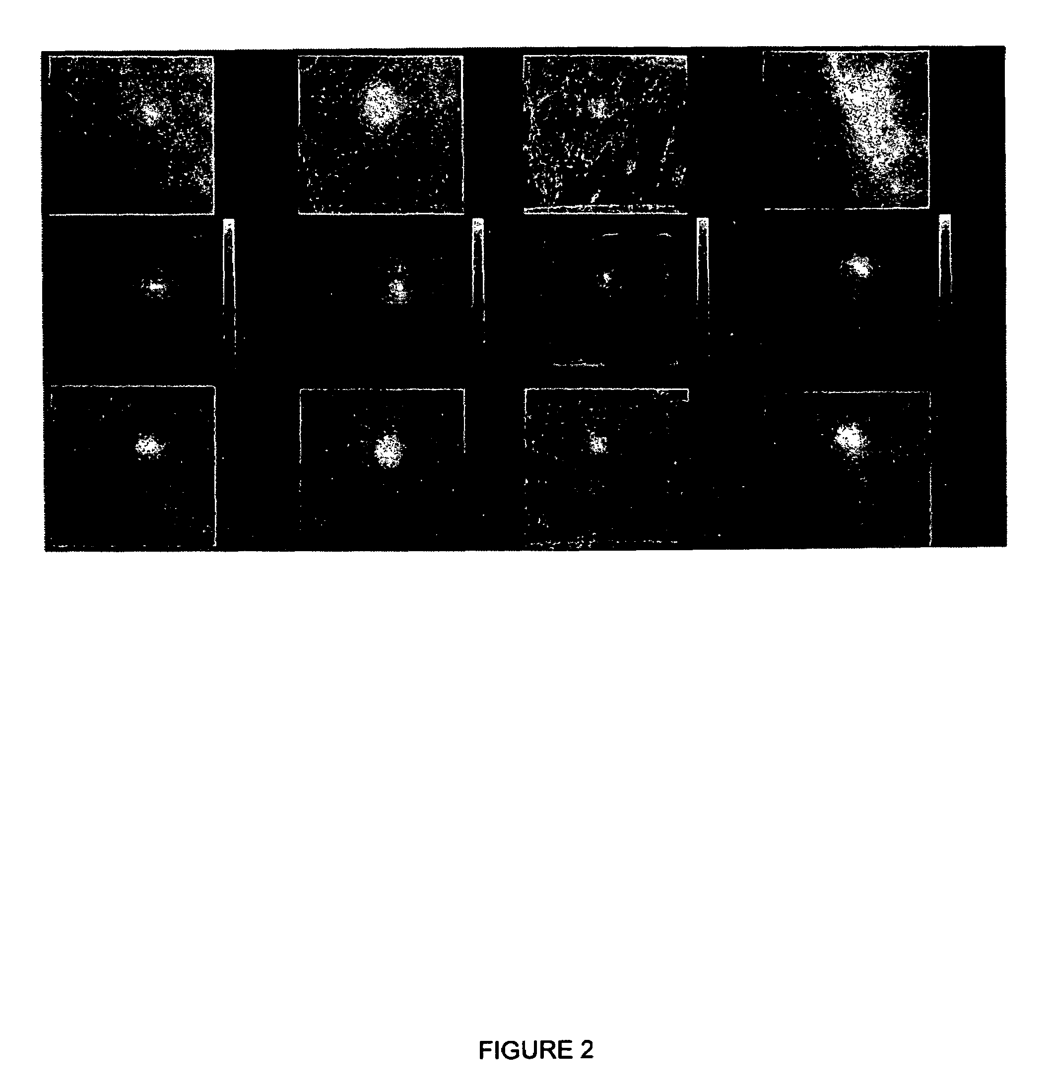
[0107]In the second experiment, about a 4.5 cm thick compressed breast phantom with a Tc99m concentration of 0.9 μCi / ml was prepared containing three lesions (two of about 8 mm diameter and one of about 6 mm diameter) containing an about 6:1 concentration over background and two 10 minute static acquisitions were obtained. A SPECT acquisition was performed with the same lesions and background solution transferred to a cylindrical (“uncompressed”) phantom with a diameter of about 9.25 cm. The SPECT acquisition angular sampling was set at about 3 degrees / step and the imaging time was set at about 30 seconds / frame. These parameters were selected to simulate about a 40 minute patient imaging time with a dual head system. Image reconstruction was obtained using a filtered back-projection technique.

[0108]In the planar imaging case, both the about 8 mm lesions and the about 6 mm lesion were visible in detector position 1 (Panel I of FIG. 10A). However, the about 6 mm lesion was not seen fr...

specific example 3

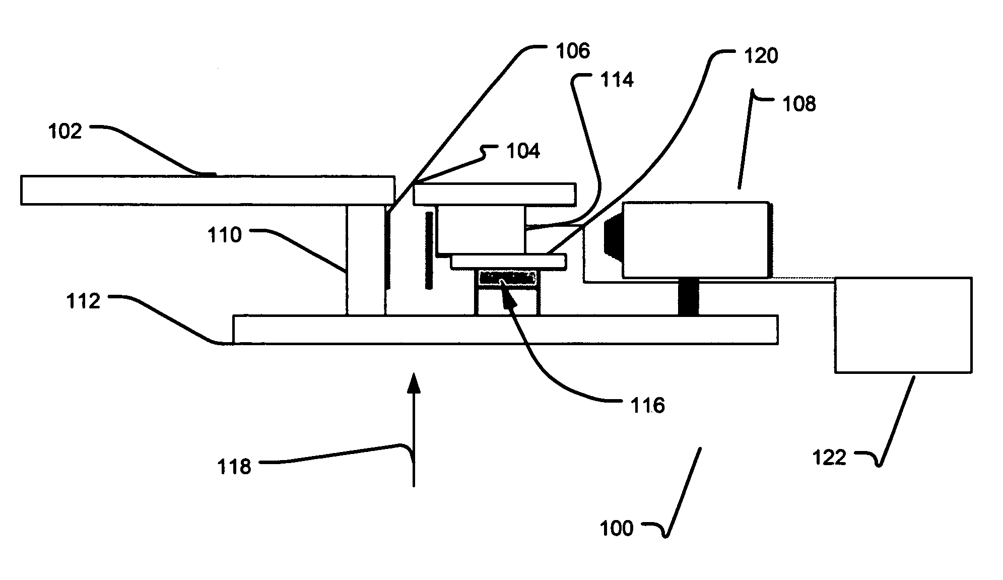
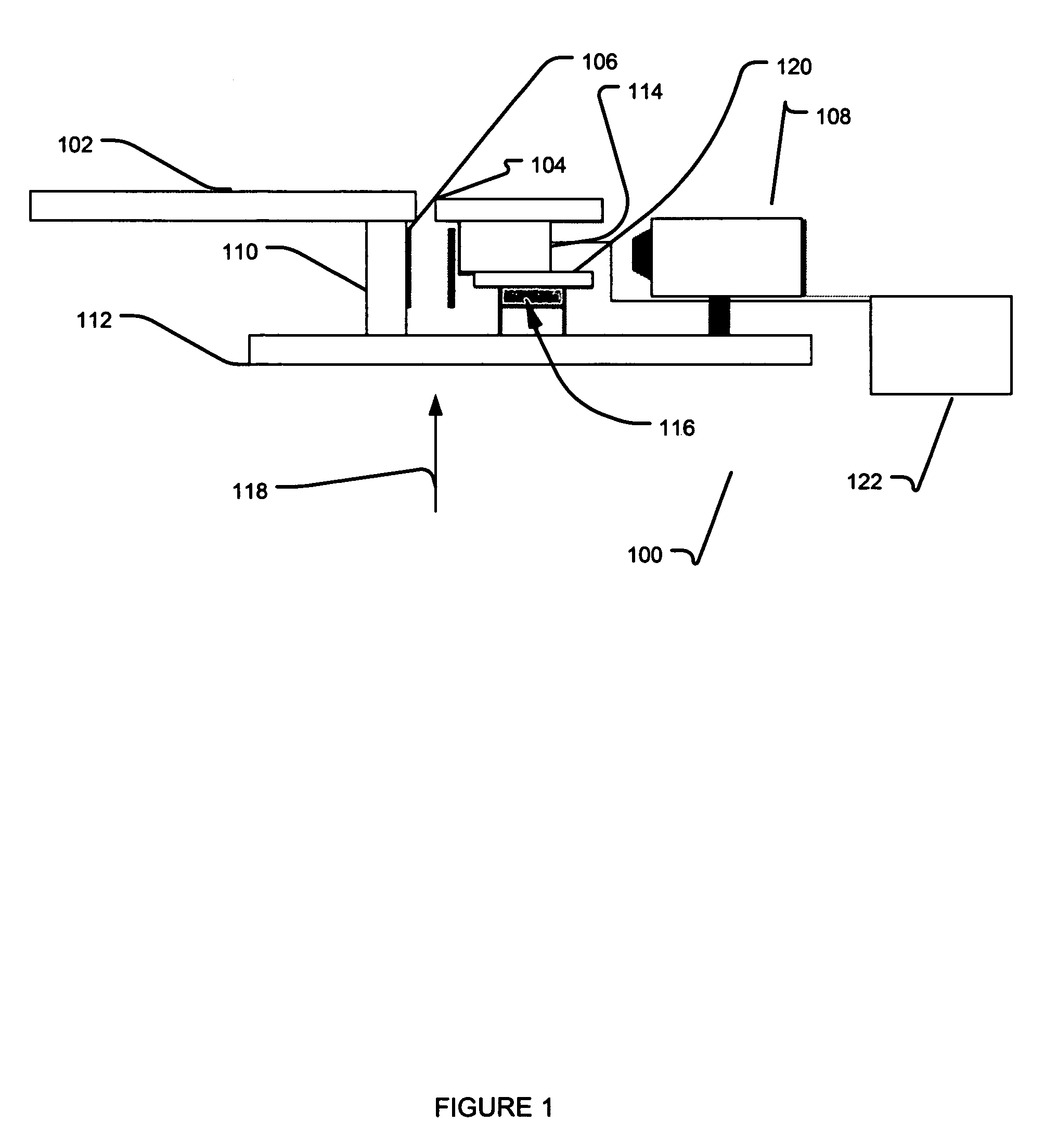
[0113]The patients enlisted in this study (N=55) were selected after a suspicious finding was reported in a routine X-ray screening mammogram. Using the mammographic films as guidance, the patients were placed on the stereotactic system table and the breast was compressed with a 5 cm×5 cm compression paddle (mean compression tissue thickness of about 5.96 cm, SD=about 1.41 cm). Scout views were obtained with the X-ray system until it was verified that the region-of-concern demonstrated in the mammogram was in the field-of-view. The mini gamma camera was then mounted to the X-ray system gantry in the needle driver position, see FIG. 1. A radiotracer was administered via venous puncture and an acquisition was initiated at the time of injection for about 10 minutes. Digital X-ray images were stored as high-resolution tiff images, and about 10-minute static gamma camera images were obtained for all patients. Additionally, dynamic data was stored in list mode for 33 of the 55 cases and r...


PUM


Abstract

The invention relates generally to biopsy needle guidance which employs an x-ray / gamma image spatial co-registration methodology. A gamma camera is configured to mount on a biopsy needle gun platform to obtain a gamma image. More particular, the spatially co-registered x-ray and physiological images may be employed for needle guidance during biopsy. Moreover, functional images may be obtained from a gamma camera at various angles relative to a target site. Further, the invention also generally relates to a breast lesion localization method using opposed gamma camera images or dual opposed images. This dual head methodology may be used to compare the lesion signal in two opposed detector images and to calculate the Z coordinate (distance from one or both of the detectors) of the lesion.

Description

CROSS-REFERENCE TO RELATED APPLICATIONS[0001]This application is related to U.S. patent application Ser. No. 11 / 538,675, entitled “A METHOD AND APPARATUS FOR LESION LOCALIZATION USING A DUAL MODALITY X-RAY / GAMMA BIOPSY SYSTEM”, and U.S. patent application Ser. No. 11 / 538,658, entitled “METHOD AND APPARATUS FOR COMBINED GAMMA / X-RAY IMAGING IN STEREOTACTIC BIOPSY”, each of which are were filed simultaneously on Oct. 4, 2006, and are incorporated by reference herein in their entirety.BACKGROUND[0002]1. Field of the Invention[0003]The invention relates generally to biopsy needle guidance by employing an x-ray / gamma image spatial co-registration methodology. Further, the invention relates to using a plurality of gamma camera images taken at different positions to identify breast lesion location. Moreover, the invention also generally relates to a breast lesion localization method using opposed gamma camera images or dual opposed images.[0004]2. Related Art[0005]X-ray imaging of the breas...

Claims


Application Information

Patent Timeline
Patent Type & Authority Patents(United States)
IPC IPC(8): A61B6/00
CPCA61B6/037A61B6/502A61B19/201A61B6/469A61B6/12A61B2019/542A61B6/583A61B2019/5408A61B2090/392A61B90/11A61B2090/3908
Inventor KEPPEL, CYNTHIAKIEPER, DOUGLAS
Owner HAMPTON UNIVERSITY
Why Eureka
Try Eureka