Patient hydration monitoring and maintenance system and method for use with administration of a diuretic

a technology for monitoring and maintaining the hydration of patients, applied in the field of patient hydration system and method, can solve the problems of patient dehydration, radiocontrast media can be toxic to patients, and patients who already suffer from various medical problems, so as to prevent dehydration, overhydration, and prevent kidney damage in patients. , the effect of preventing dehydration

Active Publication Date: 2010-07-20
MEDICAL SYST
View PDF81 Cites 40 Cited by
  • Summary
  • Abstract
  • Description
  • Claims
  • Application Information

AI Technical Summary

Benefits of technology

[0024]It is a further object of this invention to provide such a system and method which prevents kidney damage in a patient.
[0025]It is a further object of this invention to provide such a system and method which protects the patient undergoing a medical procedure, for example, a procedure involving a radiocontrast agent.
[0026]It is a further object of this invention to provide such a system and method which incorporates a balancing feature intended to prevent dehydration, overhydration, and to maintain a proper intravascular volume.
[0027]It is a further object of this invention to provide a balanced diuresis method which automatically balances fluid loss in the urine.
[0028]It is a further object of this invention to provide such a system and method which is accurate, easy to implement, and simple to operate.

Problems solved by technology

Often, the patient is dehydrated when the patient arrives at the cath lab.
Unfortunately, the radiocontrast media can be toxic to the patient especially a patient who is dehydrated at the time the radiocontrast media is injected.
A patient who already suffers from various medical problems such as diabetes or kidney problems is even more prone to medical problems due to the injection of the radiocontrast media.
It has been observed that dehydration increases the risk of radiocontrast nephropathy (RCN) when radiocontrast agents are injected into a patient during coronary and peripheral vascular catheterization procedures.
Since the presence of radiocontrast media in the urine makes it much more difficult for the kidney to concentrate urine, the work of the medulla outstrips the available oxygen supply and leads to medullary ischemia.
It is also clear that dehydration increases the risk of RCN, likely because urine (and contrast media inside the kidney) is excessively concentrated.
Hydration is commonly performed at a conservative rate, especially in patients with existing heart and kidney dysfunction, since over-hydration can result in pulmonary edema (fluid in the lungs), shortness of breath, the need for intubation, and even death.
Thus, the patients at highest risk for RCN are those least likely to receive the only proven therapy for preventing RCN (I.V. hydration) due to the unpredictability of side effects from I.V. hydration.
A major limitation to the more widespread use of the already known therapeutic, or optimal, levels of I.V. hydration is the current inability to balance the amount of fluid going into the patient to the amount of fluid being removed or excreted from the patient.
It is possible to have a nurse measure a patient's urine output frequently but this method is impractical as nurses are often responsible for the care of many patients.
During this time, the amount of I.V. hydration can be significantly less than the amount of urine produced by the kidneys and stored in the bladder, leading to dehydration.
Since many patients do not normally have such a catheter during procedures using radiocontrast media, a valid measurement of urine output is not possible.
There seems to be indisputable scientific evidence that RCN in patients with even mild baseline renal insufficiency can lead to long term complications and even increased risk of mortality.
This scientific knowledge has not yet been extended to daily clinical practice as routine monitoring of renal function post-catheterization is not usually performed and limits the identification of the known short-term clinical complications.
Many cardiologists go considerable length to protect these patients including slow, overnight hydration (an extra admission day), administration of marginally effective but expensive drugs, staging the procedure, or not performing procedures at all.
An anesthetized or sedated patient may not be able to drink.
In fact, they were used to prevent RCN based on this assumption until clinical evidence suggested that they were actually deleterious.
More recently, doubts have been raised regarding the validity of those negative clinical studies.
Weinstein et al. found that furosemide-treated subjects lost 0.7 kg on average, whereas a 1.3-kg weight gain was noted in patients randomized to hydration alone, suggesting that in furosemide-treated subjects the hydration protocol has been insufficient and patients were dehydrated by excessive diuresis.
The actual effect of diuretic is typically not known for 4 hours (until the sufficient amount of urine is collected and measured) and it is too late and too difficult to correct any imbalance.
As a result the patient can lose “net” 700 ml of body fluid and become dehydrated.
If such patient is vulnerable to renal insult, they can suffer kidney damage.
Unfortunately, there are currently no practical ways of achieving this in a clinical setting since in response to the diuretic infusion the patient's urine output changes rapidly and unpredictably.
While this can be achieved in experimental setting, this method is not possible in current clinical practice where nursing time is very limited and one nurse is often responsible for monitoring the care of up to ten patients.
In addition, frequent adjustments and measurements of this kind often result in a human error.
Some patients, such as heart failure patients, may be overloaded with fluid.
Nurses, however, are often very busy and it is possible that a patient, once given a diuretic, could expel urine to the point the patient becomes hydrated.
There is no known prior art system which achieves automatic balanced hydration in a patient.

Method used


Image

  • Patient hydration monitoring and maintenance system and method for use with administration of a diuretic
  • Patient hydration monitoring and maintenance system and method for use with administration of a diuretic
  • Patient hydration monitoring and maintenance system and method for use with administration of a diuretic

Examples


Embodiment Construction

[0043]Aside from the preferred embodiment or embodiments disclosed below, this invention is capable of other embodiments and of being practiced or being carried out in various ways. Thus, it is to be understood that the invention is not limited in its application to the details of construction and the arrangements of components set forth in the following description or illustrated in the drawings. If only one embodiment is described herein, the claims hereof are not to be limited to that embodiment. Moreover, the claims hereof are not to be read restrictively unless there is clear and convincing evidence manifesting a certain exclusion, restriction, or disclaimer.

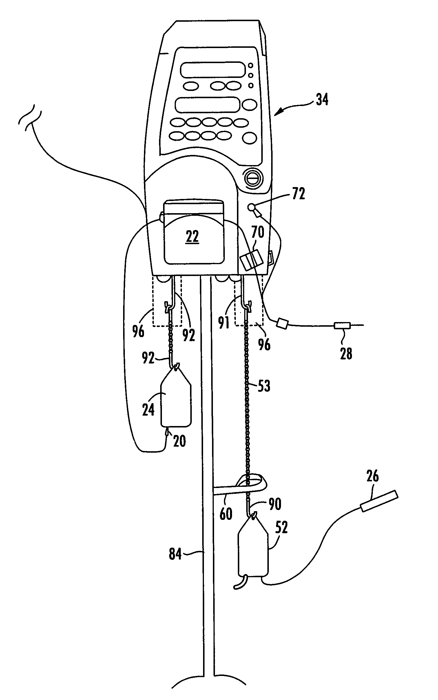
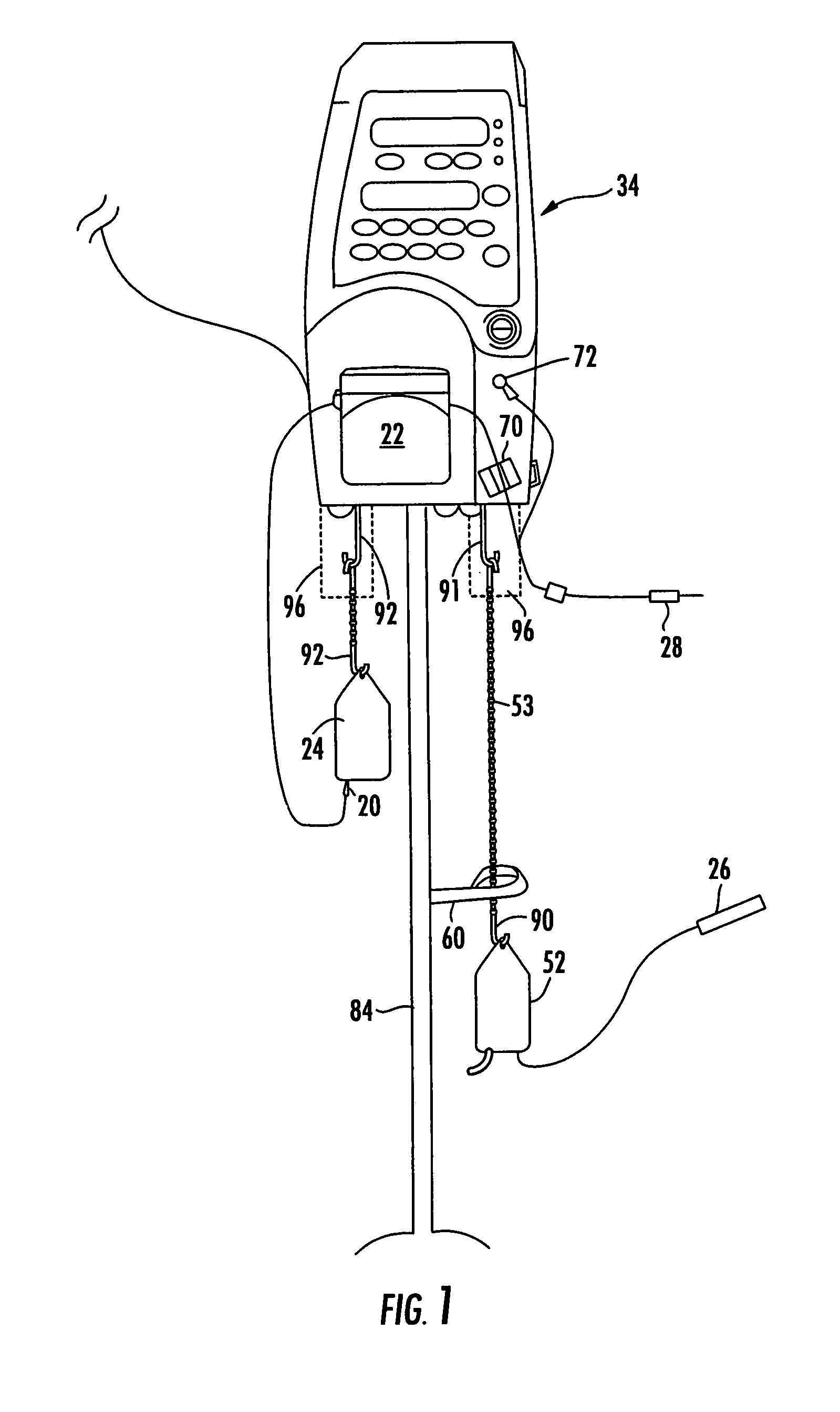
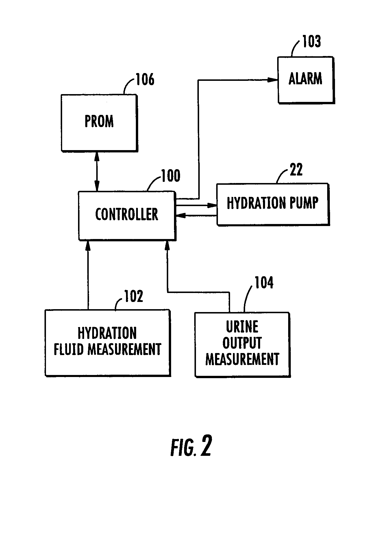
[0044]One preferred example of a patient hydration system according to this invention includes unit 34, FIG. 1 typically mounted on IV pole 84. Unit 34 has programmable controller electronics therein. There is an infusion subsystem including pump 22 responsive to source of infusion fluid 24 for infusing a patient with hydra...


PUM


Abstract

A patient hydration system with an infusion subsystem responsive to a source of hydration fluid for infusing the patient with hydration fluid. A urine output measurement subsystem determines the amount of urine output by the patient. A setting is received corresponding to an amount of urine to be output by the patient. When or after the urine output measurement subsystem indicates the set amount of urine has been output by the patient, the infusion subsystem is controlled to administer hydration fluid to the patient based on the patient's further urine output.

Description

RELATED APPLICATIONS[0001]This application is a continuation-in-part application of U.S. patent application Ser. No. 10 / 936,945, filed Sep. 9, 2004, entitled “Patient Hydration System and Method”. This application is also related to co-pending application Ser. Nos. 11 / 408,851; 11 / 408,391; 11 / 409,171; and 11 / 580,354 all of which are incorporated herein by this reference.FIELD OF THE INVENTION[0002]This invention relates to a patient hydration system and method.BACKGROUND OF THE INVENTION[0003]The “cath lab” in a hospital is where a patient is injected with a radiocontrast media, imaged, diagnosed, and often operated on. Typically, a cardiologist refers the patient to the cath lab and the patient is instructed not to eat or drink the night before. In the case of a patient suffering a heart attack, the patient may be transferred directly to the cath lab.[0004]Often, the patient is dehydrated when the patient arrives at the cath lab. The patient is prepped and the radiocontrast media in...

Claims


Application Information

Patent Timeline
Patent Type & Authority Patents(United States)
IPC IPC(8): A61M31/00A61M1/00
CPCA61B5/201A61B5/208A61B5/4839A61M5/007A61M5/142G01G17/04A61M5/16895A61M5/1723A61M5/365A61M2205/18A61M2205/3355A61M2205/3393
Inventor GELFAND, MARKRUDKO, ROBERT I.TAUSCHER, MARK R.LEVIN, HOWARD R.HALPERT, ANDY
Owner MEDICAL SYST
Why Eureka
Try Eureka