Multi-threshold peripheral equalization method and apparatus for digital mammography and breast tomosynthesis

a technology of peripheral equalization and digital mammography, applied in the field of image processing, can solve problems such as unsatisfactory artifacts in images, and achieve the effects of improving image reading and interpretation efficiency, enhancing intensity, and enhancing the peripheral area of breasts

Active Publication Date: 2010-07-27
THE GENERAL HOSPITAL CORP
View PDF8 Cites 6 Cited by
  • Summary
  • Abstract
  • Description
  • Claims
  • Application Information

AI Technical Summary

Benefits of technology

[0006]In accordance with the present invention, it has been recognized that it would be desirable to improve the efficiency of image reading and interpretation by equalizing the intensity of the peripheral and central areas of a breast image such that the entire image can be viewed (and thus assessed by a reader) at one window-level setting.
[0007]A peripheral equalization (PE) method for digital mammography and breast tomosynthesis includes identifying areas of the breast corresponding to at least a central part and a peripheral part, using a fixed pixel intensity to process pixel data associated with the central part of the breast and utilizing multiple thresholds to process the pixel data associated with the peripheral part of the breast. With this tiered arrangement, an image having a peripheral area with enhanced intensity is provided. The image may be generated via mammography or tomosynthesis. By utilizing a fixed pixel intensity to process pixel data associated with the central part of the breast and utilizing multiple thresholds to process the pixel data associated with the peripheral part of the breast, it is possible to provide two-dimensional and three-dimensional presentations of the breast having an enhanced peripheral area of the breast without also introducing substantial and distracting “segmentation lines” (or boundary effect artifacts) into the image. Thus, a reader of the image can view and assess the breast image at one window-level setting.
[0008]In accordance with a further aspect of the present invention, a technique for segmenting and enhancing images includes using multiple thresholds to generate multiple images and averaging corresponding pixel values between each of the images to produce an image having smooth transitions between a central region and peripheral regions of the breast image. With this tiered arrangement, a segmentation and peripheral enhancement technique which provides enhancement of the peripheral area of the breast without also introducing substantial and distracting “segmentation lines” (or a “boundary effect” artifacts) into the image is provided. Thus, the technique of the present invention eliminates the so-called “boundary effect” typically observed in images if only a single threshold is used in the segmentation.
[0009]In accordance with a still further aspect of the present invention, a method of processing a mammographic image includes converting a projection map of an image into a density map of the image, performing a transform on the density map, filtering the transformed data, performing an inverse transform on the filtered data to provide a new image and segmenting and enhancing the new image using multiple thresholds to process image data corresponding to at least a peripheral region of a breast in the image to provide an image having peripheral equalization. With this particular arrangement, a technique which provides peripheral equalization in digital mammography data sets and images is provided. By using multiple thresholds, the technique which provides enhancement of a peripheral area of the breast without also introducing substantial and distracting “segmentation lines” (or a “boundary effect” artifacts) into the image is provided. Thus, the technique of the present invention eliminates the so-called “boundary effect” typically observed in images if only a single threshold is used in the segmentation.
[0010]A method of processing tomographic image data comprising selecting a subset of the tomographic image data which corresponds to a slice of the image, performing a transform on the selected subset of image data, filtering the transformed data, performing an inverse transform on the filtered data to provide a new image and segmenting and enhancing at least a portion of the subset of the tomographic image data corresponding to a peripheral region of a breast in the new image using multiple thresholds to provide an image having peripheral equalization. With this particular arrangement, a technique which utilizes multiple thresholds to provide a tomographic image having enhanced peripheral areas of the breast without also introducing substantial and distracting “segmentation lines” (or a “boundary effect”) into the image is provided. It should be appreciated that a three-dimensional (3-D) tomosynthesis reconstruction can be provided from a plurality of two-dimensional (2-D) images. Thus, the peripheral equalization (PE) technique for a 3-D volume is substantially similar to that for a 2-D image in that the peripheral equalization technique can be applied to each image slice of a 3-D volume. For example, if a 3-D tomosynthesis volume contains fifty 2-D image slices, one approach would be to perform peripheral equalization on each of the 50 slices. Alternatively, the technique can be applied to a subset of the 2-D images which make up the 3-D volume (e.g. the peripheral equalization technique can be applied to one out of every N 2-D image slices). Thus, the second approach is to compute the PE ratio for only certain slices (as opposed to every slice) and then use the PE ratio for neighboring slices. For example, a PE ratio can be computed for slice three and then used for slices one, two, three, four and five. A next PE ratio can then be computed for slice eight and used for slices six, seven, eight, nine and ten. This continues until each slice has a PE ratio applied thereto. This approach works well in those image datasets in which neighbor slices contain similar structures and PE ratios tend to be relatively close. This second approach can reduce the amount of time and / or resources necessary for computations.

Problems solved by technology

These approaches, however, result in undesirable artifacts within the image.
In particular, prior art techniques result in the image having so-called segmentation lines and characteristic “overshoot” banding which are distracting and thus undesirable to a reader (e.g. a radiologist or other medical professional) of the image.

Method used


Image

  • Multi-threshold peripheral equalization method and apparatus for digital mammography and breast tomosynthesis
  • Multi-threshold peripheral equalization method and apparatus for digital mammography and breast tomosynthesis
  • Multi-threshold peripheral equalization method and apparatus for digital mammography and breast tomosynthesis

Examples


Embodiment Construction

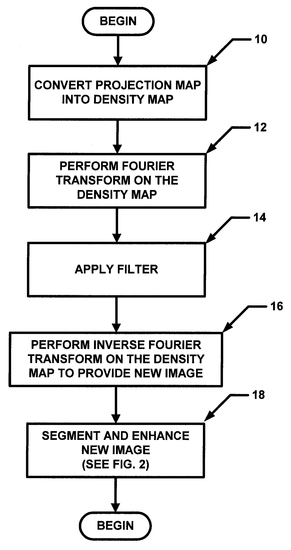
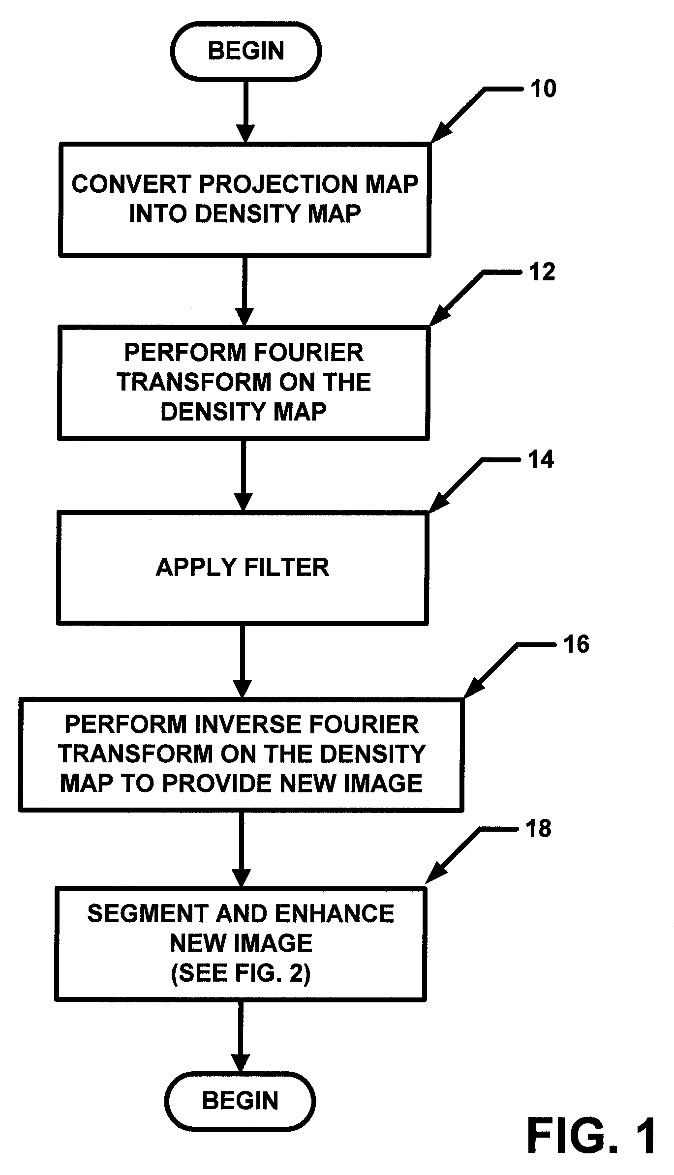
[0019]In general overview, the general concept described herein is to estimate the normalized thickness profile (NTP) of a breast from an image (e.g. a mammogram image) and enhance the peripheral area. This can accomplished by dividing the NTP from the mammogram. In one particular but exemplary embodiment, a projection mammogram was first segmented into “breast” and “background” regions using a threshold value computed using the Otsu technique. A segmentation image (SI) was generated in which pixels were assigned a first value (e.g. value of one) in a breast region and a second value (e.g. a value of zero) in background region. The projection was then converted into an attenuation image (AI). A two-dimensional (2D) low-pass filter was applied to the AI in the spatial frequency domain to obtain a blurred image (BI), which primarily reflected variations in breast thickness. The low-pass filter used had the following filter characteristic:

F(fx,fy)=1 / {[(1−|fx| / fc)^128]*[(1−|fy| / fc)^128]...


PUM


Abstract

A peripheral equalization (PE) method and apparatus for compensating for thickness reduction in outer edges of the breast in a mammogram (i.e. a two-dimensional image) while keeping the central area substantially unchanged. The PE method and apparatus can also be applied to three dimensional (tomosynthesis) images of a breast. The peripheral equalization is achieved by segmenting the image of the breast into at least two regions and using a multi-threshold technique to process the data in at least one of the two regions.

Description

CROSS-REFERENCE TO RELATED APPLICATIONS[0001]This application claims the benefit of U.S. Provisional Application No. 60 / 711,023 filed on Aug. 24, 2005, under 35 U.S.C. §119(e), which application is hereby incorporated herein by reference in its entirety.STATEMENTS REGARDING FEDERALLY SPONSORED RESEARCH[0002]Not applicable.FIELD OF THE INVENTION[0003]This invention relates generally to image processing and more particularly to image processing of mammography and breast tomosynthesis images.BACKGROUND OF THE INVENTION[0004]As is known in the art, in two-dimensional (2-D) mammography and three-dimensional (3-D) breast tomosynthesis images, the peripheral area of the breast tends to have lower intensity than the central area of the breast. This is due at least in part to the thickness of a compressed breast decreasing from the central area to the peripheral area. Thus, to assess an image of the entire breast, a radiologist (or other medical professional or reader of the image) typically...

Claims


Application Information

Patent Timeline
Patent Type & Authority Patents(United States)
IPC IPC(8): G06K9/00A61B6/00
CPCG06T5/10G06T7/0081G06T5/007G06T2207/10112G06T2207/20012G06T2207/20056G06T2207/20216G06T2207/30068G06T2207/20148G06T7/11G06T7/136
Inventor WU, TAOMOORE, RICHARD H.KOPANS, DANIEL B.
Owner THE GENERAL HOSPITAL CORP
Why Eureka
Try Eureka