

Nuclear medical diagnostic device
a diagnostic device and nuclear technology, applied in the field of nuclear medical diagnostic devices, can solve the problems of adverse impact on image quality, inaccurate tomogram obtained by the pet device, medical diagnostic devices, etc., and achieve the effect of accurate assignment, high-quality tomogram and easy determination of optimal parameters
- Summary
- Abstract
- Description
- Claims
- Application Information
AI Technical Summary
Benefits of technology
Problems solved by technology
Method used
Image

Examples
first embodiment
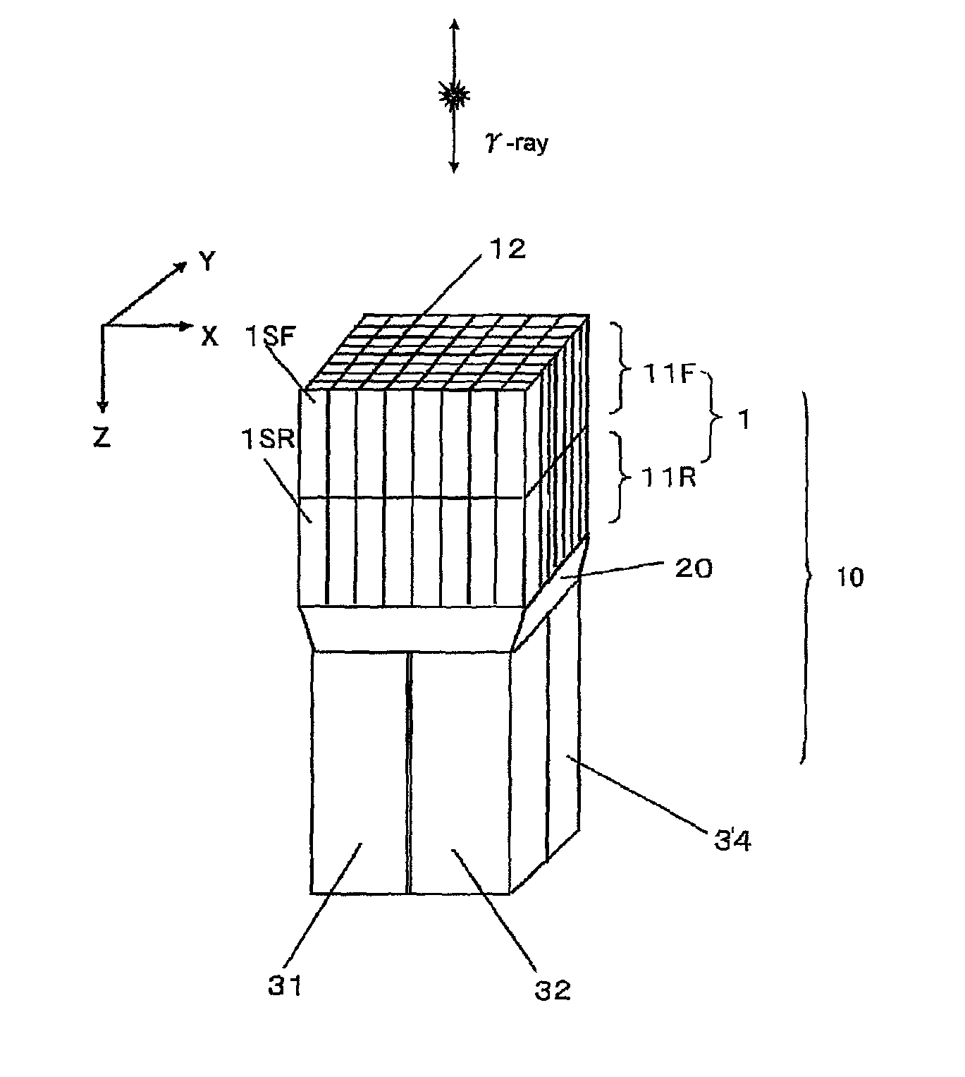
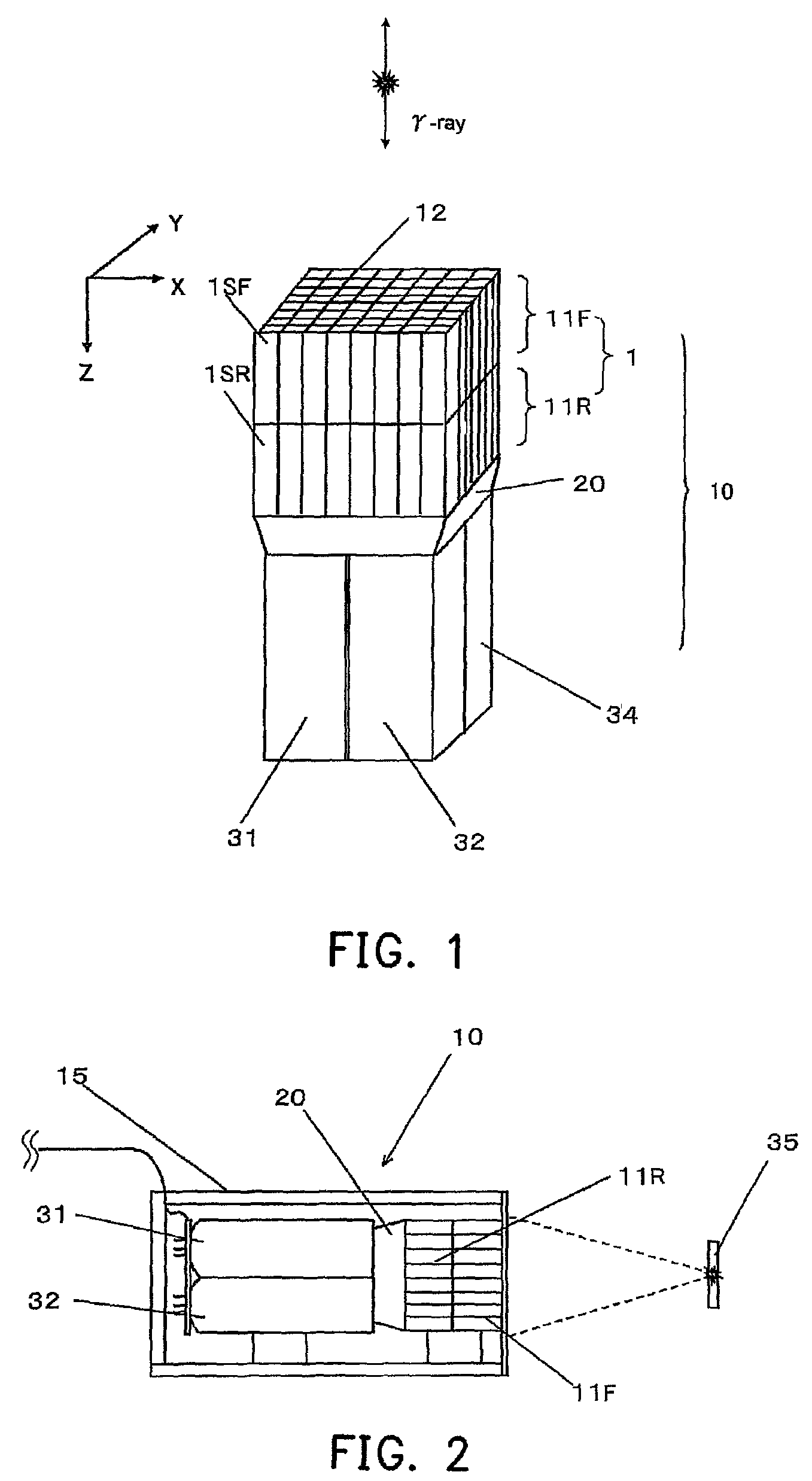
[0043]The detailed structure of a γ-ray detector according to the first embodiment of the present invention is shown in the drawings and is illustrated in the following. FIG. 1 is an outside view of a γ-ray detector 10 having scintillator arrays of a two-stage structure of the present invention. As shown in FIG. 1, in the γ-ray detector 10, a scintillator block 1 is arranged by being divided in a γ-ray incident depth direction, that is, the radiation detector 10 is a depth of interaction (DOI) γ-ray detector having three-dimensionally arranged scintillators. The DOI γ-ray detector of this embodiment has scintillator arrays of a two-stage structure.
[0044]The γ-ray detector 10 of this embodiment is approximately formed by four parts. The first part is a scintillator array 11F having scintillators 1SF that are two-dimensionally and compactly arranged and have a luminescence pulse with a shorter attenuation time. The scintillators 1SF are divided by appropriately sandwiching a light ref...
second embodiment
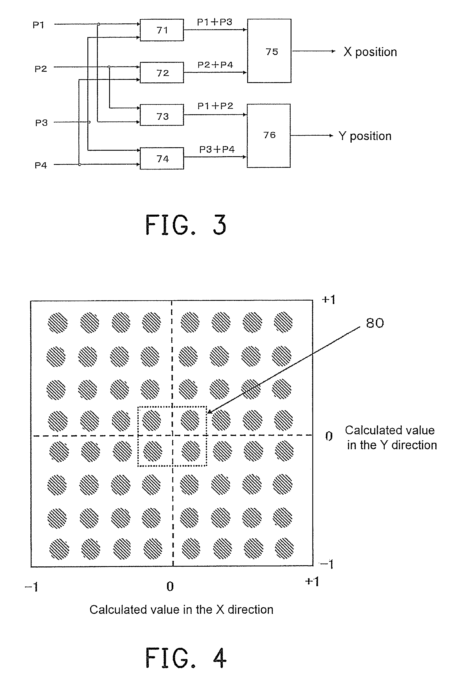
[0066]The detailed structure of the γ-ray detector according to the second embodiment of the present invention is shown in the drawings and is illustrated in the following. This embodiment is used for the situation in which the optimal values are more strictly decided. The scintillator array identification mechanism defines the first signal count ratio, which is R1=(N1−N1b) / (N2−N2b) and R1′=(N2′−N2b) / (N1′−N1b), obtained during the first inspection stage of the γ-ray detector single unit, and defines the parameters T1, T2, and K when the values of R and R′ are equal and become the maximum as the optimal values. Until now, the process is the same as that of the first embodiment (FIGS. 6 to 8).
[0067]As shown in FIG. 11, during a second inspection stage of the γ-ray detector single unit, the γ-ray detector 10 is disposed in the dark box 15, the optimal parameters T1, T2, and K decided above are input to the processing circuit for inspection 16. In this state, the Ri ray source 35 is arr...
third embodiment
[0075]The detailed structure of the γ-ray detector according to the third embodiment of the present invention is shown in the drawings and is illustrated in the following. The scintillator array identification mechanism defines the first signal count ratio, which is R1=(N1−N1b) / (N2−N2b) and R1′=(N2′−N2b) / (N1′−N1b), obtained during the first inspection stage of the γ-ray detector single unit, and defines the parameters T1, T2, and K when the values of R and R′ are equal and become the maximum as the optimal values. Until now, the process is the same as that of the first embodiment (FIGS. 6 to 8).
[0076]Then, as shown in FIG. 14, during a second inspection stage of the γ-ray detector single unit, the γ-ray detector 10 is disposed in the dark box 15, the optimal parameters T1, T2, and K decided above are input to the processing circuit for inspection 16. In this state, the Ri ray source 35 is arranged on a position at a distance 1 relative to the γ-ray detectors 10, and irradiates the γ...
PUM
 Abstract
Description
Claims
Application Information