Intraocular lens system

a lens system and intraocular technology, applied in intraocular lenses, medical science, prosthesis, etc., can solve the problems of corneal refractive surgery, lack of post-operative refractive accuracy, and current procedures and methods used by refractive surgeons may not meet the total refractive needs of patients,

Active Publication Date: 2010-10-12
EMMETROPIA
View PDF74 Cites 76 Cited by
  • Summary
  • Abstract
  • Description
  • Claims
  • Application Information

AI Technical Summary

Benefits of technology

[0037]It is an additional aspect of the present invention to provide a multi-component intraocular lens system with foldable components in order to minimize trauma to the eye. Trauma is minimized by allowing the use of a delivery system for the foldable lens which requires an incision smaller than the unfolded diameter of the foldable lens.
[0042]Further, the present invention includes a feature wherein the haptic of the mid lens has projections extending anteriorly and posteriorly that capture the top lens (circular configuration) and retain the top and mid lens (circular configuration) as an optical assembly. The dual direction extending projections allow the surgeon or manufacturer the ability to orient the components independently at each of 360° orientations. Each of the circular components (i.e., the mid lens and top lens) can be used to correct sphere cylinder presbyopia, spherical aberrations, and higher order aberrations.
[0043]Also, the present invention includes an embodiment wherein the mid and top lenses are integrated into a single component such that an adhesive or other such joining mechanism is not necessary to join the mid and top lenses together.
[0045]The intraocular lens system of the present invention allows assembly without the use of special equipment or techniques for securing the top and mid lenses together.

Problems solved by technology

Current procedures and methods used by refractive surgeons may not satisfy the total refractive needs of the patient.
Particularly, the most commonly performed refractive surgical procedures, such as, for example, cataract extraction with intraocular lens implantation, in addition to the most recently popularized corneal refractive surgical procedures, such as eximer laser photoblation, exhibit limitations.
One reason for the limitations is the lack of post-operative refractive accuracy.
The lack of post-operative refractive accuracy renders the commonly known refractive surgical procedures uncompetitive with currently available non-surgical alternatives for patients, for example, glasses and contact lenses.
Difficulties arise in treating presbyopia because typically once a person manifests symptoms of presbyopia, the symptoms worsen as the person ages.
Conventional techniques for replacing an intraocular lens each time the patient's vision deteriorated do not always present a practical or cost-effective approach.
Patients suffering from eye trauma or other eye afflictions may have the iris or other portions of the eye distorted, destroyed, or discolored.
Such visualization, although not outside the scope of a surgeon skilled in the art, increases the degree of difficulty of the procedure, thus increasing the chance that a surgical error or other problem will arise in the surgical procedure, leading to unwanted complications.
Furthermore, analyses of current corneal refractive technologies indicate the presence of a significant amount of preexisting or naturally occurring post-operative, as well as preoperative, image distortion (optical aberration) or degradation, particularly under low light conditions, such as when driving at night.
Due to the practical limits of performing intraocular surgery, as well as the biological and physical behavior of the human eye during and after various types of intraocular surgery, predictability at the + / −0.25 D accuracy level with a single surgical procedure is difficult to achieve as a practical matter.
Furthermore, factors such as biometry errors, variable wound healing, and capsular contraction around the intraocular lenses contribute to decreasing the likelihood of achieving the desired refractive accuracy.
The engulfment typically makes the removal of the lenses and their haptics more difficult.
Exchange of iris fixated anterior chamber lenses does not typically guarantee precise position or orientation.
Posterior chamber lenses similarly cannot be removed because of posterior capsule fibrosis.
However, the surgical implantation procedure itself may create additional optical aberrations which cannot be anticipated preoperatively and thus the primary lens implant cannot account for these optical aberrations.
However, the design of current intraocular lenses typically makes such a procedure difficult and impractical.
Furthermore, after a period of time with normal healing, it becomes physically dangerous and / or nearly impossible to the patient to have the implanted lens removed once the eye tissue takes hold on the capsular fixation holes of the lens.
Therefore, such an argument is not realistic, practical, or safe.
A single component intraocular lens, which in general is not designed to be removed and with only two optical surfaces, cannot accurately allow for compensation of sphere, cylinder, cylindrical axis, and all forms of optical aberrations that may be discovered after the initial implantation.
The easy exchangeability of optical elements is critical because the actual surgery of implanting the lens in the first place, as well as variances in the manner in which the eye heals after implantation, potentially create distortions which may not stabilize for several months after the operation.
Therefore, the ability to measure and to compensate for the distortion(s) optimally takes place several months after surgery and cannot typically be predicted prior thereto.
Since the same surgical wound is used for both the primary and secondary operations, additional distortion due to wound healing would not be anticipated as a result of the second operation.
From an optical standpoint, any time incisions are made to the cornea, the cornea loses some of its natural globularity due to imperfections caused by the incisions and the resultant trauma.
The imperfections in the cornea lead to induced astigmatism, or optical aberrations caused by irregularities in the shape of the cornea.

Method used


Image

  • Intraocular lens system
  • Intraocular lens system
  • Intraocular lens system

Examples


Embodiment Construction

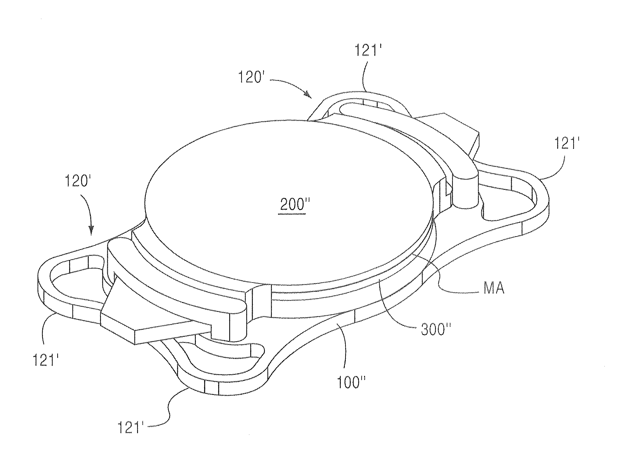
[0073]It should be noted that according to the preferred embodiments of the present invention, the fully assembled or end appearance of the base lens 100′ of the present invention compared to the base lens 100 described above and illustrated in FIGS. 12A, 12B and 13 is substantially similar to the base lens 100 disclosed in the '875 application. Therefore, a detailed description of many of the common features of the base lens 100′ with the base lens 100 is omitted herefrom in order to avoid redundancy.

[0074]However, it should be noted that the base lens 100′ of the present invention differs from the above-described base lens 100 in that the base lens 100′ is initially manufactured to be in three components that are to be assembled together and haptics 120′ in the base lens 100′ are configured to be a plate shape haptic 120′ as opposed to the C-loop haptic 120 in the above-described base lens 100. The plate shaped hepatic 120′ (FIG. 24) is configured to have ears 121′, 121′ which are...


PUM


Abstract

A multi-component intraocular lens implanted in an optical system of a human eye, including one or more foldable removable components, each component being foldable. One component acts as a base lens, including a flange with a slot. Another component acts an optical assembly that may include a top lens joined to or integrated with a mid lens. The top lens, the mid lens or the optical assembly may include at least one projection that engages the slot of the base lens. A medical adhesive may be applied to an outer circumferential surface of the top lens to join the top lens to the mid lens or may be applied to a top surface of the top lens opposing a bottom surface of the mid lens.

Description

CROSS-REFERENCE TO RELATED APPLICATION[0001]This application is a continuation-in-part application of application Ser. No. 11 / 698,875 filed Jan. 29, 2007, the entire contents of which are incorporated herein by reference.BACKGROUND OF THE INVENTION[0002]1. Field of the Invention[0003]This invention relates to a method for correcting the optical system of an eye using an intraocular lens system. Particularly, this invention relates to a method of correcting focusing abnormalities and optical aberrations measured by wave front or similar technology to quantify optical aberrations in the optical system of the eye, using a laser, or other apparatus and / or methods of fabricating or modifying a lens, for the optical system of an eye having a foldable, interchangeable intraocular lens system provided therein.[0004]2. Description of Related Art[0005]The field of refractive surgery has evolved rapidly during the past few decades. Current procedures and methods used by refractive surgeons may...

Claims


Application Information

Patent Timeline
Patent Type & Authority Patents(United States)
IPC IPC(8): A61F2/16
CPCA61F2/1648A61F2250/0059A61F2220/0033A61F2220/005A61F2002/169053
Inventor WERBLIN, THEODORE P.
Owner EMMETROPIA
Why Eureka
Try Eureka