

Compression paddle membrane and tensioning apparatus for compressing tissue for medical imaging purposes
a compression paddle and tissue technology, applied in the field of probe interface assembly, can solve the problems of inconvenient compression, adverse effects on ultrasound image quality, inconvenient compression, etc., and achieve the effect of reducing the burden on the radiologist to correlate an x-ray image to an ultrasound, and improving the accuracy of the compression paddl
- Summary
- Abstract
- Description
- Claims
- Application Information
AI Technical Summary
Benefits of technology
Problems solved by technology
Method used
Image


Examples
Embodiment Construction
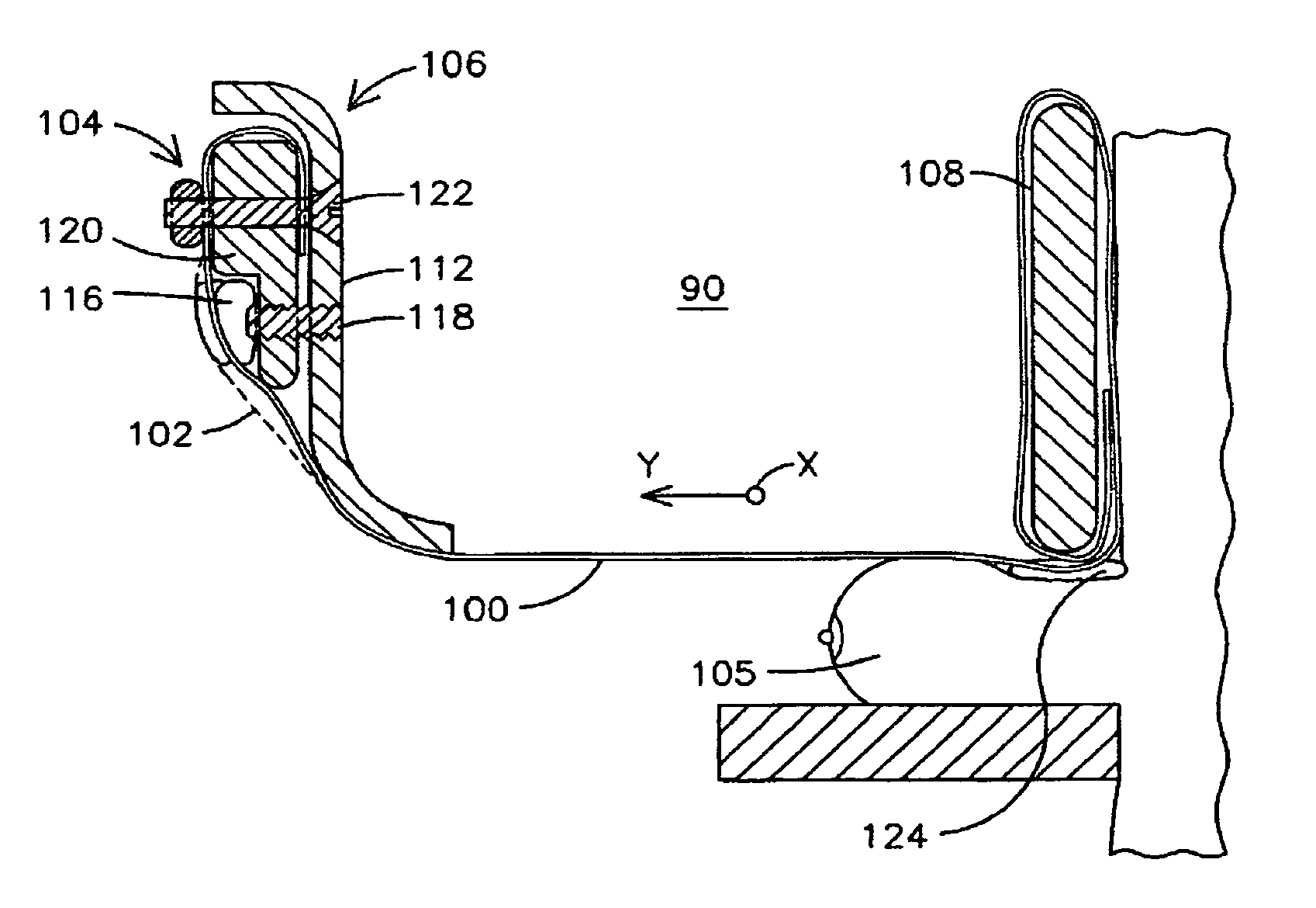
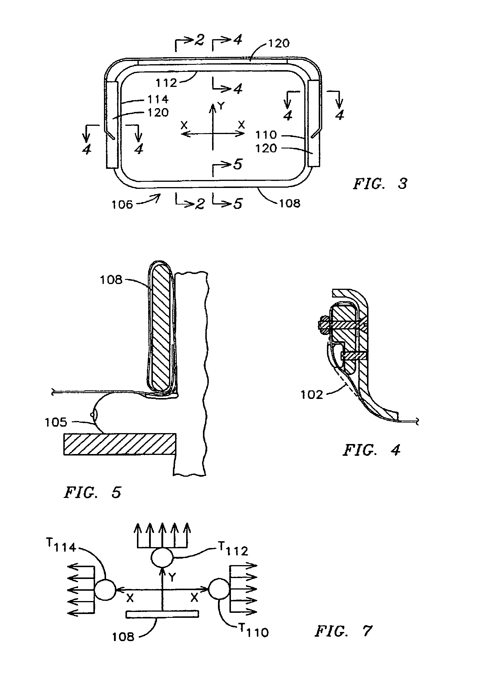
[0016]The inventors of the present invention have recognized an innovative means for compressing tissue to be scanned for medical imaging purposes, such as during an automated ultrasound breast scan that may be combined with an X-ray mammogram. Aspects of the present invention enable accurate, reproducible ultrasound images reducing distortion and attenuation, which may be introduced as a consequence of combining the ultrasound scanning with X-ray mammography.
[0017]In one exemplary embodiment, a thin polymeric membrane may be used to compress the breast tissue. The membrane thinness (e.g., 90 is utilized to apply tensile forces that pulls the membrane taut and prevents it from excessive deflection. In one exemplary embodiment, this tensioning apparatus enables the membrane to apply compression loads up to ˜30 dkN with deformations comparable and even less than those achieved using a conventional rigid plastic paddle, e.g., 5 mm maximum deflection at the plate center for 20 dkN compr...
PUM
 Abstract
Description
Claims
Application Information