
Endoscopic apparatus with integrated multiple biopsy device

a biopsy device and endoscope technology, applied in the field of medical devices, can solve the problems of reducing the flexibility of the scope, hand-assembling, and high cost of the conventional endoscop

Inactive Publication Date: 2010-12-07
BOSTON SCI SCIMED INC
View PDF595 Cites 78 Cited by
  • Summary
  • Abstract
  • Description
  • Claims
  • Application Information

AI Technical Summary

Benefits of technology

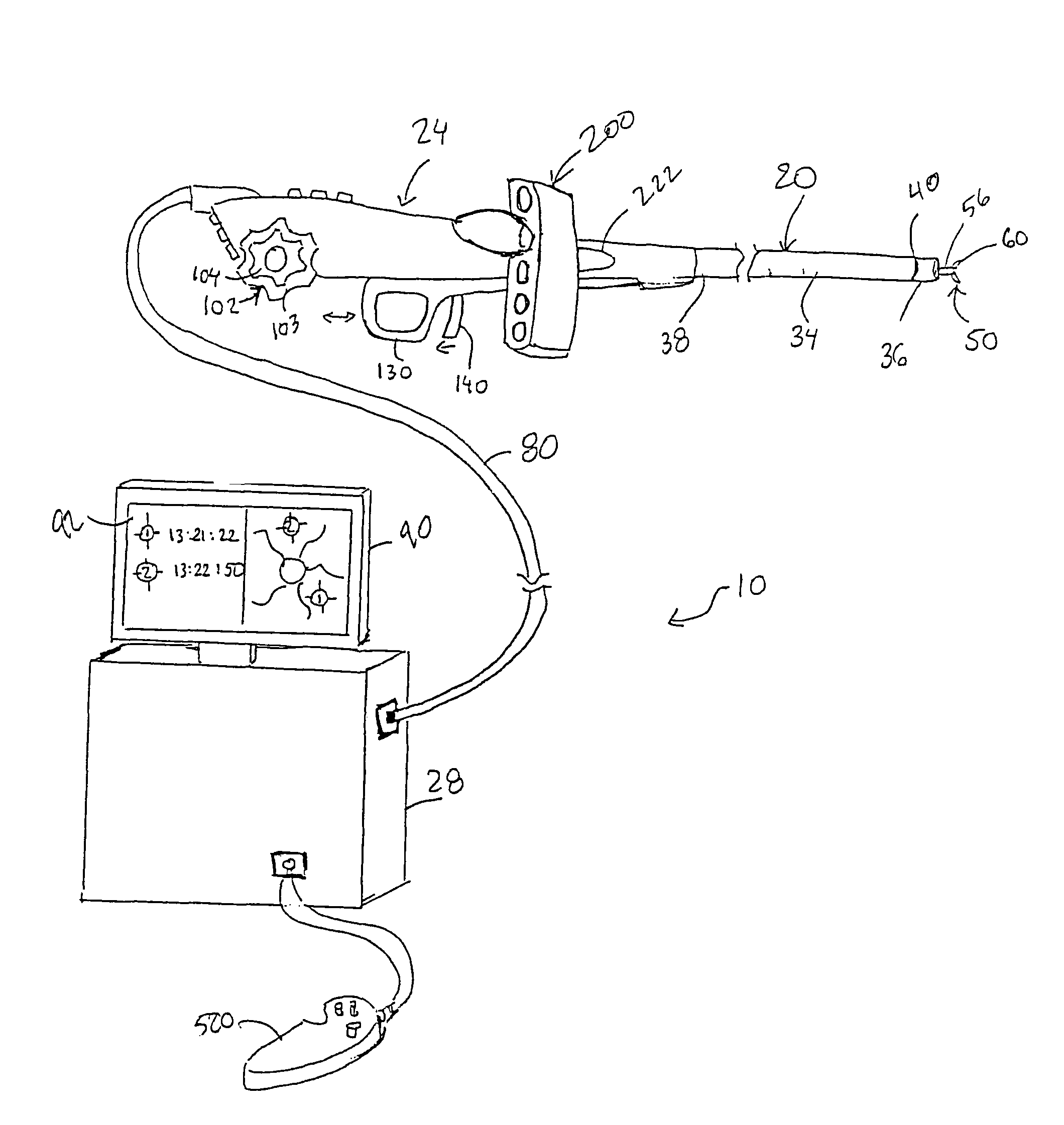
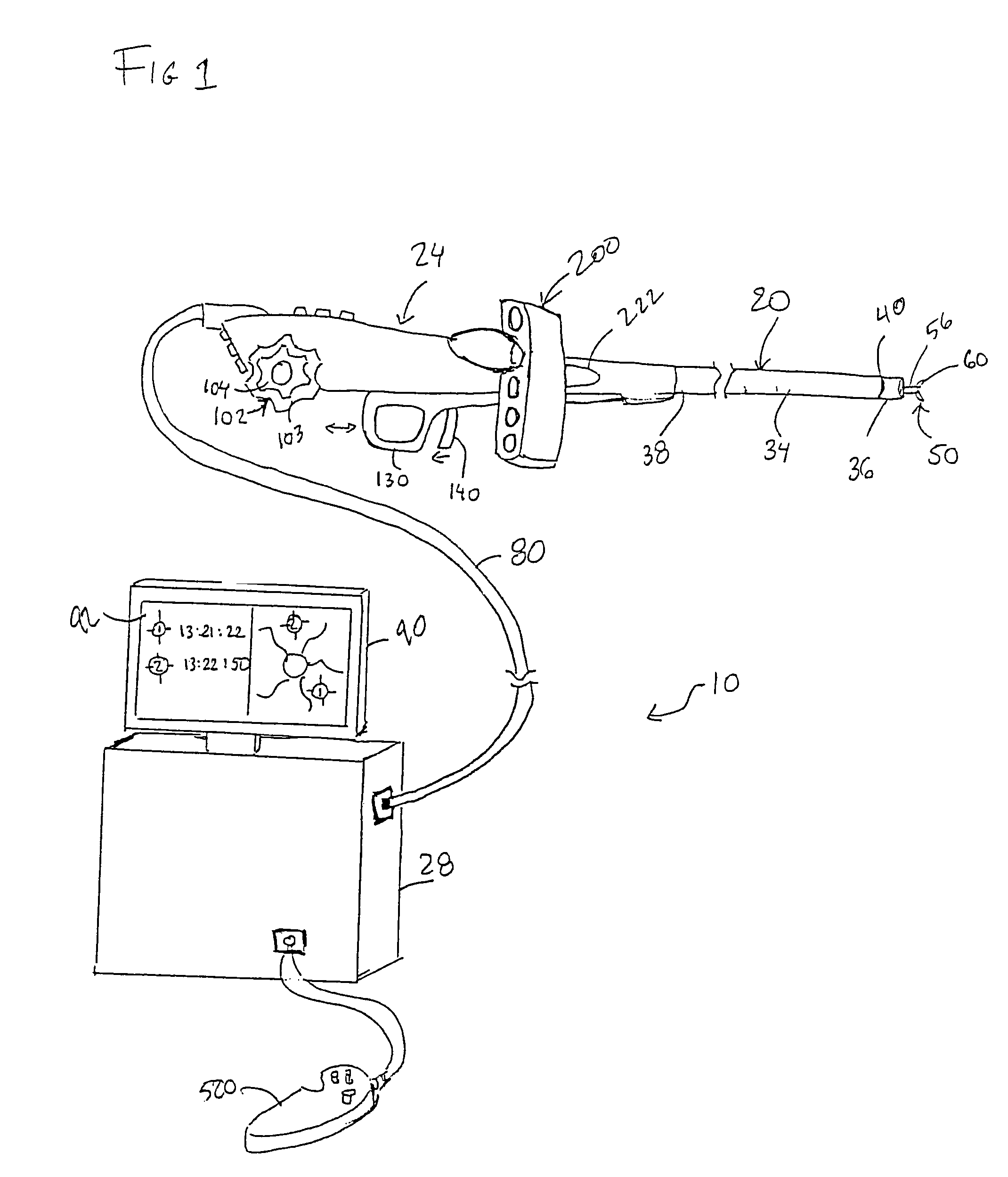
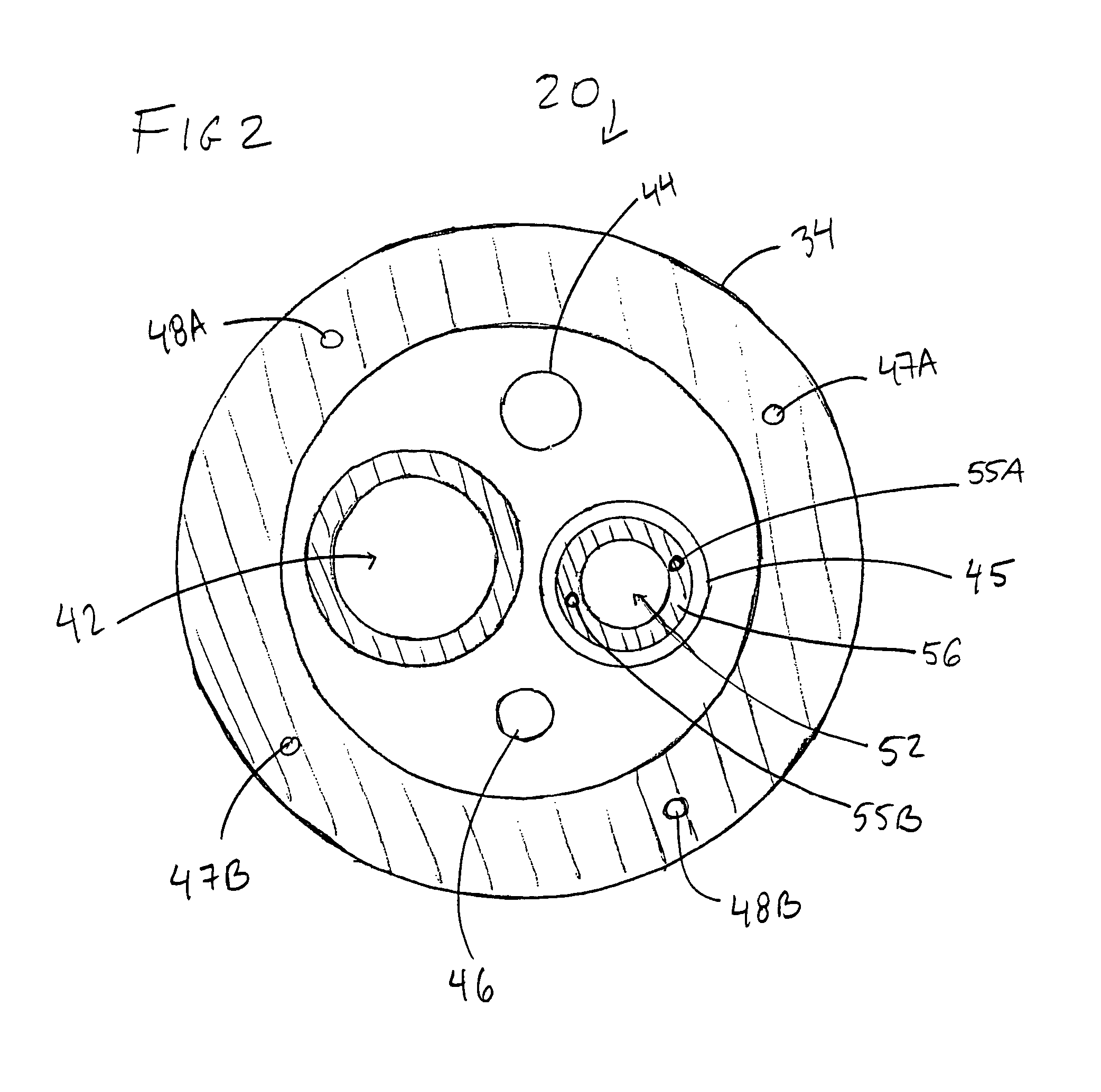
The present invention is an imaging endoscope with biopsy forceps disposed in the distal end. The endoscope shaft has a biopsy sample lumen for receiving biopsy samples obtained with the forceps. The handle has a sample collection apparatus with one or more sample chambers for collecting the samples. The sample chambers can be located in the handle or in a removable cassette. The invention also includes a control unit with a tracking system and a digital monitor for recording and labeling biopsy information. The technical effects of the invention include improved biopsy forceps functionality and efficient sample collection and management.

Problems solved by technology

Conventional endoscopes are expensive, hand assembled, medical devices costing in the range of $25,000 for an endoscope, and much more for the associated operator console.
Conventional endoscopes are generally built of sturdy materials, which decreases the flexibility of the scope and thus can decrease patient comfort.
Furthermore, conventional endoscopes are complex and fragile instruments that frequently need expensive repair as a result of damage during use or during a disinfection procedure.
One medical device that has not previously been inexpensive enough to be considered truly disposable is the endoscope, such as a colonoscope, bronchoscope, gastroscope, duodenoscope, etc.
The process of taking a biopsy presents several challenges, including accurate targeting of the biopsy site, precise and accurate penetration of the biopsy site, preservation of the biopsy tissue upon extraction, and tracking the source of the biopsy sample.
To obtain multiple samples with a conventional biopsy system, biopsy tools must be repeatedly inserted and the sample retrieved, thereby compounding the already awkward procedure.
In addition, conventional biopsy systems are not equipped with a sample retrieval device for receiving and cataloging multiple samples, and such systems do not provide a method by which the precise location of the biopsy site can be tracked and correlated with the biopsy sample.

Method used


Image

  • Endoscopic apparatus with integrated multiple biopsy device
  • Endoscopic apparatus with integrated multiple biopsy device
  • Endoscopic apparatus with integrated multiple biopsy device

Examples


Embodiment Construction

[0026]To address the problems associated with obtaining multiple biopsy samples using conventional endoscope systems and others, the present invention is an imaging endoscope having an elongated shaft with a proximal and distal end with biopsy forceps capable of taking multiple biopsy samples disposed within the distal end. The present invention provides many advantages over conventional endoscope systems and biopsy devices. For example, the present invention provides ease of use such that a single operator may obtain multiple biopsy samples without withdrawing the endoscope or the biopsy tool. Other advantages include, but are not limited to, the ability to capture a plurality of biopsy samples into individual sterile containers, the ability to map the location coordinates of the biopsy site, and the use of a programmable firing mechanism for obtaining precise and high quality tissue samples.

[0027]The various embodiments of the endoscope described herein may be used with both reusa...


PUM


Abstract

An imaging endoscope comprising a shaft having a proximal end adapted to be secured to a handle, and a distal end having a biopsy forceps disposed therein. The biopsy forceps includes one or more end-effector elements that are actuated with a control cable that may be connected to the handle. The endoscope shaft includes a biopsy sample lumen that is configured to receive a biopsy sample obtained from the forceps assembly. A sample collection apparatus is attached to the handle to capture multiple biopsy samples. In some embodiments, the endoscope is a single-use endoscope.

Description

FIELD OF THE INVENTION[0001]The present invention relates to medical devices in general and to biopsy devices in particular.BACKGROUND OF THE INVENTION[0002]It has become well established that there are major health benefits from regular endoscopic examinations of a patient's internal structures such as the alimentary canals and airways, e.g., the esophagus, stomach, lungs, colon, uterus, urethra, kidney, and other organ systems. Endoscopes are also commonly used to perform surgical, therapeutic, diagnostic, or other medical procedures under direct visualization. A conventional imaging endoscope used for such procedures includes an illuminating mechanism such as a fiber optic light guide connected to a proximal source of light, and an imaging means such as an imaging light guide to carry an image to a remote camera, or eye piece, or a miniature video camera within the endoscope itself. In addition, most endoscopes include one or more working channels through which medical devices su...

Claims


Application Information

Patent Timeline
Patent Type & Authority Patents(United States)
IPC IPC(8): A61B5/00
CPCA61B10/06A61B1/018A61B10/0096A61B2010/0225A61B2017/00362
Inventor HOFFMAN, DAVID W.ROWLAND, CHRISTOPHER
Owner BOSTON SCI SCIMED INC
Learn More