
HX2004-6 polypeptide expressed in cancerous cells

a cancerous cell and polypeptide technology, applied in the field of polynucleotides and polypeptides, to achieve the effect of facilitating cell identification

Inactive Publication Date: 2011-01-11
NOVARTIS VACCINES & DIAGNOSTICS INC
View PDF23 Cites 0 Cited by
  • Summary
  • Abstract
  • Description
  • Claims
  • Application Information

AI Technical Summary

Benefits of technology

"The present invention provides methods and compositions for detecting, identifying, and targeting cancerous cells. The invention is based on the discovery of genes that are differentially expressed in cancer cells, and the use of these genes for diagnosis, therapy, and drug discovery. The invention also provides polynucleotides and polypeptides that are differentially expressed in cancer cells, as well as antibodies that specifically bind to these proteins. These molecules can be used for various diagnostic, therapeutic, and drug discovery purposes. The invention also features methods for identifying agents that inhibit the growth of cancerous cells by contacting them with a candidate agent and assessing its effect on TTK activity. Overall, the invention provides useful tools for identifying and targeting cancerous cells, which can aid in the development of effective treatments for cancer."

Problems solved by technology

However, it is likely that other, non-genetic factors are also significant in the etiology of the disease.

Method used


Image

  • HX2004-6 polypeptide expressed in cancerous cells
  • HX2004-6 polypeptide expressed in cancerous cells
  • HX2004-6 polypeptide expressed in cancerous cells

Examples


example 1

Source of Patient Tissue Samples

[0647]Normal and cancerous tissues were collected from patients using laser capture microdissection (LCM) techniques, which techniques are well known in the art (see, e.g., Ohyama et al. (2000) Biotechniques 29:530-6; Curran et al. (2000) Mol. Pathol. 53:64-8; Suarez-Quian et al. (1999) Biotechniques 26:328-35; Simone et al. (1998) Trends Genet 14:272-6; Conia et al. (1997) J. Clin. Lab. Anal. 11:28-38; Emmert-Buck et al. (1996) Science 274:998-1001). Table 1 provides information about each patient from which the samples were isolated, including: the Patient ID and Path ReportID, numbers assigned to the patient and the pathology reports for identification purposes; the anatomical location of the tumor (AnatomicaLoc); The Primary Tumor Size; the Primary Tumor Grade; the Histopathologic Grade; a description of local sites to which the tumor had invaded (Local Invasion); the presence of lymph node metastases (Lymph Node Metastasis); incidence of lymph no...

example 2

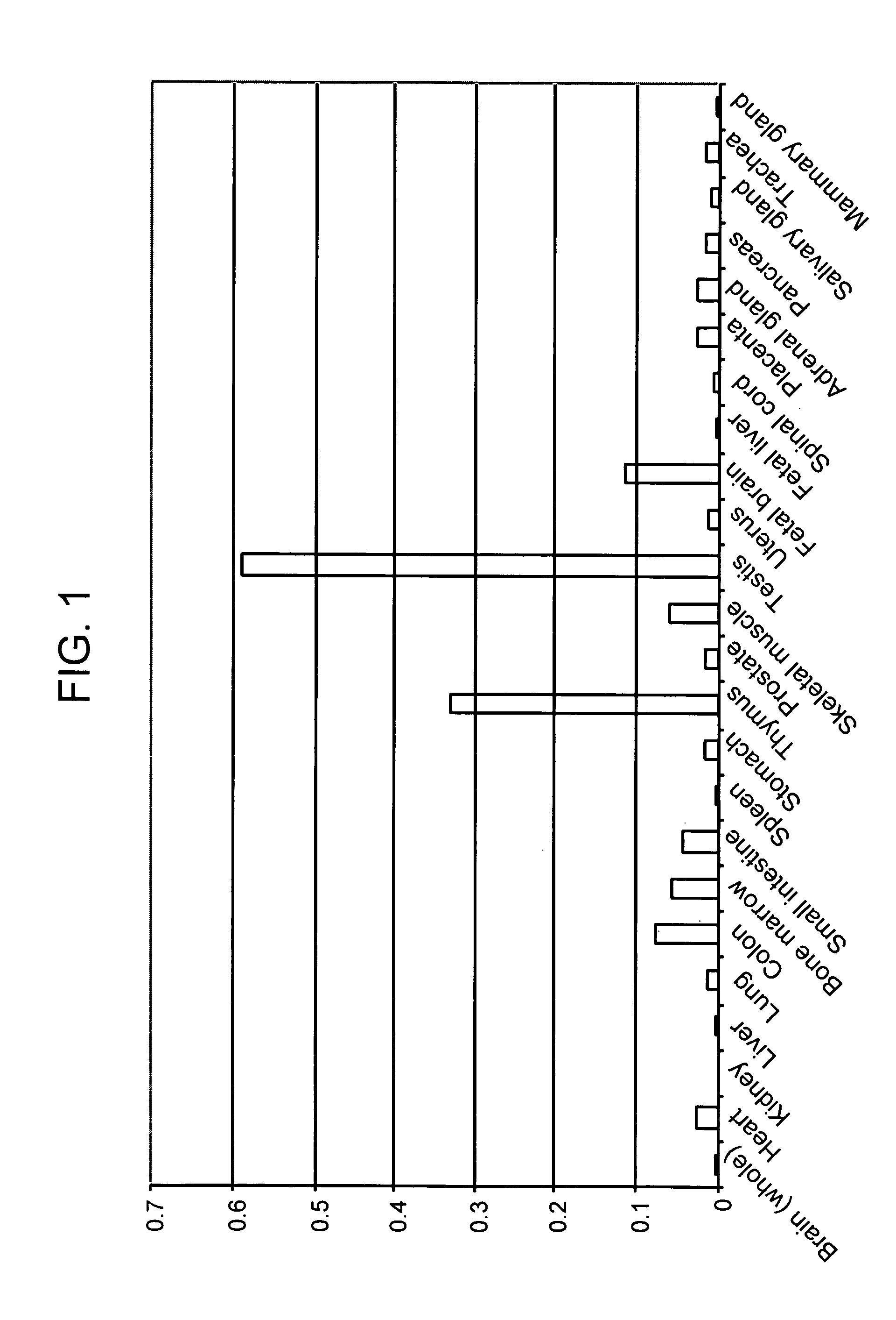
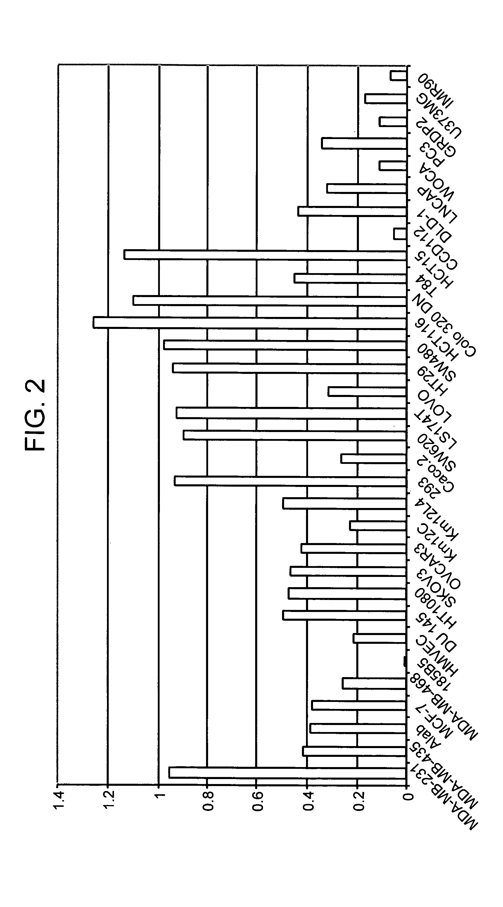
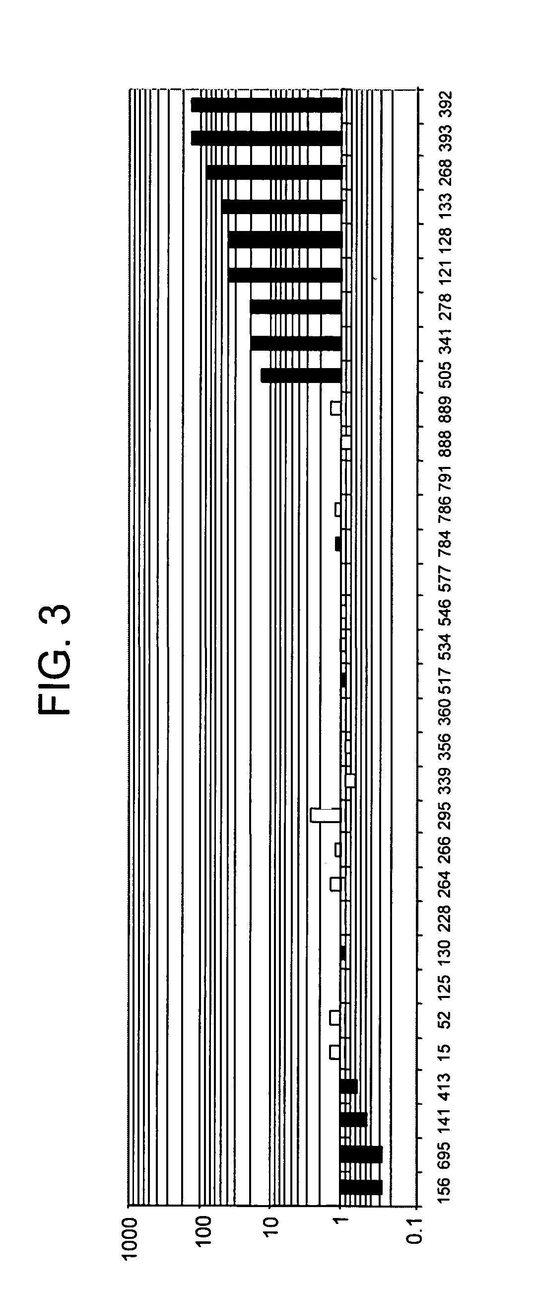
Differential Expression of TTK

[0649]cDNA probes were prepared from total RNA isolated from the patient cells described in Example 1. Since LCM provides for the isolation of specific cell types to provide a substantially homogenous cell sample, this provided for a similarly pure RNA sample.

[0650]Total RNA was first reverse transcribed into cDNA using a primer containing a T7 RNA polymerase promoter, followed by second strand DNA synthesis. cDNA was then transcribed in vitro to produce antisense RNA using the T7 promoter-mediated expression (see, e.g., Luo et al. (1999) Nature Med 5:117-122), and the antisense RNA was then converted into cDNA. The second set of cDNAs were again transcribed in vitro, using the T7 promoter, to provide antisense RNA. Optionally, the RNA was again converted into cDNA, allowing for up to a third round of T7-mediated amplification to produce more antisense RNA. Thus the procedure provided for two or three rounds of in vitro transcription to produce the fina...

example 3

Hierarchical Clustering and Stratification of Colon Cancers Using Differential Expression Data

[0660]Differential expression patterns from Example 2 were analyzed by applying hierarchical clustering methods to the data sets (see Eisen et al. (1998) PNAS 95:14863-14868). In short, hierarchical clustering algorithms are based on the average-linkage method of Sokal and Michener (Sokal, R R & Michener, C D (1958) Univ. Kans. Sci. Bull. 38, 1409-1438), which was developed for clustering correlation matrixes. The object of this algorithm is to compute a dendrogram that assembles all elements into a single tree. For any set of n genes, an upper-diagonal similarity matrix is computed which contains similarity scores for all pairs of genes. The matrix is scanned to identify the highest value (representing a similar pair of genes). Using this technique, four groups of differential expression patterns were identified and assigned to clusters.

[0661]Application of hierarchical clustering to the d...


PUM


Abstract

The present invention provides polynucleotides, as well as polypeptides encoded thereby, that are differentially expressed in cancer cells. These polynucleotides are useful in a variety of diagnostic and therapeutic methods. The present invention further provides methods of reducing growth of cancer cells. These methods are useful for treating cancer.

Description

FIELD OF THE INVENTION[0001]The present invention relates to polynucleotides and polypeptides that are differentially expressed in cancer cells, and uses thereof.BACKGROUND OF THE INVENTION[0002]Cancer, like many diseases, is not the result of a single, well-defined cause, but rather can be viewed as several diseases, each caused by different aberrations in informational pathways, that ultimately result in apparently similar pathologic phenotypes. Identification of polynucleotides that correspond to genes that are differentially expressed in cancerous, pre-cancerous, or low metastatic potential cells relative to normal cells of the same tissue type, provides the basis for diagnostic tools, facilitates drug discovery by providing for targets for candidate agents, and further serves to identify therapeutic targets for cancer therapies that are more tailored for the type of cancer to be treated.[0003]Identification of differentially expressed gene products also furthers the understandi...

Claims


Application Information

Patent Timeline
Patent Type & Authority Patents(United States)
IPC IPC(8): C12P21/00C12N15/09C07K14/00C12P21/02C07K14/435A61K38/00C07K14/47C07K14/72C12N9/12C12Q1/68G01N33/574G01N33/68
CPCC07K14/47C07K14/723C12N9/1205C12Q1/6886C12Y207/01037G01N33/5005G01N33/5008G01N33/5011G01N33/57415G01N33/57419G01N33/57496G01N33/68G01N33/6842A01K2217/05A01K2217/075A61K38/00A61K2039/505C12Q2600/112C12Q2600/136C12Q2600/158G01N2500/00
Inventor REINHARD, CHRISTOPHJEFFERSON, ANNE BENNETTCHAN, VIVIEN W.KAUFMANN, JOERGXIN, HONGKENNEDY, GIULIA C.HARROWE, GREGKHOJA, HAMIDUDDINSHYAMALA, VENKATAKRISHNA
Owner NOVARTIS VACCINES & DIAGNOSTICS INC
Learn More