Methods and systems of using exosomes for determining phenotypes

a technology of exosomes and phenotypes, applied in the field of methods and systems of using exosomes for determining phenotypes, can solve the problems of invasive methods, complication risks for patients, and readouts that lack the requisite sensitivity, and achieve greater sensitivity or specificity.

Inactive Publication Date: 2011-03-01
CARIS LIFE SCI LUXEMBOURG HLDG
View PDF233 Cites 90 Cited by
  • Summary
  • Abstract
  • Description
  • Claims
  • Application Information

AI Technical Summary

Benefits of technology

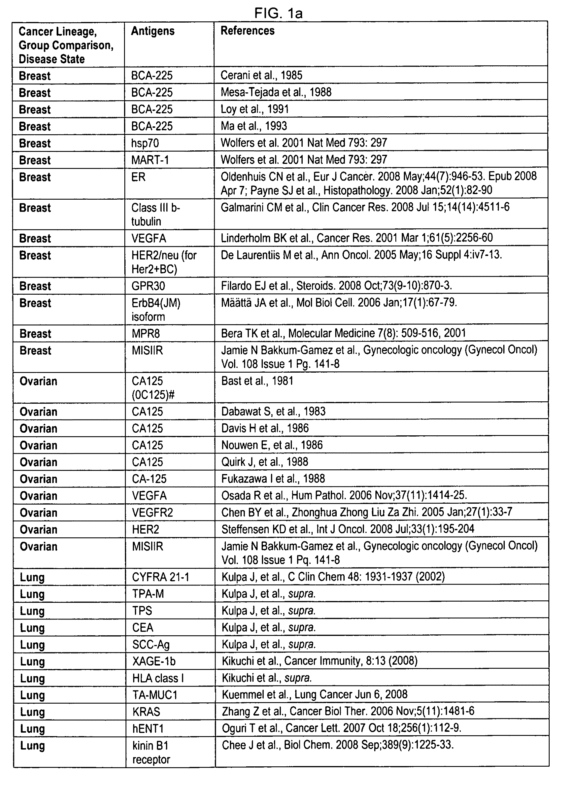
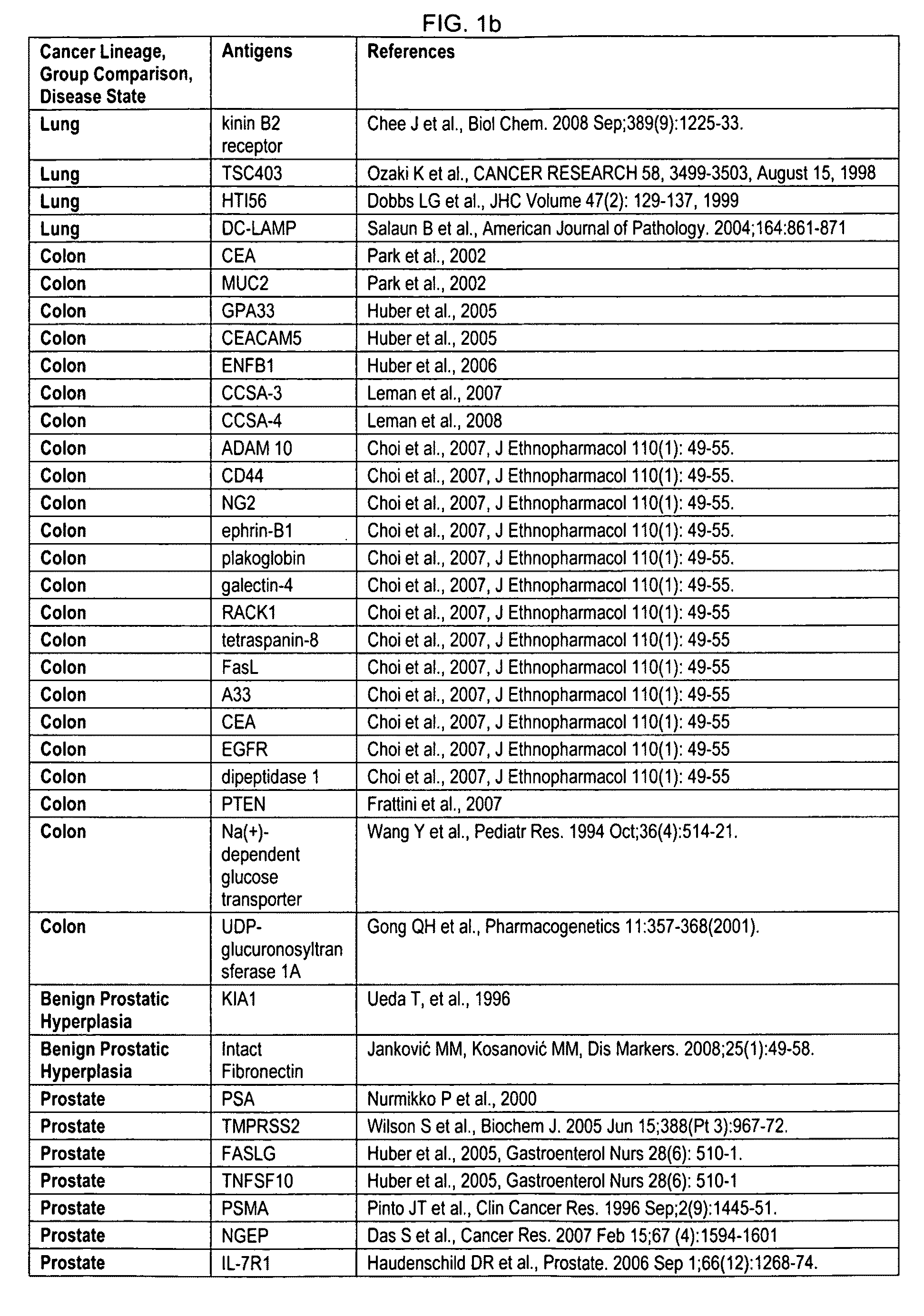
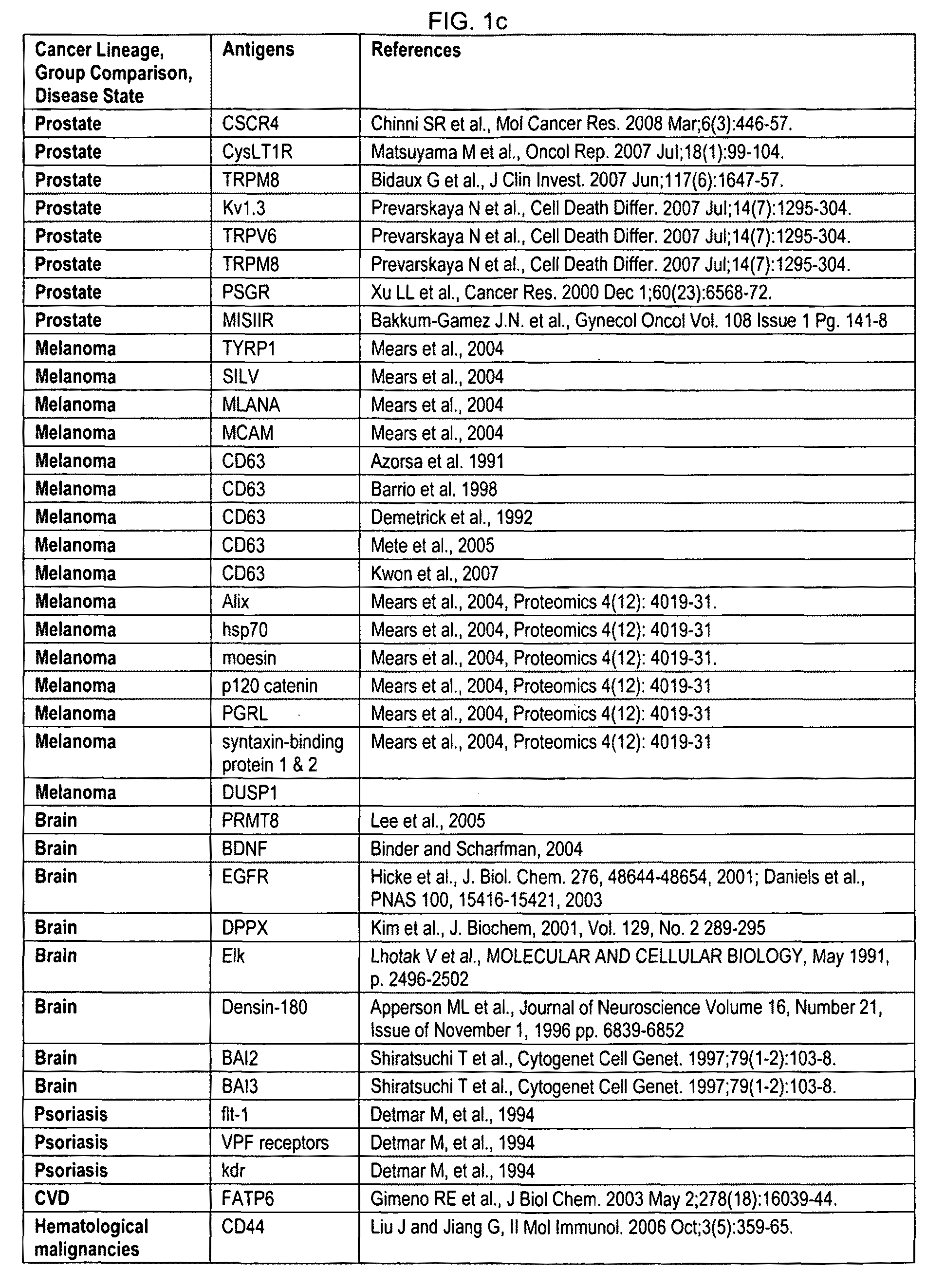
[0010]One or more biomarkers of an exosome can be assessed for characterizing a phenotype. The biomarker can be a nucleic acid, peptide, protein, lipid, antigen, carbohydrate or proteoglycan, such as DNA or RNA. The RNA can be mRNA, miRNA, snoRNA, snRNA, rRNAs, tRNAs, siRNA, hnRNA, or shRNA. The biomarker can be an antigen selected from FIG. 1, or a biomarker selected from a table listed in FIG. 3-60. One or more biomarkers can be assessed and used to characterize a phenotype. The bio-signature can comprise one or more miRNAs selected from the group consisting of: miR-9, miR-629, miR-141, miR-671-3p, miR-491, miR-182, miR-125a-3p, miR-324-5p, miR-148b, and miR-222. The bio-signature can be used to characterize a phenotype, such as prostate cancer. Other biomarkers can be selected from the group consisting of: CD9, PSCA (prostate stem cell antigen), TNFR, CD63, MFG-E8, EpCam, Rab, CD81, STEAP, PCSA (prostate cell surface antigen), PSM (or PSMA, prostate specific membrane antigen), 5T4, CD59, CD66, CD24 and B7H3. Detecting a plurality of biomarkers can provide greater sensitivity or specificity as compared to detecting less than a plurality of biomarkers.
[0011]Methods of multiplexing, or mu...

Problems solved by technology

Methods to obtain these tissues of interest for analysis are often invasive, costly and pose complication risks for the patient.
Furthermore, use of bodily fluids to isolate or detect biomarkers often significantly dilutes a bioma...

Method used


Image

  • Methods and systems of using exosomes for determining phenotypes
  • Methods and systems of using exosomes for determining phenotypes
  • Methods and systems of using exosomes for determining phenotypes

Examples


example 1

Purification of Exosomes from Prostate Cancer Cell Lines

[0880]Prostate cancer cell lines are cultured for 3-4 days in culture media containing 20% FBS (fetal bovine serum) and 1% P / S / G. The cells are then pre-spun for 10 minutes at 400×g at 4° C. The supernatant is kept and centrifuged for 20 minutes at 2000×g at 4. The supernatant containing exosomes can be concentrated using a Millipore Centricon Plus-70 (Cat # UFC710008 Fisher).

[0881]The Centricon is pre washed with 30 mls of PBS at 1000×g for 3 minutes at room temperature. Next, 15-70 mls of the pre-spun cell culture supernatant is poured into the Concentrate Cup and is centrifuged in a Swing Bucket Adapter (Fisher Cat #75-008-144) for 30 minutes at 1000×g at room temperature.

[0882]The flow through in the Collection Cup is poured off. The volume in the Concentrate Cup is brought back up to 60 mls with any additional supernatant. The Concentrate Cup is centrifuged for 30 minutes at 1000×g at room temperature until all of the cell...

example 2

Purification of Exosomes from VCaP and 22Rv1

[0885]Exosomes from Vertebral-Cancer of the Prostate (VCaP) and 22Rv1, a human prostate carcinoma cell line, derived from a human prostatic carcinoma xenograft (CWR22R) were collected by ultracentrifugation by first diluting plasma with an equal volume of PBS (1 ml). The diluted fluid was transferred to a 15 ml falcon tube and centrifuged 30 minutes at 2000×g 4° C. The supernatant (˜2 mls) was transferred to an ultracentrifuge tube 5.0 ml PA thinwall tube (Sorvall #03127) and centrifuged at 12,000×g, 4° C. for 45 minutes.

[0886]The supernatant (˜2 mls) was transferred to a new 5.0 ml ultracentrifuge tubes and filled to maximum volume with addition of 2.5 mls PBS and centrifuged for 90 minutes at 110,000×g, 4° C. The supernatant was poured off without disturbing the pellet and the pellet resuspended with 1 ml PBS. The tube was filled to maximum volume with addition of 4.5 ml of PBS and centrifuged at 110,000×g, 4° C. for 70 minutes.

[0887]The...

example 3

Plasma Collection and Exosome Purification

[0888]Blood is collected via standard veinpuncture in a 7 ml K2-EDTA tube. The sample is spun at 400 g for 10 minutes in a 4° C. centrifuge to separate plasma from blood cells (SORVALL Legend RT+ centrifuge). The supernatant (plasma) is transferred by careful pipetting to 15 ml Falcon centrifuge tubes. The plasma is spun at 2,000 g for 20 minutes and the supernatant is collected.

[0889]For storage, approximately 1 ml of the plasma (supernatant) is aliquoted to a cryovials, placed in dry ice to freeze them and stored in −80° C. Before exosome purification, if samples were stored at −80° C., samples are thawed in a cold water bath for 5 minutes. The samples are mixed end over end by hand to dissipate insoluble material.

[0890]In a first prespin, the plasma is diluted with an equal volume of PBS (example, approximately 2 ml of plasma is diluted with 2 ml of PBS). The diluted fluid is transferred to a 15 ml Falcon tube and centrifuged for 30 minut...


PUM

PropertyMeasurementUnit
Diameteraaaaaaaaaa
Diameteraaaaaaaaaa
Diameteraaaaaaaaaa

Abstract

Exosomes can be used for detecting biomarkers for diagnostic, therapy-related or prognostic methods to identify phenotypes, such as a condition or disease, for example, the stage or progression of a disease. Cell-of-origin exosomes can be used in profiling of physiological states or determining phenotypes. Biomarkers or markers from cell-of-origin specific exosomes can be used to determine treatment regimens for diseases, conditions, disease stages, and stages of a condition, and can also be used to determine treatment efficacy. Markers from cell-of-origin specific exosomes can also be used to identify conditions of diseases of unknown origin.

Description

CROSS-REFERENCE[0001]This application claims the benefit of U.S. Provisional Application Nos. 61 / 114,045, filed Nov. 12, 2008; 61 / 114,058, filed Nov. 12, 2008; 61 / 114,065, filed Nov. 13, 2008; 61 / 151,183, filed Feb. 9, 2009; 61 / 278,049, filed Oct. 2, 2009; 61 / 250,454, filed Oct. 9, 2009; and 61 / 253,027 filed Oct. 19, 2009, of which each is incorporated herein by reference in its entirety. This application is also related to U.S. application Ser. No. 12 / 609,847, filed Oct. 30, 2009, which claims the benefit of U.S. Provisional Application Nos. 61 / 109,742, filed Oct. 30, 2008; 61 / 112,571, filed Nov. 7, 2008; 61 / 114,045, filed Nov. 12, 2008; 61 / 114,058, filed Nov. 12, 2008; 61 / 114,065, filed Nov. 13, 2008; 61 / 151,183, filed Feb. 9, 2009; 61 / 278,049, filed Oct. 2, 2009; 61 / 250,454, filed Oct. 9, 2009, and 61 / 253,027 filed Oct. 19, 2009, each of which is incorporated herein by reference in its entirety.BACKGROUND[0002]A critical need for disease detection, prognostic prediction, monitori...

Claims


Application Information

Patent Timeline
IPC IPC(8): G01N33/53
CPCC12Q1/6886G01N33/5023G01N33/54326G01N33/57419G01N33/57434C12Q2600/112C12Q2600/16C12Q2600/178C12Q1/6811G01N33/574G01N33/57484G01N33/6848G01N2800/50G01N2800/52C12Q2525/205C12Q2525/207G01N33/57488G01N33/68
Inventor KLASS, MICHAELKUSLICH, CHRISTINEPOSTE, GEORGE
Owner CARIS LIFE SCI LUXEMBOURG HLDG
Why Eureka
Try Eureka