
Polysaccharide biomaterials and methods of use thereof

a polysaccharide and biomaterial technology, applied in the field of body treatment compositions, can solve the problems of affecting so as to improve the quality of blood vessels, improve the cost, and improve the effect of materials

Active Publication Date: 2012-01-24
BIOINTERACTIONS
View PDF57 Cites 10 Cited by
  • Summary
  • Abstract
  • Description
  • Claims
  • Application Information

AI Technical Summary

Benefits of technology

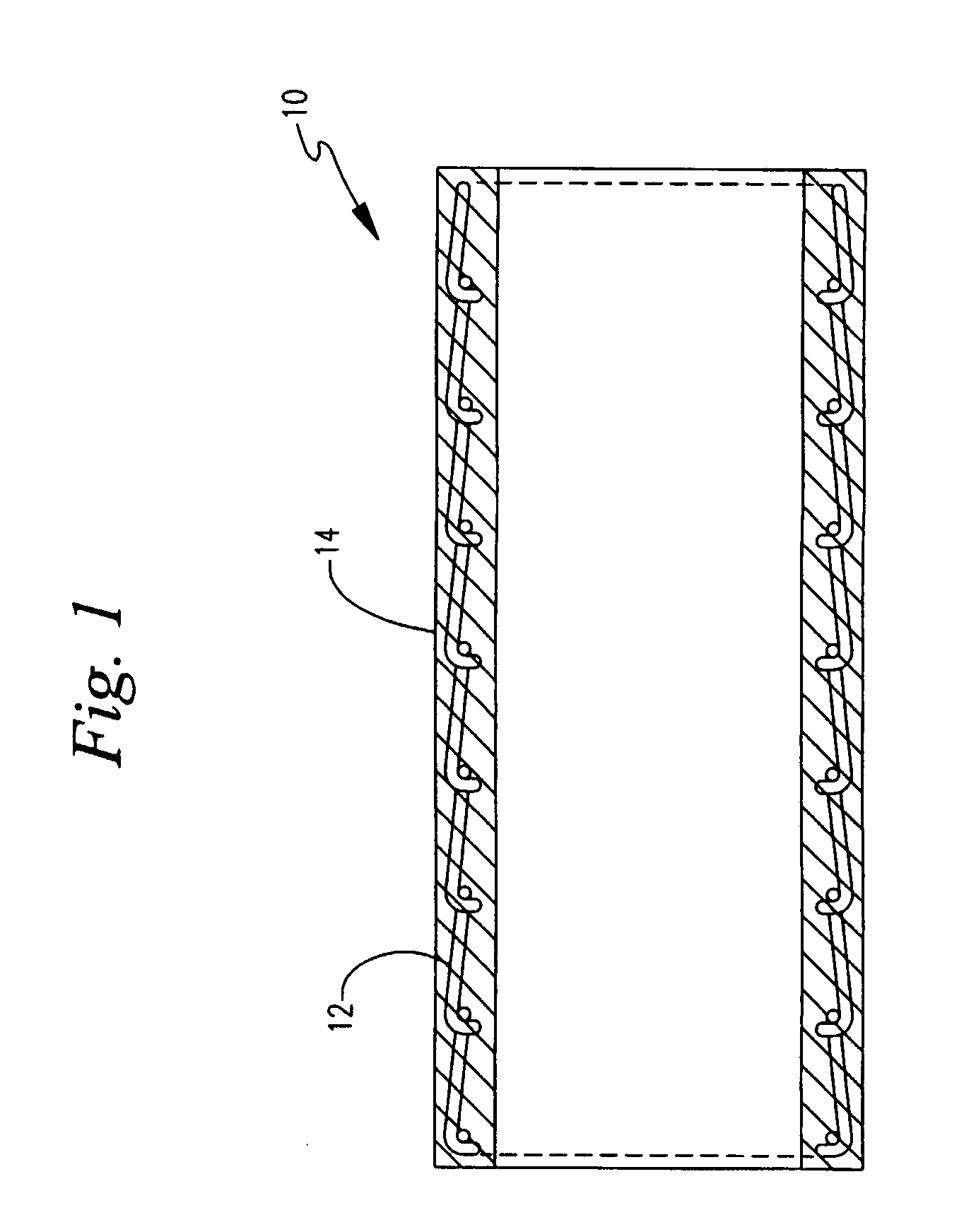
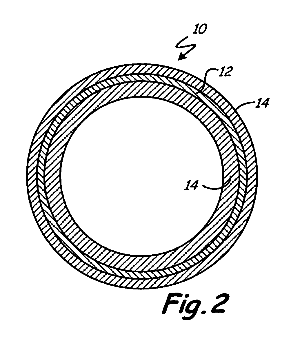
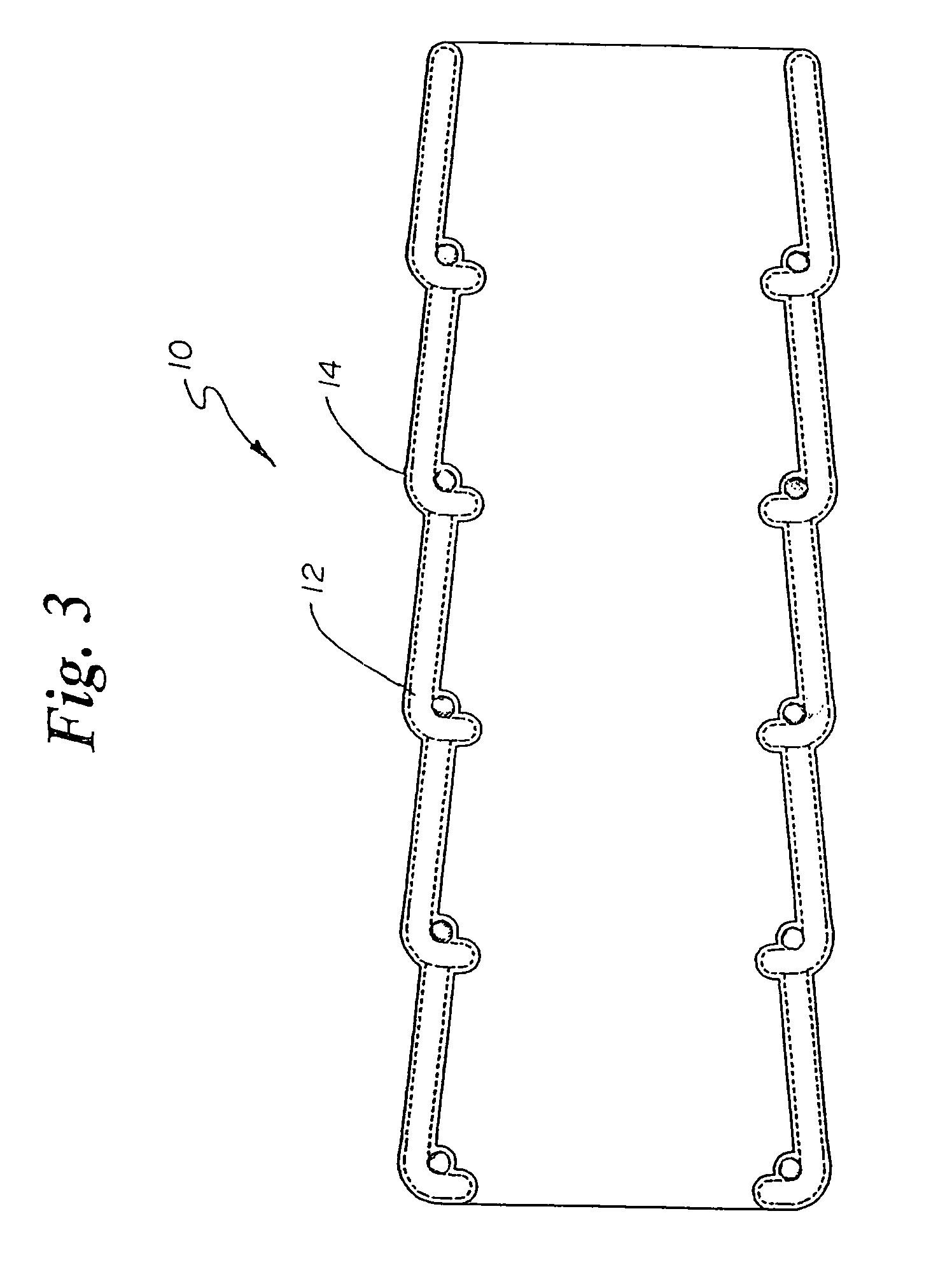
The present invention provides a new type of material that combines the benefits of polysaccharides and hydrogels, making it suitable for use in various medical applications. The material can be made into a three-dimensional crosslinked hydrogel that has a hollow cylinder structure, which can be used as a graft for small diameter vascular grafts. The material can also be used to encapsulate medical devices, providing a protective coating. The invention also provides a method for making the material and a coating on a medical device using the material. The invention has various advantages, such as being strong, flexible, and having good biocompatibility.

Problems solved by technology

The blood clot can also grow on the inside of the vessel and block or severely restrict the blood flow.
This approach, however, does not work for small diameter blood vessels because the small diameter tubes are blocked when the blood clots onto the walls.
There are no currently known materials and techniques for manufacturing small diameter vascular grafts made of synthetic materials.
Unfortunately, there is a great need for such grafts.
The blood drains poorly from the leg and amputation can result.
Although hydrogels tend to elicit less blood clotting than other synthetic materials, hydrogels have not previously been successfully used to make a small diameter vascular graft.
Coating the inside of a synthetic material tube with heparin tends to increase the amount of time that the tube remains open to blood flow but, to date, small diameter vascular grafts coated with heparin have failed to resist blockage by blood clots for a medically useful length of time.

Method used


Image

  • Polysaccharide biomaterials and methods of use thereof
  • Polysaccharide biomaterials and methods of use thereof
  • Polysaccharide biomaterials and methods of use thereof

Examples


example 1

Heparin Complex

[0142]5 g sodium heparin (Celsus Laboratories, Inc, USP lyophilized from porcine intestinal mucosa) was dissolved in 80 ml de-ionized water and allowed to stir for 1 hour in a 250 ml beaker.

[0143]8 g benzalkonium chloride (Aldrich Chemical Company, Inc) was allowed to dissolve in 80 ml de-ionized water with gentle warming (40-50° C.) on a magnetic stirrer hotplate for 1 hour and then allowed to reach room temperature.

[0144]To the above vigorously stirred solution of sodium heparin, the benzalkonium chloride solution was added. A white precipitate immediately formed and the suspension was further stirred for 1 minute. The precipitate was filtered through a Whatman qualitative filter paper (grade 1).

[0145]The white precipitate was collected from the filter paper and re-suspended in 400 ml deionized water and allowed to stir for 20 minutes. The suspension was filtered as above and resuspended in 400 ml de-ionized water and filtered again. The precipitate was once again s...

example 2

[0148]3 g 4,4′-methylenebis (phenyl isocyanate) (MDI) (Aldrich Chemical Company, Inc) was dissolved in 100 ml anhydrous tetrahydrofuran (THF). Polyurethane tubing was dipped in to the above solution for 30 seconds and was then allowed to stir-dry at 60° C. for 1 hour.

[0149]5 g of dry crystals of complexed heparin from Example 1 were dissolved in 100 ml anhydrous dichloromethane (DCM). The MDI coated polyurethane tubing was dipped into the DCM solution of complexed heparin for 30 seconds and then allowed to air-dry for 2 hours.

[0150]The tubing was dyed with 0.075% w / v pH 8.5 toludine blue aqueous solution for 30 seconds and washed with de-ionized water. A very faint purple color due to the complexed heparin was observed. The tubing was then immersed in 25% w / v solution of sodium chloride at 40° C. for 30 minutes. The tubing was washed with de-ionized water and again dyed. An intense dark purple color due to complexed heparin was observed.

[0151]In a similar experiment where the polyur...

example 3

1. Heparin Methacrylate (Methacryloyl Chloride)

[0152]5 g of dry crystals of complexed heparin from Example 1 were dissolved in 100 ml anhydrous DCM in a 250 ml quickfit conical flask. To this was added 0.1265 g (1.25×10⁻³ moles) triethylamine.

[0153]Methacryloyl chloride (Aldrich Chemical Company, Inc) was distilled under reduced pressure to obtain a very pure sample. 0.1306 g (1.25×10⁻³ moles) of the above distilled methacryloyl chloride was dissolved in 30 ml anhydrous DCM and placed in a stoppered quickfit pressure equalizing funnel above the vigorously stirred solution of DCM containing the complexed heparin. The methacryloyl chloride solution was added drop-wise over a period of 30 minutes to the complexed heparin solution in DCM.

[0154]The DCM was rotary evaporated and the complexed heparin methacrylate was dried in a vacuum oven at 40° C. for 2 hours.

[0155]Complexed methacrylate was characterized by 1H and 13C.

[0156]0.2 g of the above complexed heparin methacrylate was dissolve...


PUM

PropertyMeasurementUnit
diameteraaaaaaaaaa
diameteraaaaaaaaaa
temperatureaaaaaaaaaa

Abstract

The invention includes a medical hydrogel made from polymerized polysaccharide macromers. The macromers are preferably polysaccharides decorated with polymerizable groups, for example, methacrylates. The macromers may also be made into polymers of at least two macromers polymerized together. These polymers are preferably multi-armed or high-molecular weight and used for medical uses, for example, making coatings on medical devices. Macromers of N-vinylpyrrolidone are also disclosed herein.

Description

RELATED APPLICATIONS[0001]The present application claims priority to U.S. provisional patent application Ser. No. 60 / 301,176, entitled “Polysaccharide Biomaterials and Methods of Use Thereof”, filed Jun. 26, 2001, which is hereby incorporated herein by reference.FIELD OF INVENTION[0002]The invention relates to body treating compositions, especially those formed as a matrix. More specifically, the invention includes hydrogel matrices of polymerized polysaccharides, especially heparin, so as to create a hydrogel, especially for vascular graft applications.BACKGROUND OF THE INVENTION[0003]Many synthetic materials have been medically used in the body, including polyester (e.g., DACRON™), polyethylene (e.g., milk jugs), and fluorocarbons (e.g., TEFLON™), and metals. A patient's body responds by treating a synthetic material as an invader, although it responds only mildly in some medical applications; for example, a metal hip implant is generally well tolerated. One common response to an ...

Claims


Application Information

Patent Timeline
Patent Type & Authority Patents(United States)
IPC IPC(8): A61F2/82A61K31/715A61K38/14A61K47/30A61L27/00A61L27/20A61L27/28A61L27/34A61L27/50A61L27/52A61L33/00A61L33/08A61L33/10C08B37/00
CPCA61L27/20A61L27/34A61L27/507A61L27/52A61L33/0011A61L33/0064C08B37/0063C08B37/0069C08B37/0075C08L39/06D10B2403/0122D10B2509/06D04B21/205
Inventor LUTHRA, AJAY K.SANDHU, SHIVPAL S.HUDSON, JOHN O.
Owner BIOINTERACTIONS
Learn More