Radio guided seed localization of imaged lesions

a radioguided, imaged technology, applied in radiation therapy, therapy, etc., can solve the problems of difficult current methods of breast cancer treatment with radioactive seed deployment, and achieve the effect of easy transportability and minimal or no migration

Active Publication Date: 2012-02-14
UNIV OF SOUTH FLORIDA
View PDF43 Cites 10 Cited by
  • Summary
  • Abstract
  • Description
  • Claims
  • Application Information

AI Technical Summary

Benefits of technology

[0016]It is also desirable for the implanted seed to maintain position once placed within tissues. Thus, it is a further object of the subject invention to provide an improved seed with minimal or no migration tendencies.
[0017]Current methods of treatment of breast cancer with radioactive seed deployment are difficult because there is no way to distribute radioactive seeds in a stable array capable of providing consistent radiation delivery for the period of treatment, which usually lasts from 6 to 12 months. Spatial orientation of seeds can change minute by minute depending on a variety of factors, including, for example, whether the patient is wearing a brassiere, activity or position of the patient, compression of the breast, etc. Thus, it is a further object of the subject invention to provide a means for deploying radioactive seeds in a stable array. More specifically, the invention provides a method for consistent, precise delivery of radiation to lesions.
[0018]It is a further object of the present invention to provide a new and improved portable kit for use in localizing and excising lesions that is easily transportable and includes in one container the various instruments necessary in the localization and excision of lesions.
[0020]According to the present invention, a primary object is to provide a safe, disposable seed-implantation tool that is inexpensive to manufacture and easy to use.

Problems solved by technology

Current methods of treatment of breast cancer with radioactive seed deployment are difficult because there is no way to distribute radioactive seeds in a stable array capable of providing consistent radiation delivery for the period of treatment, which usually lasts from 6 to 12 months.

Method used


Image

  • Radio guided seed localization of imaged lesions
  • Radio guided seed localization of imaged lesions
  • Radio guided seed localization of imaged lesions

Examples


example 1

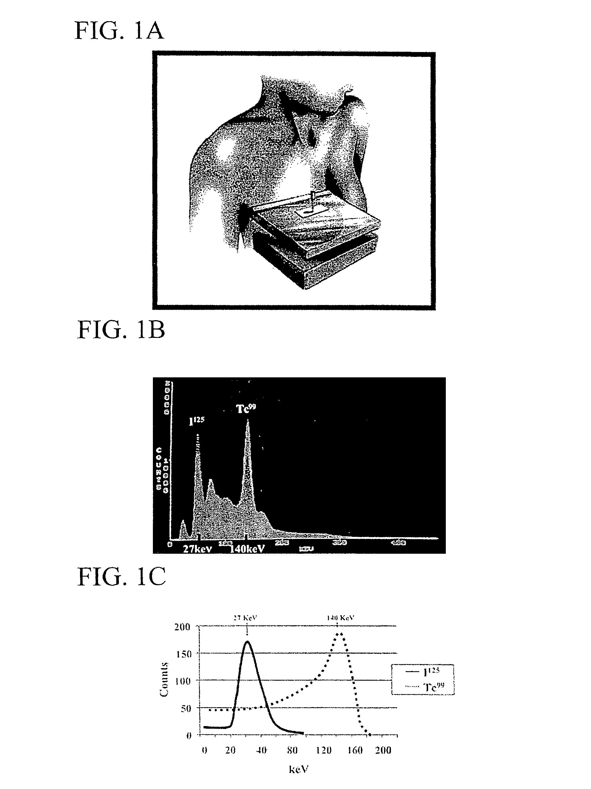
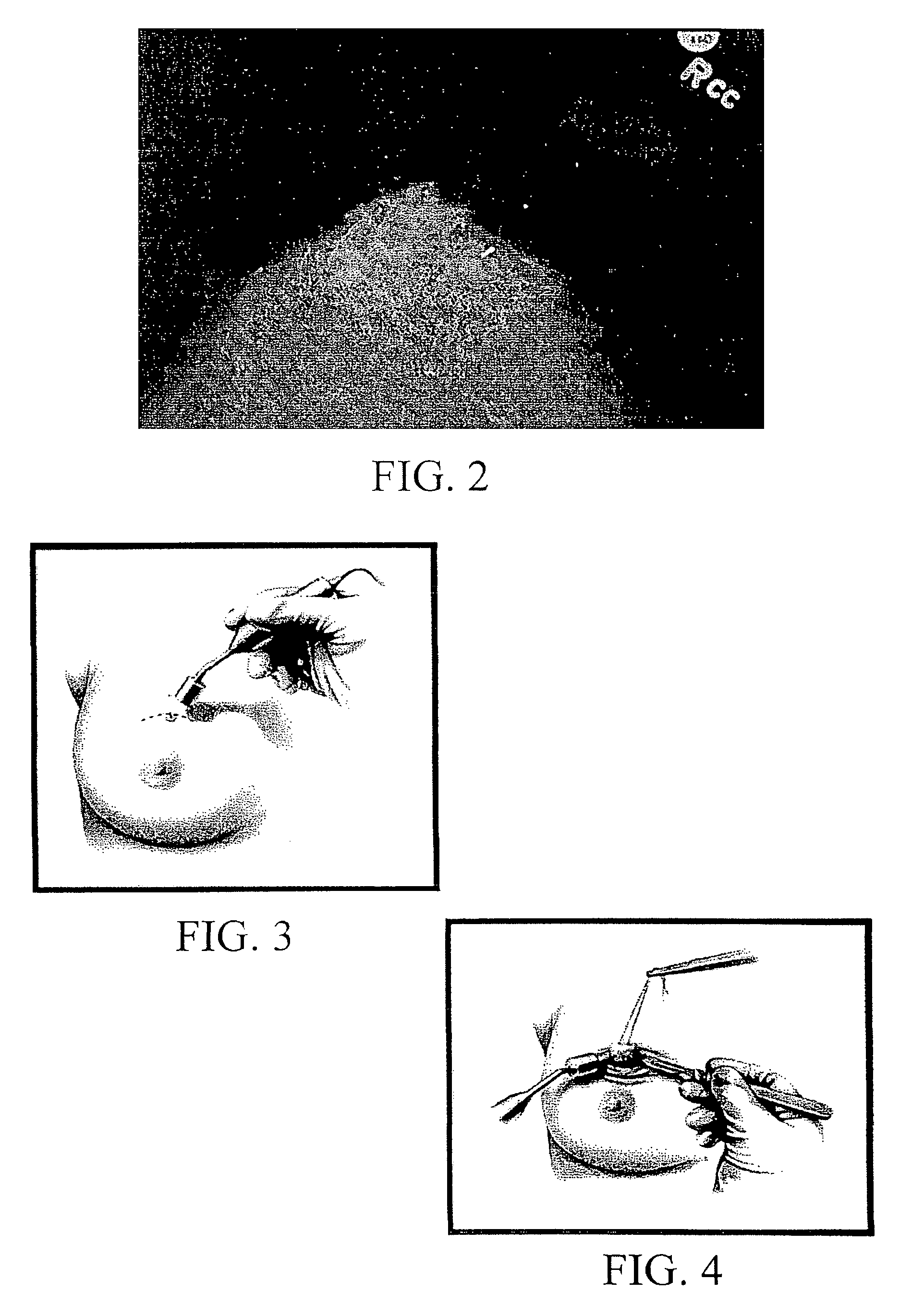
[0107]Mammographic placement of a single 125-I seed by the radiologist is performed. Once the patient is placed in the mammographic device and the location of the lesion is determined, the skin is cleansed. Local anesthetic is injected at the site of the placement. An 18-gauge needle with sterile bone wax occluding the tip is loaded with a single 125-I seed. The needle is placed into the breast tissue under mammographic guidance to the suspicious lesion. A stilette is placed into the bore of the needle displacing the seed through the tip. The needle and stilette apparatus are removed from the breast tissue. The seed location is confirmed to be at the lesion with mammography and films are taken. The patient is then taken to the operating room.

[0108]The patient is prepared and draped in the normal sterile fashion. A sterile sleeve is placed over a Neoprobe™ Gamma counter. A hand-held probe, which is properly windowed and calibrated to detect I125, is then run across the skin surface o...

example 2

[0112]Needle localized breast biopsy (NLBBx) has been the standard for diagnosis of nonpalpable lesions for the past 20 years. Low dose radioactive seed localization (RSL) can be used in conjunction with a hand held gamma detector (HHGD) to localize nonpalpable breast lesions and accurately remove the radiographic lesion with reduced operative time (OT) and -tissue volume (TV).

Methods

[0113]A titanium seed containing 0.05-0.1 mCi of I125 is placed with mammographic or ultrasound guidance localizing the suspicious breast lesion. The HHGD is used to externally locate the seed. The incision is placed directly over the seed / lesion. The HHGD directs the excision and verifies seed / lesion removal. A specimen radiograph (S-X-ray) was performed to confirm the seed / lesion removal. Variables included OT, TV, surgeon retrieval success (SRS), and pathologist retrieval success (PRS). Success of identification of the seed / lesion by the surgeon and pathologist were assessed prior to S-X-ray utilizin...

example 3

Materials and Methods

[0117]Patients were recruited from the Comprehensive Breast Center who had been referred for suspicious mammographically detected lesions requiring NLBB. Variables analyzed included the size and weight of the specimen, total time in the operating room, surgeon retrieval success, and cumulative radiation exposure to the surgeon, radiologist and pathologist.

[0118]The technique involves placing a titanium seed containing 0.05-0.1 mCi of I-125 into an 18-gauge needle with sterile bone wax occluding the tip. The apparatus is placed into the breast parenchyma under radiographic guidance (mammography or ultrasound). A stilette is placed into the needle displacing the seed through the tip localizing the lesion. The seed localization is confirmed to be at the lesion with mammography.

[0119]After surgical preparation, the sterile sheathed HHGD is utilized to identify location of the seed / lesion by counts of radioactivity. After administration of a local anesthetic, the spe...


PUM


Abstract

A method of removing lesions by implanting a radioactive seed at the location of the lesion, locating the lesion with the radioactive seed, and removing the lesion with the radioactive seed. A kit is also provided by the present invention for use in removing lesions by implanting a radioactive seed at the location of the lesion, locating the lesion with the radioactive seed, and removing the lesion with the radioactive seed. Other embodiments of the invention include: a disposable needle preloaded with radioactive seed(s); and radioactive seeds comprising hooks to ensure proper localization of the seed(s) in the patient.

Description

CROSS-REFERENCE TO RELATED APPLICATIONS[0001]This application is a continuation-in-part application of U.S. Ser. No. 11 / 363,227, filed Feb. 27, 2006 now abandoned; which is a continuation application of U.S. Ser. No. 10 / 292,377, filed Nov. 12, 2002, now abandoned; which is a continuation of U.S. Ser. No. 09 / 413,293, Oct. 6, 1999, now U.S. Pat. No. 6,496,717; which claims the benefit of U.S. Provisional Application No. 60 / 103,169, filed Oct. 6, 1998.BACKGROUND OF THE INVENTION[0002]1. Technical Field[0003]The present invention relates to methods of localizing lesions. More specifically, the present invention relates to methods of localizing lesions using radioactive seeds. The invention also relates to techniques and methods for reducing or eliminating migration of radioactive seeds after placement.[0004]2. Background Art[0005]Localization of non-palpable lesions for biopsy or excision during surgery is a necessary procedure. Several techniques are currently available. As described h...

Claims


Application Information

Patent Timeline
Patent Type & Authority Patents(United States)
IPC IPC(8): A61M36/00
CPCA61N5/1027
Inventor COX, CHARLES E.DAUWAY, EMILIA L.
Owner UNIV OF SOUTH FLORIDA
Why Eureka
Try Eureka