Catheter guidance through a calcified aortic valve

a catheter and aortic valve technology, applied in balloon catheters, medical science, surgery, etc., can solve the problems of limiting physical activity, fatigue, palpitation, dyspnea or anginal pain, and affecting the normal operation of the body,

Active Publication Date: 2012-03-13
SPEYSIDE MEDICAL LLC
View PDF240 Cites 28 Cited by
  • Summary
  • Abstract
  • Description
  • Claims
  • Application Information

AI Technical Summary

Problems solved by technology

Patient with cardiac disease resulting in slight limitation of physical activity.
Ordinary physical activity results in fatigue, palpitation, dyspnea, or anginal pain.
Patients with cardiac disease resulting in marked limitation of physical activity.
Patients with cardiac disease resulting in inability to carry on any physical activity without discomfort.
If any physical activity is undertaken, discomfort is increased.
Access to these sites generally include a thoracotomy or a sternotomy for the patient and include a great deal of recovery time.
Although valve repair and replacement can successfully treat many patients with valvular insufficiency, techniques currently in use are attended by significant morbidity and mortality.

Method used


Image

  • Catheter guidance through a calcified aortic valve
  • Catheter guidance through a calcified aortic valve
  • Catheter guidance through a calcified aortic valve

Examples


Embodiment Construction

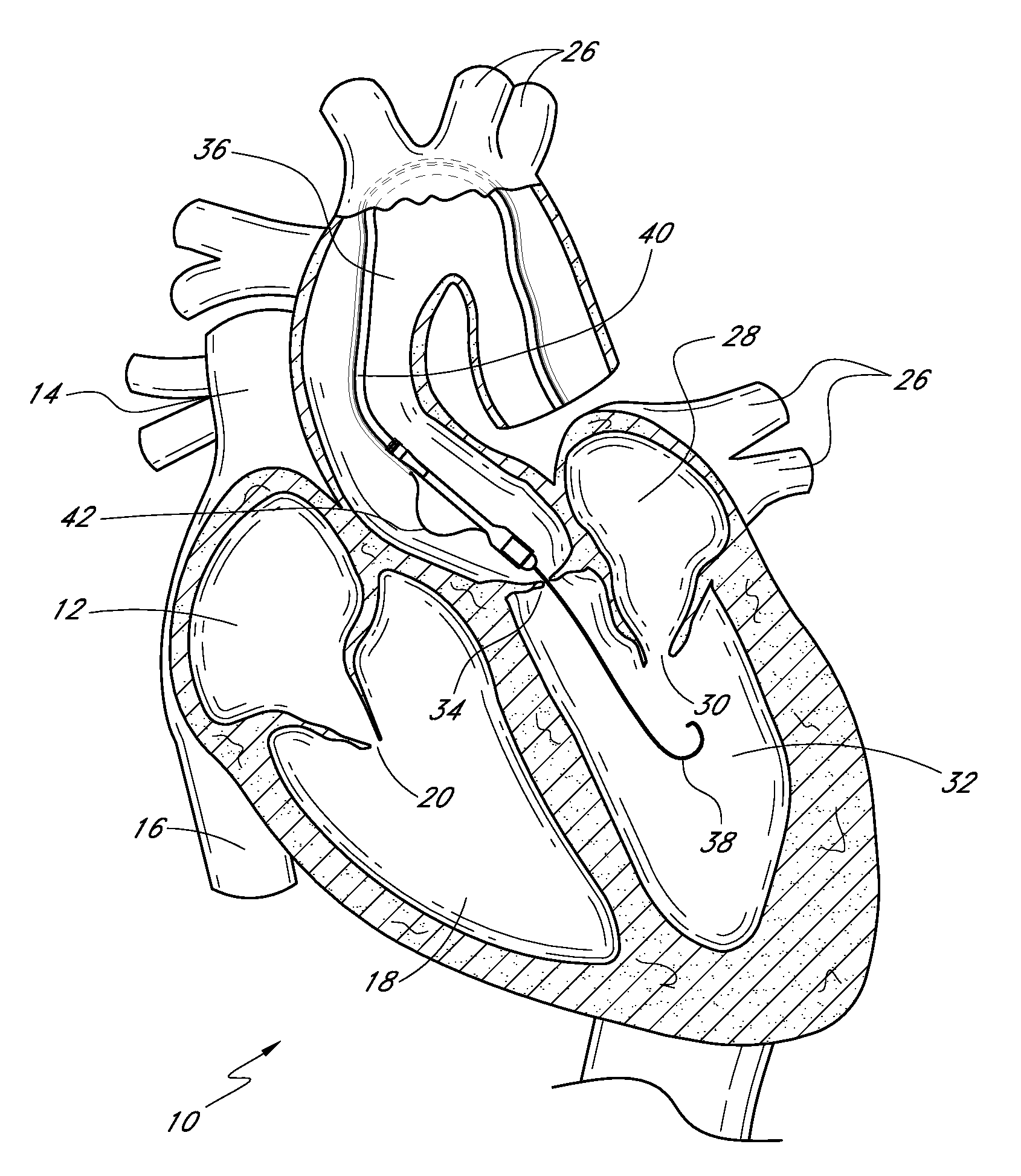
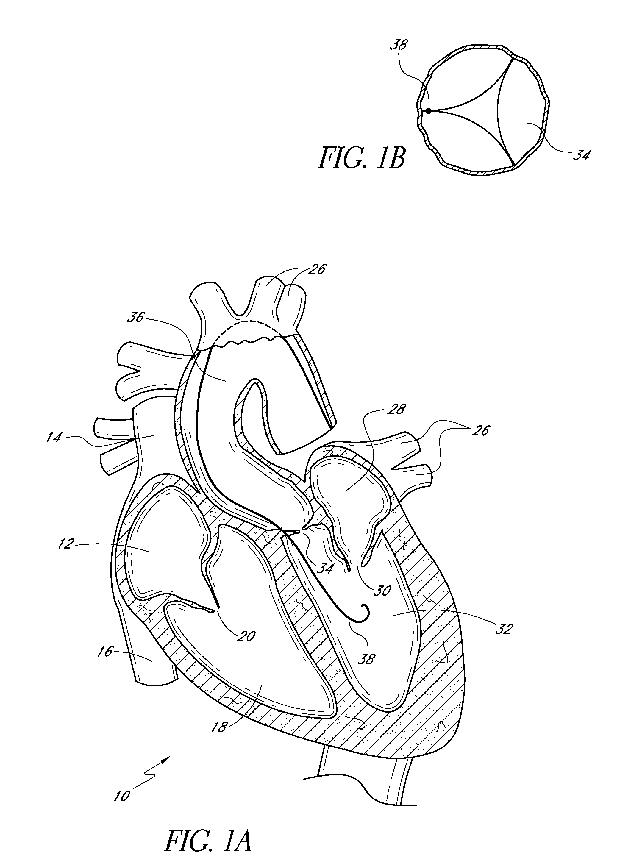
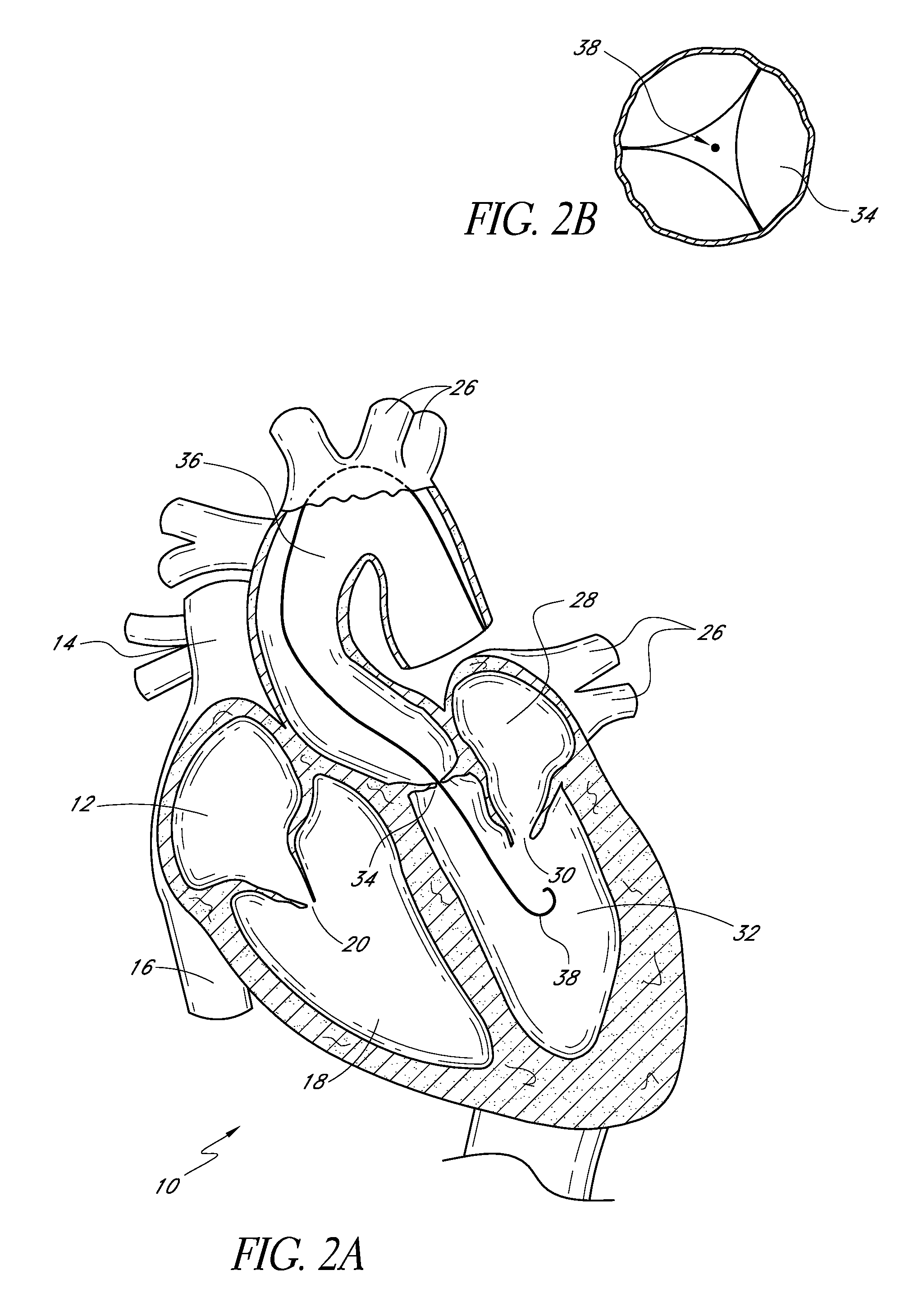
[0042]FIG. 1A is a schematic cross-sectional illustration of the anatomical structure and major blood vessels of a heart 10. Deoxygenated blood is delivered to the right atrium 12 of the heart 10 by the superior and inferior vena cava 14, 16. Blood in the right atrium 12 is allowed into the right ventricle 18 through the tricuspid valve 20. Once in the right ventricle 18, the heart 10 delivers this blood through the pulmonary valve to the pulmonary arteries and to the lungs for a gaseous exchange of oxygen. The circulatory pressures carry this blood back to the heart 10 via the pulmonary veins 26 and into the left atrium 28. Filling of the left ventricle 32 occurs as the mitral valve 30 opens allowing blood to be drawn into the left ventricle 32 for expulsion through the aortic valve 34 and on to the body extremities through the aorta 36. When the heart 10 fails to continuously produce normal flow and pressures, a disease commonly referred to as heart failure occurs.

[0043]One cause ...


PUM


Abstract

A method of treating a patient with a calcified aortic valve includes introducing a guide wire into a blood vessel. The guide wire is advanced through the aorta and the aortic valve. A balloon dilatation catheter is introduced into the blood vessel over the guide wire. The balloon dilatation catheter includes an elongate body, a distal portion, a guide wire lumen, an inflation lumen, and a dilatation balloon. The balloon dilatation catheter further includes at least one deflection wire lumen, and at least one deflection wire residing in the at least one deflection wire lumen and having a distal end attached to the distal portion. The balloon dilatation catheter is advanced over the guide wire through the aorta. The deflection wire is pushed towards the distal portion so that the deflection wire bows outward. The balloon dilatation catheter is advanced through the aortic valve, and the dilatation balloon is inflated.

Description

CROSS-REFERENCE TO RELATED APPLICATIONS[0001]This application claims the priority benefit to U.S. Provisional No. 60 / 862,187 filed Oct. 19, 2006, the entirety of which is hereby incorporated by reference herein.BACKGROUND OF THE INVENTION[0002]1. Field of the Invention[0003]The present invention relates to medical methods and devices, and, in particular, to methods and devices for guiding a catheter through an aortic valve.[0004]2. Description of the Related Art[0005]According to recent estimates, more than 79,000 patients are diagnosed with aortic and mitral valve disease in U.S. hospitals each year. More than 49,000 mitral valve or aortic valve replacement procedures are performed annually in the U.S., along with a significant number of heart valve repair procedures.[0006]The circulatory system is a closed loop bed of arterial and venous vessels supplying oxygen and nutrients to the body extremities through capillary beds. The driver of the system is the heart providing correct pr...

Claims


Application Information

Patent Timeline
Patent Type & Authority Patents(United States)
IPC IPC(8): A61M31/00
CPCA61F2/2418A61M25/0147A61M25/1011A61M29/00A61M25/10A61F2250/0003A61F2220/0075A61M2025/1047A61F2/2412A61F2/2445A61M29/02A61M2029/025
Inventor LASHINSKI, RANDALL T.
Owner SPEYSIDE MEDICAL LLC
Why Eureka
Try Eureka