
Method for co-culture of human embryonic stem cells and fibroblast feeder cells using a polyester membrane

a technology of fibroblast feeder cells and polyester membrane, which is applied in the field of co-culture of fibroblast feeder cells, can solve the problems of reducing the cell survival of feeder cells to 5 to 7 days, difficult to culture and manipulate human embryonic stem cells, and improper mass-cultur

Active Publication Date: 2012-09-04
MODERN CELL & TISSUE TECH
View PDF8 Cites 9 Cited by
  • Summary
  • Abstract
  • Description
  • Claims
  • Application Information

AI Technical Summary

Benefits of technology

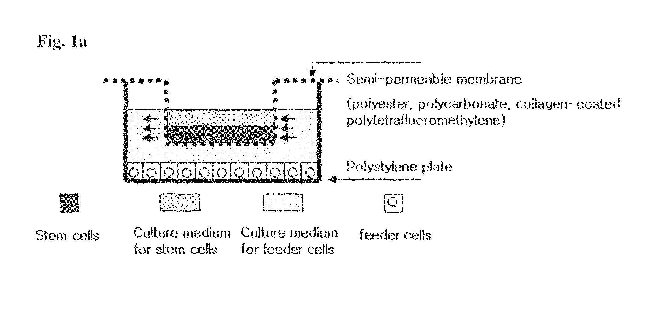
"The present invention provides a method for co-culturing stem cells and feeder cells using a polymer membrane with pores. The stem cells remain undifferentiated while the feeder cells are cultured under optimized conditions. The method can be used with different types of stem cells and feeder cells, and the membrane can be made of polyester. The diameter of the pores ranges from 0.2 to 0.5 μm and the porosity of the membrane ranges from 2.0 E+06 to 6.0 E+06. The stem cells can be embryonic stem cells and the feeder cells can be pretreated with serum or not. The method can also be used for stem cell sub-culturing and transfer."

Problems solved by technology

The human embryonic stem cell is more difficult to be cultured and manipulated than the mouse stem cell.
Either, it is improper to be mass-cultured, even if necessary to develop therapeutic agents by performing a gene or in vitro manipulation.
In addition, this procedure reduces the cell survival of feeder cells to 5 to 7 days, since treating mitomycin or irradiating.
Besides, feeder cells may not be safe due to mitomycin.
Furthermore, stem cells are inevitably mixed with feeder cells when being sub-cultured, because 2 kinds of cells are cultivated on the same culture plate.
This contamination causes a serious problem in case of clinical applications.

Method used


Image

  • Method for co-culture of human embryonic stem cells and fibroblast feeder cells using a polyester membrane
  • Method for co-culture of human embryonic stem cells and fibroblast feeder cells using a polyester membrane
  • Method for co-culture of human embryonic stem cells and fibroblast feeder cells using a polyester membrane

Examples


example 1

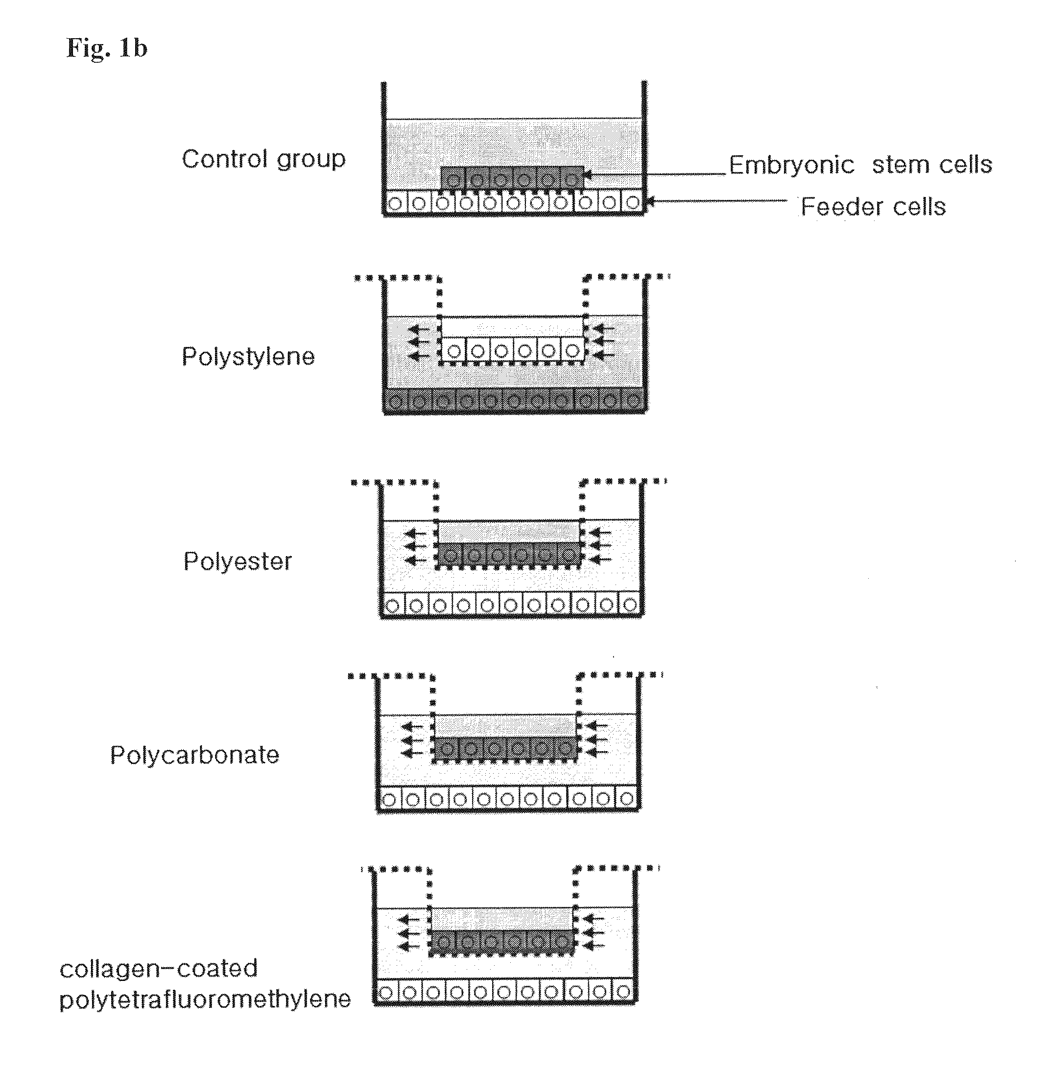
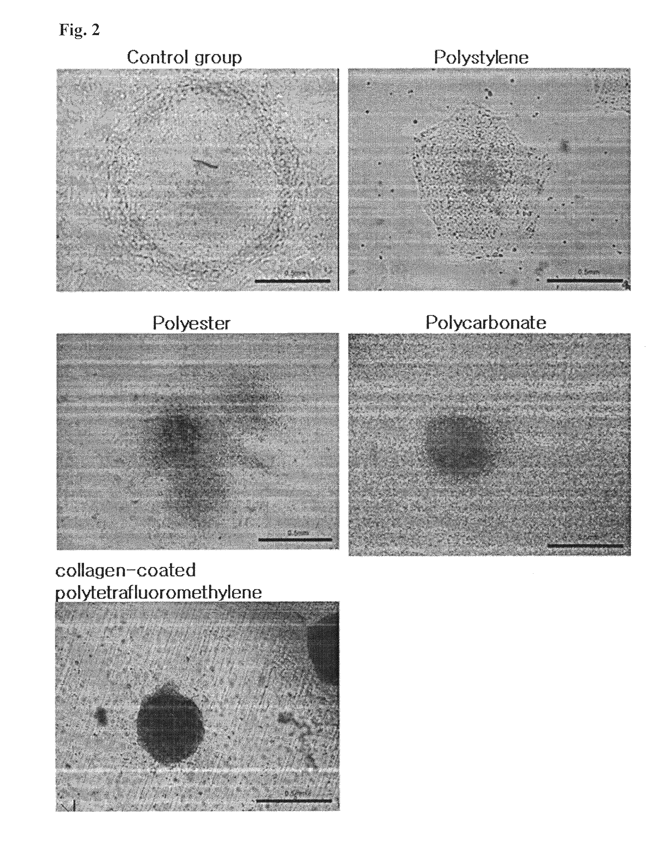
Cultivation of Embryonic Stem Cells in Control Group

[0056]Mouse embryonic fibroblasts were extracted from 13.5 day-pregnant mice (CFI, C57BL6) and primarily cultured. The resulting cells were treated with 10 μg / ml of mitomycin (Mitomycin C, Sigma, Cat. No. M-4287) as a cytostatic agent for one and a half hours to be used for feeder cells. Then, the resultants were inoculated in 4×10⁴ / cm2 of cell density and on the next day, embryonic stem cells (HSF6) were seeded. In the composition of culture media, 3.069 g / l sodium bicarbonate (Sigma, USA, Cat. No. S5761), 2 mM L-glutamine (Sigma, Cat. No. S8540), 1% penicillin (50 U / ml; Sigma, Cat. No. P4687) / streptomysin (50 ug / ml; Sigma, USA, Cat. No. S1277), 20% Knock-Out serum replacement (SR; Invitrogen BRL, Cat. No. 10828-028), 4 ng / ml Basic Fibroblasts Growth Factor (bFGF; Invitrogen BRL, Cat. No. 13256-029) were added to basic DMEM / F12 medium (GIBCO, USA, Cat. No. 12500-062). The stem cells were cultivated at 37° C. in 5% CO2 incubator wh...

example 2

Co-Culture of Embryonic Stem Cells Using Semi-Permeable Membrane

[0057]Co-culture was performed by using general polystylene plates and commercially available transwells purchased from Corning Co. Ltd. Above all, mouse embryonic fibroblasts were treated with 10 μg / ml of mitomycin (Mitomycin C, Sigma, Cat. No. M-4287) as a cytostatic agent for one and a half hours to be used for feeder cells. Otherwise without mitomycin, the fibroblasts were inoculated in 5×10³ / cm2 of cell density. Then on the next day, fresh culture media for embryonic stem cells was poured on the upper space and equilibriated. After monitoring equilibrium between upper and lower media, embryonic stem cells (HSF6) were seeded onto a membrane. In the composition of culture media, DMEM media (GIBCO, USA, Cat. No.) was blended with 3.7 g / l sodium bicarbonate (Sigma, USA, Cat. No. S5761), 2 mM L-glutamine (Sigma, Cat. No. S8540), penicillin (50 U / ml; Sigma, Cat. No. P4687) / streptomycin (50 ug / ml; Sigma, USA, Cat. No. S12...

example 3

Alkaline Phosphatase Staining

[0058]Human embryonic stem cells were selected by using alkaline phosphatase staining. Stained colonies of stem cells were judged to remain indifferent and counted. Clear colonies without staining were judged to be differentiated and calculated. In order to perform alkaline phosphatase staining, NBT / BCIP (Roche, Germany, Cat. No. 1 681 451) solution was added to Tris-Cl (pH 9.5) buffer in 99:1 of ratio and reacted. Then, coloring reaction was monitored (See FIGS. 3 and 8; Experimental results 1 and 2).


PUM

PropertyMeasurementUnit
diameteraaaaaaaaaa
diameteraaaaaaaaaa
pHaaaaaaaaaa

Abstract

The present invention relates to a method for co-culture of stem cells using feeder cells, more particularly to a method for culturing stem cells by using a membrane having a number of pores to separate stem cells and feeder cells. In the present invention, the culture condition of stem cells optimized is provided, in which stem cells and feeder cells are cultivated independently in separate spaces while permeating essential substances selectively. The stem cells prepared in the present invention continue to remain indifferent and be supported by feeder cells until needing being sub-cultured. In addition, the stem cells even for therapeutic use can be obtained without any contaminant since not pretreated with a cytostatic agent such as mitomycin or irradiated. Therefore, the method for co-culturing stem cells by using a membrane of the present invention can be widely used for clinical applications.

Description

[0001]This application is U.S. National Phase of International Application PCT / KR2007 / 002597, filed May 29, 2007 designating the U.S., and published in English as WO 2008 / 020675 on Feb. 21, 2008, which claims priority to Korean Patent Application No. 10-2006-0077478, filed Aug. 17, 2006.TECHNICAL FIELD[0002]The present invention relates to a method for co-culture of stem cells using feeder cells, more particularly to a method for culturing stem cells by using a membrane having a number of pores to separate stem cells from feeder cells. The present invention provides an optimal condition for culturing stem cells, in which 2 kinds of cells can be cultivated independently in separate spaces while permeating essential substances selectively.BACKGROUND ART[0003]In 1981, mouse embryonic stem cells have been first cultured in a test tube. In 1988, this method of in vitro culture has been combined with a gene targeting technique so as to first make a transformed mouse having a targeted gene...

Claims


Application Information

Patent Timeline
Patent Type & Authority Patents(United States)
IPC IPC(8): C12N5/02C12N5/071C12N5/00C12N5/0735
CPCC12N5/0606C12N2501/115C12N2533/30C12N2502/13C12N5/00
Inventor PARK, HYUN SOOKLEE, SUN RAY
Owner MODERN CELL & TISSUE TECH
Learn More