
Radiographic image detection apparatus

a radiographic image and detection apparatus technology, applied in the field of radiographic image detection apparatus, can solve the problems of high radiation dose to the patient in the width direction, high cost of the apparatus, and shift of image-overlapping areas, so as to improve the image quality of the synthesis image, accurately detect the inclination angle, and accurately correct the inclination of the radiographic image

Inactive Publication Date: 2012-09-25
FUJIFILM CORP
View PDF6 Cites 12 Cited by
  • Summary
  • Abstract
  • Description
  • Claims
  • Application Information

AI Technical Summary

Benefits of technology

This solution reduces the exposure dose of radiation to the patient by enabling accurate image correction and positioning using the collimator image, improving image quality and reducing unnecessary radiation exposure during long-size radiography.

Problems solved by technology

However, if an accurate stage that does not shift nor rotate even if the radiographic image detector moves for such a long distance is adopted, the cost of the apparatus becomes very high.
Especially, a rotation error (inclination) with respect to an axis perpendicular to a detection plane (surface) of the radiographic image detector causes shifting of image-overlapping areas.
Hence, there is a problem that the exposure dose of radiation to the patient with respect to the width direction becomes high.

Method used


Image

  • Radiographic image detection apparatus
  • Radiographic image detection apparatus
  • Radiographic image detection apparatus

Examples


Embodiment Construction

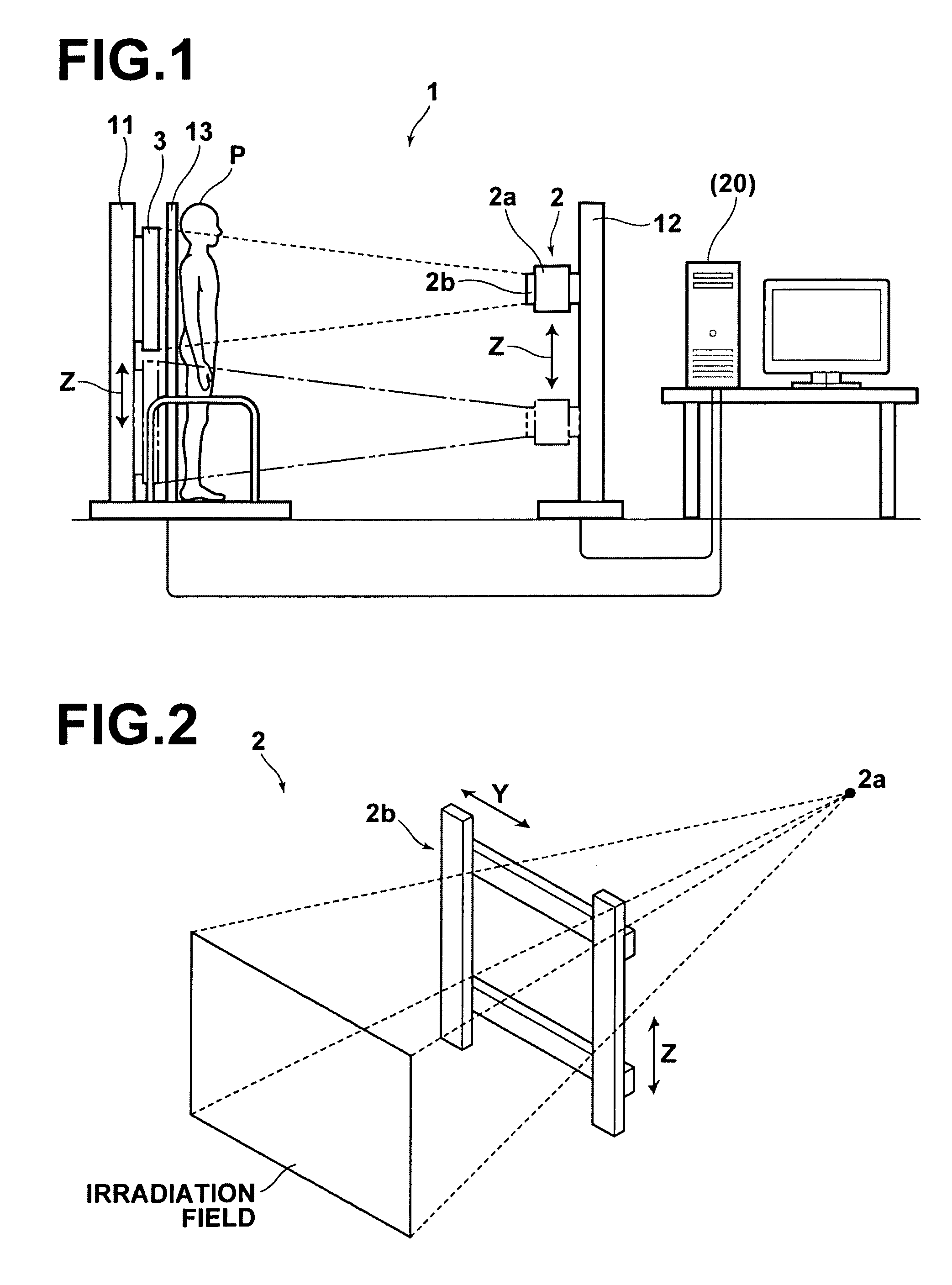
[0040]Hereinafter, embodiments of the present invention will be described in detail with reference to drawings. FIG. 1 is a schematic diagram illustrating a side view of a radiographic image obtainment system according to an embodiment of the present invention. A radiographic image detection apparatus 1 illustrated in FIG. 1 can perform two kinds of radiography, namely, so-called long-size radiography (longitudinal radiography) and ordinary radiography. In the long-size radiography, radiography is performed a plurality of times to obtain radiographic images of different regions of a subject. In the ordinary radiography, radiography is performed on a predetermined region of the subject. When the long-size radiography is performed, a screen 13 is attached to the radiographic image detection apparatus 1, and when the ordinary radiography is performed, the screen 13 is removed from the radiographic image detection apparatus 1.

[0041]The radiographic image detection apparatus 1 includes a...


PUM


Abstract

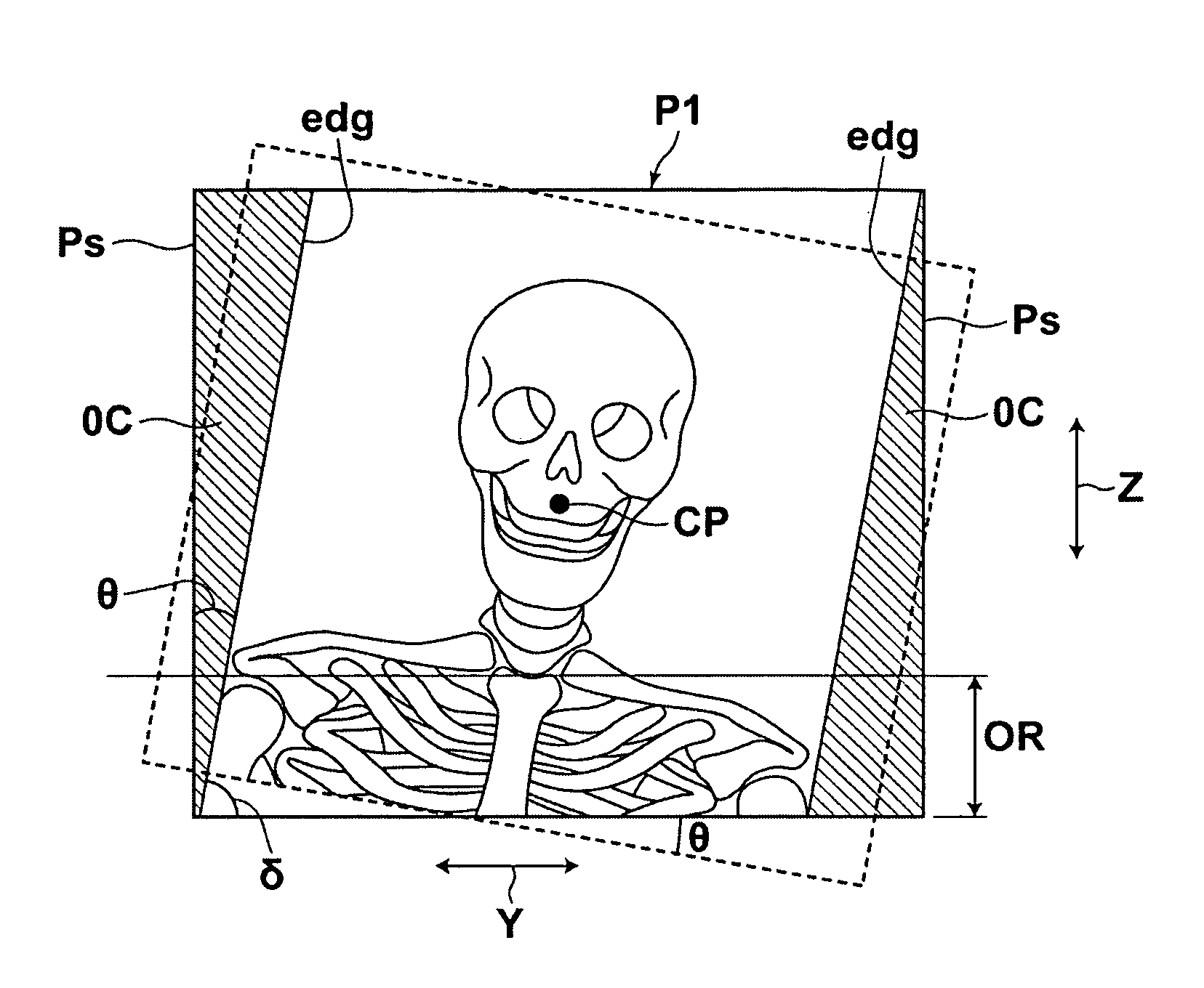
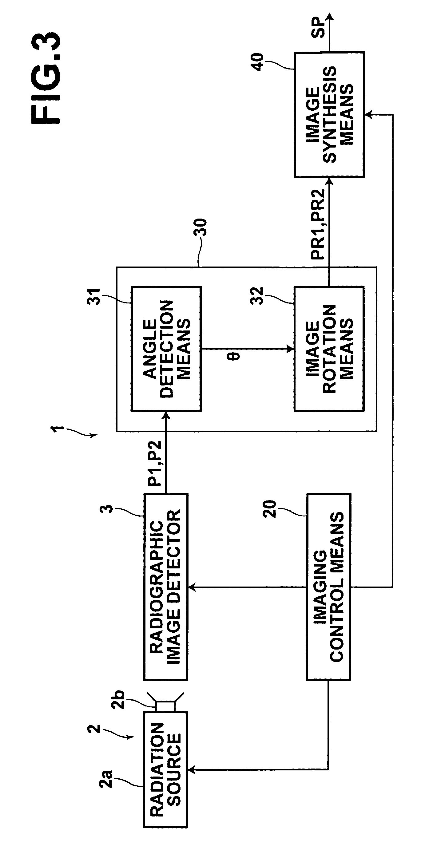
In a radiographic image detection apparatus that can perform long-size radiography on a region of a patient that is larger than the detection range of a radiographic image detector, radiography is performed so that a collimator image is included in a radiographic image in such a manner that the collimator image is present in the vicinity of a side edge of the radiographic image. Further, the inclination of each radiographic image caused by inclination of the radiographic image detector at the time of radiography is corrected based on the collimator image. Further, a synthesis image is produced by combining the plurality of radiographic images the inclination of which has been corrected.

Description

CROSS-REFERENCE TO RELATED APPLICATIONS[0001]The present application claims priority from Japanese Patent Application No. 2008-244056, filed September 24, 2008, the contents of which are herein incorporated by reference in their entirety.BACKGROUND OF THE INVENTION[0002]1. Field of the Invention[0003]The present invention relates to a radiographic image detection apparatus that can perform so-called long-size radiography, in which a plurality of radiographic images are combined to produce a single long-size radiographic image.[0004]2. Description of the Related Art[0005]Conventionally, in medical fields and the like, long-size radiography (longitudinal radiography) is performed in some cases. In the long-size radiography, a radiographic image detector is moved with respect to a subject (patient) to obtain radiographic images at a plurality of positions, and the obtained radiographic images are combined. Therefore, in the long-size radiography, it is necessary that the radiographic i...

Claims


Application Information

Patent Timeline
Patent Type & Authority Patents(United States)
IPC IPC(8): A61B6/03
CPCA61B6/00A61B6/06A61B6/469A61B6/5241G06T7/0028G06T2200/32G06T2207/10116G06T2207/30004G06T7/33
Inventor OOGAMI, SATORU
Owner FUJIFILM CORP
Learn More