Piezo micro-markers for ultrasound medical diagnostics

a micro-marker and ultrasound technology, applied in the field of medical imaging, can solve the problems of troublesome image of metal objects such as biopsy needles, and the inability to easily see small medical devices made of plastics or polymers in ultrasound images

Active Publication Date: 2012-10-09
ARIZONA STATE UNIVERSITY
View PDF7 Cites 46 Cited by
  • Summary
  • Abstract
  • Description
  • Claims
  • Application Information

AI Technical Summary

Problems solved by technology

Small medical devices made from plastics or polymers are often not easily seen in ultrasound images because their acoustic properties are similar to those of the surrounding tissue.
Metal objects such as biopsy needles can also be troublesome to image because they specularly reflect ultrasound.

Method used


Image

  • Piezo micro-markers for ultrasound medical diagnostics
  • Piezo micro-markers for ultrasound medical diagnostics
  • Piezo micro-markers for ultrasound medical diagnostics

Examples


Embodiment Construction

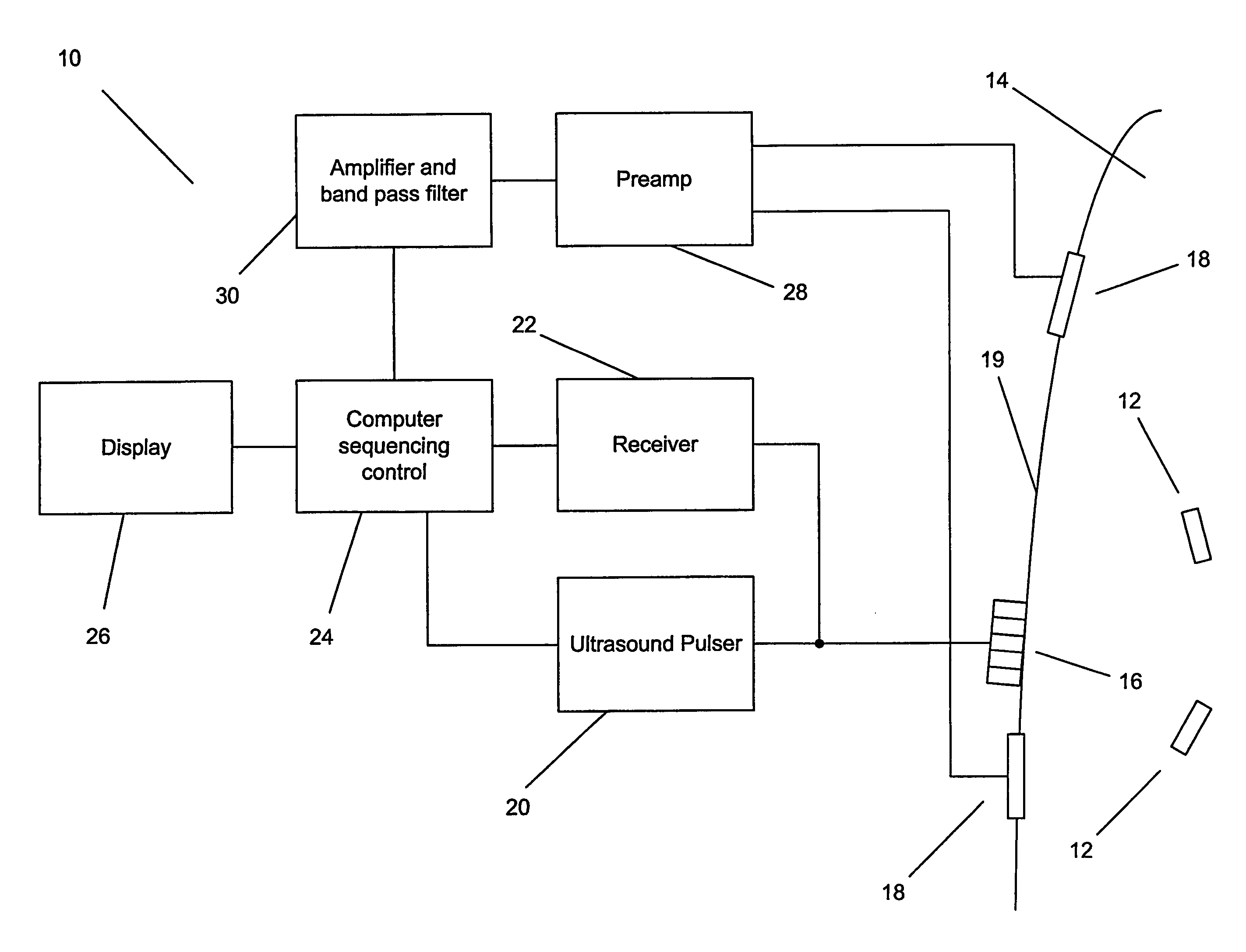
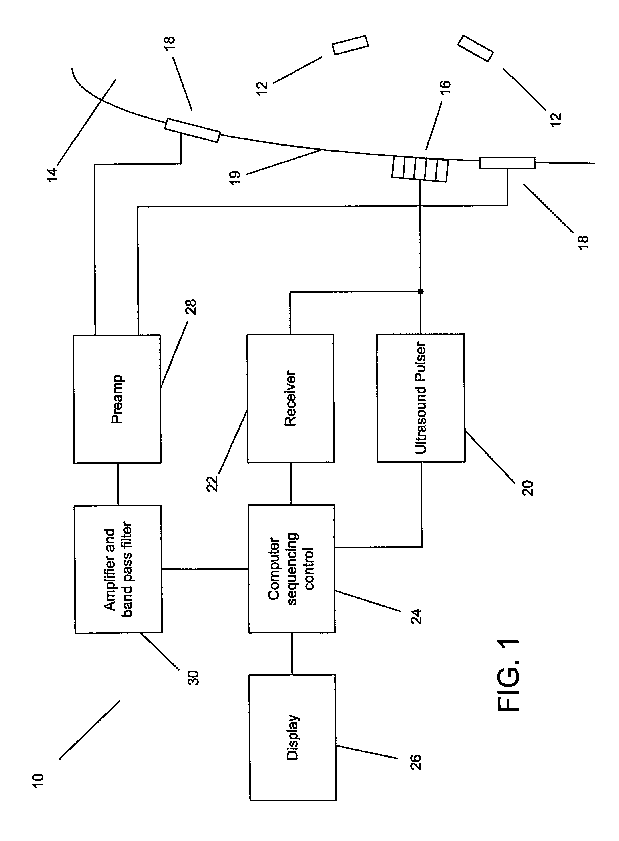
[0015]Turning now to the drawings, embodiments of the present invention include piezoelectric markers that generate electric fields in response to excitation by ultrasound pressure waves. The generated electric fields can be detected using electrodes to provide positional information. In several embodiments, the positional information can be combined with information from ultrasound reflections to provide an ultrasound image of a subject's body that includes the piezoelectric markers, which would otherwise be difficult to observe.

[0016]An embodiment of an imaging system in accordance with the present invention is illustrated in FIG. 1. The imaging system 10 includes at least one piezoelectric marker 12 embedded inside a subject's body 14. An ultrasound transducer array 16 is positioned external to the subject's body to direct ultrasound pressure waves into the subject's body and electrodes 18 are attached to the surface 19 of the subject's body. The ultrasound transducer array is co...


PUM


Abstract

An imaging system is disclosed that uses piezoelectric markers. The piezoelectric fields in combination with ultrasound reflections can be used to construct an image of an otherwise difficult to detect feature within a subject's body. In one embodiment, the invention includes a piezoelectric marker, including at least one piece of piezoelectric material, an ultrasound transducer connected to an ultrasound pulser and a receiver, a computer sequencing control connected to the receiver and the ultrasound pulser, a display connected to the computer sequencing control and electrodes connected to the computer sequencing control via amplification circuitry.

Description

BACKGROUND[0001]The present invention relates generally to medical imaging and more specifically to the imaging of foreign objects such as medical devices that are inserted into the body of a subject.[0002]Image contrast in conventional medical ultrasound results from differences in tissue acoustic properties. Small medical devices made from plastics or polymers are often not easily seen in ultrasound images because their acoustic properties are similar to those of the surrounding tissue. Metal objects such as biopsy needles can also be troublesome to image because they specularly reflect ultrasound. An alternative to ultrasound imaging is X-ray radiography. X-ray radiography is routinely used to position catheters or locate implanted markers, but involves radiation that can be ionizing.SUMMARY OF TIE INVENTION[0003]Embodiments of the present invention use piezoelectric materials to enable the imaging of foreign objects in the body of a subject using ultrasound. One aspect of the in...

Claims


Application Information

Patent Timeline
Patent Type & Authority Patents(United States)
IPC IPC(8): A61B8/14A61BA61B8/00A61B8/08A61B8/12
CPCA61B8/0833A61B8/481
Inventor TOWE, BRUCE
Owner ARIZONA STATE UNIVERSITY
Why Eureka
Try Eureka