Apparatus and method for non-contact electrical impedance imaging

a non-contact, electrical impedance imaging technology, applied in the field of apparatus and method for electrical impedance imaging, can solve the problems of unrepeatable image production, unexpected and unknown electrical impedances, preventing the accurate detection and monitoring of the development of, for example, non-healthy tissue, etc., to achieve high repeatability of measurements, prevent fluid impedance variation introducing errors, and improve image resolution

Active Publication Date: 2012-10-23
WANG WEI DR
View PDF42 Cites 83 Cited by
  • Summary
  • Abstract
  • Description
  • Claims
  • Application Information

AI Technical Summary

Benefits of technology

[0010]One advantage of having a continuum of fluid intervene between each electrode and the object instead of having the electrodes directly contact the object is that not only does it substantially standardise the impedance between each electrode and the object, but it also achieves “perfect” contact between electrode and object based on “sizeless” ions. This allows for more reliable imaging.
[0012]An advantage of using a fluid comprising ions is that the conductive mechanism between the electrode and the object is ‘electrode-ions-object’ based on “sizeless” ions. This conduction mechanism provides a “perfect” contact between the electrode and the object with known “half-cell” potentials once the electrode material is chosen. As the conduction mechanism is not a typical “electrode-skin interface” it does not suffer the disadvantages associated with any “contact” based “electrode-skin interface” namely an unknown effective contact area which provides an unknown contact impedance including associated capacitance from the “electrode-skin interface”. The electrode-ion-object conduction mechanism is not dependent upon the contact area between the electrode and the object. This allows the use of smaller electrodes, which allows a greater number of electrodes to be used to image an object, which in turn provides greater resolution in the image produced.
[0013]The temperature of the fluid is thermostatically controlled at a fixed temperature close to human body temperatures. An advantage of maintaining the temperature at a constant value is that the impedance between each electrode and the object remains constant over time. This prevents variation in the impedance of the fluid introducing errors.
[0015]The algorithm used to image an object may take as an input an indication of the volume of the housing. The volume is associated with a particular known arrangement of electrodes, and knowledge of this arrangement can be used in the algorithm to improve the accuracy of the image produced. To be able to reconstruct a high-quality image by a selected imaging reconstruction algorithm, it is required to know the exact correct positions of each electrode and its relation to the object to be imaged so that the errors between the ideal electrode positions used by the algorithms and actual positions of the electrodes will be significantly minimized. Furthermore, to be able to reconstruct a highly repeatable image for diagnosis, it is necessary for the object to be in a similar geometry and three dimensional position for each measurement so that the image collected from a different time and even from a different hospital with the same model of EIM device can be highly comparable. To achieve this, the same volume and therefore the same electrode arrangement may be used in a series of distinct and separate imaging procedures for the same patient, so that the images produced in each procedure are comparable. This improves the repeatability of an imaging procedure and allows the development of unhealthy tissue in a breast, for example, to be accurately detected and monitored over time. In particular, it allows a technician or operator to be able to distinguish two separate areas of healthy and unhealthy tissues and monitor their development.

Problems solved by technology

A most common problem associated with impedance imaging is that there are unexpected and unknown electrical impedances present other than those provided by the object to be imaged.
This can introduce significant unpredictable errors into the produced image and can therefore result in an unrepeatable image being produced.
This will prevent the accurate detection and monitoring of the development of, for example, non-healthy tissue.

Method used


Image

  • Apparatus and method for non-contact electrical impedance imaging
  • Apparatus and method for non-contact electrical impedance imaging

Examples


Embodiment Construction

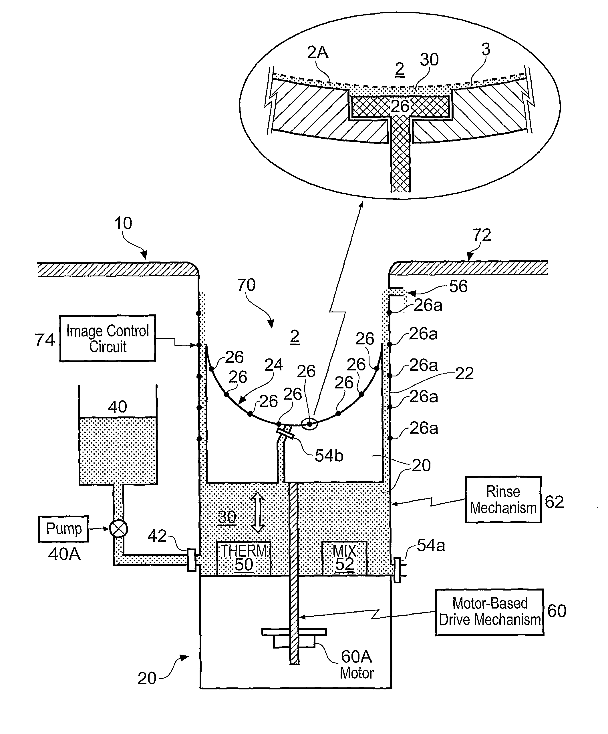
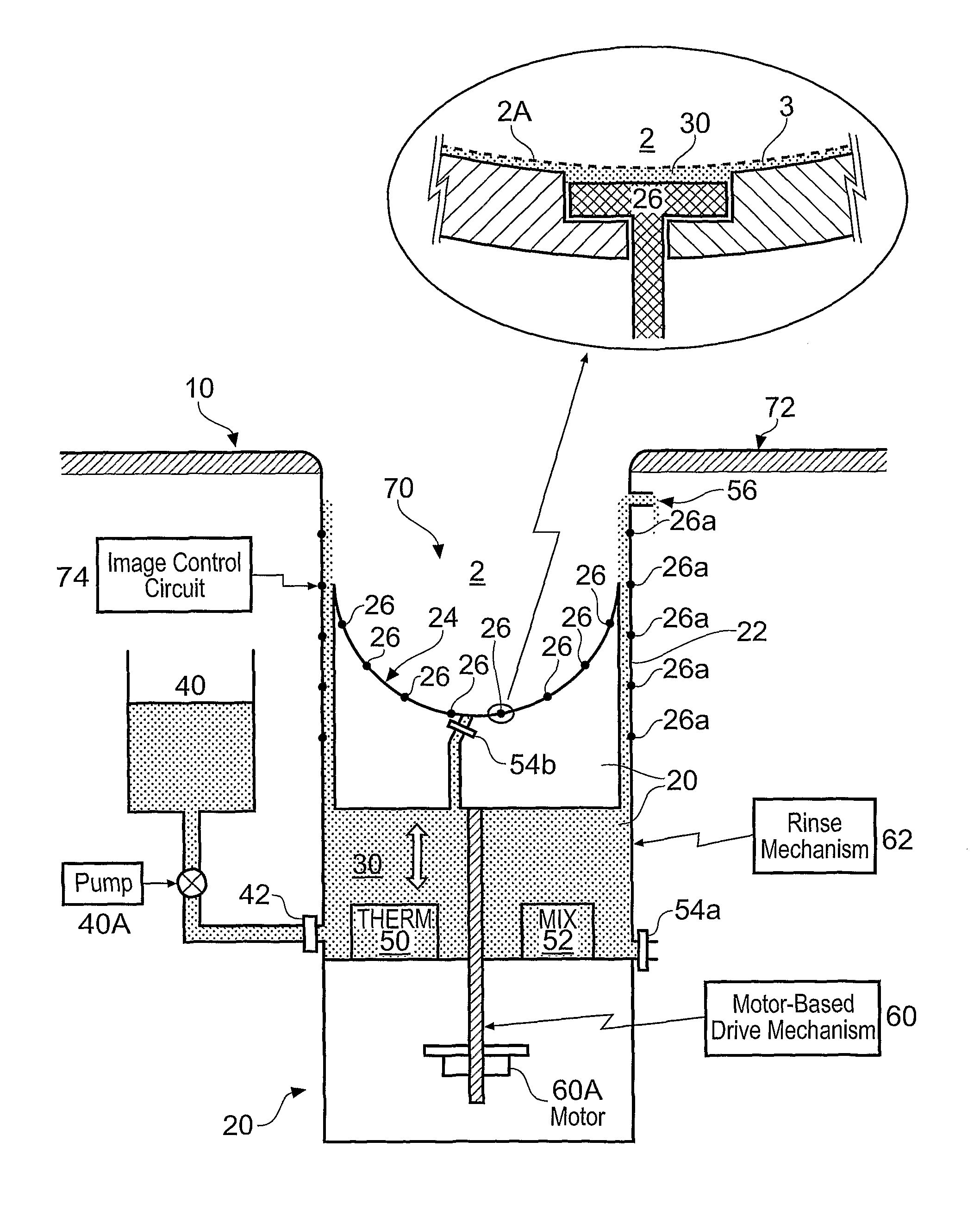
[0019]FIG. 1 illustrates an apparatus 10 for electrical impedance imaging of an object 2. The apparatus 10 comprises: a housing 20 having a cavity 70 for containing a fluid 30 in which an object 2 to be imaged is at least partially immersed; and a plurality of electrodes 26, 26A positioned at the periphery of the cavity 70. In use, each of the electrodes 26, 26A does not contact the object 2 to be imaged but is electrically coupled to the object 2 via the fluid 30 in which the object 2 is immersed.

[0020]The apparatus 10 described in the following paragraphs has been adapted for use in electrical impedance imaging of a human breast, particularly the female breast.

[0021]The female lies on a table 72 and places her breast into a cavity 70 that is already filled with fluid 30 or is subsequently filled with fluid 30. The cavity 70 has a variable volume and is sized to match the size of the breast 2 to be imaged.

[0022]The cavity 70 is defined by a housing 20 which comprises a cylindrical ...


PUM


Abstract

A method of electrical impedance imaging using multiple electrodes in which each of the multiple electrodes does not contact the object to be imaged but is electrically coupled to the object via electrically conductive fluid in which the object is at least partially immersed.

Description

CROSS REFERENCE TO RELATED APPLICATIONS[0001]This application is the U.S. National Stage of International Application Number PCT / GB2006 / 002887 filed on Aug. 3, 2006 which was published in English on Feb. 15, 2007 under International Publication Number WO 2007 / 017634.FIELD OF THE INVENTION[0002]Embodiments of the present invention relate to an apparatus and method for electrical impedance imaging.BACKGROUND TO THE INVENTION[0003]Electrical impedance mammography (EIM), or Electrical impedance imaging (EII), also referred to as electrical impedance tomography (EIT), electrical impedance scanner (EIS) and applied potential tomography (APT), is an imaging technique that is particularly used in medical applications.[0004]The technique images the spatial distribution of electrical impedance inside an object, such as the human body. The technique is attractive as a medical diagnostic tool because it is non-invasive and does not use ionizing radiation as in X-ray tomography or the generation...

Claims


Application Information

Patent Timeline
Patent Type & Authority Patents(United States)
IPC IPC(8): A61B5/05A61B5/053
CPCA61B5/0536A61B2562/046A61B2562/0209A61B5/053
Inventor WANG, WEI
Owner WANG WEI DR
Why Eureka
Try Eureka