
Methods and apparatuses for image guided medical procedures

a technology of image guided medical procedures and methods, applied in the direction of image enhancement, instruments, catheters, etc., can solve the problems of impractically rapid patient and/or equipment position changes, particularly bothersome problems, and noticeable and annoying time delays between displayed images, so as to facilitate the adjustment of the rotational orientation of the catheter, accurately position the needle, and reduce or even out artifacts

Active Publication Date: 2012-11-06
ABBOTT CARDIOVASCULAR
View PDF180 Cites 222 Cited by
  • Summary
  • Abstract
  • Description
  • Claims
  • Application Information

AI Technical Summary

Benefits of technology

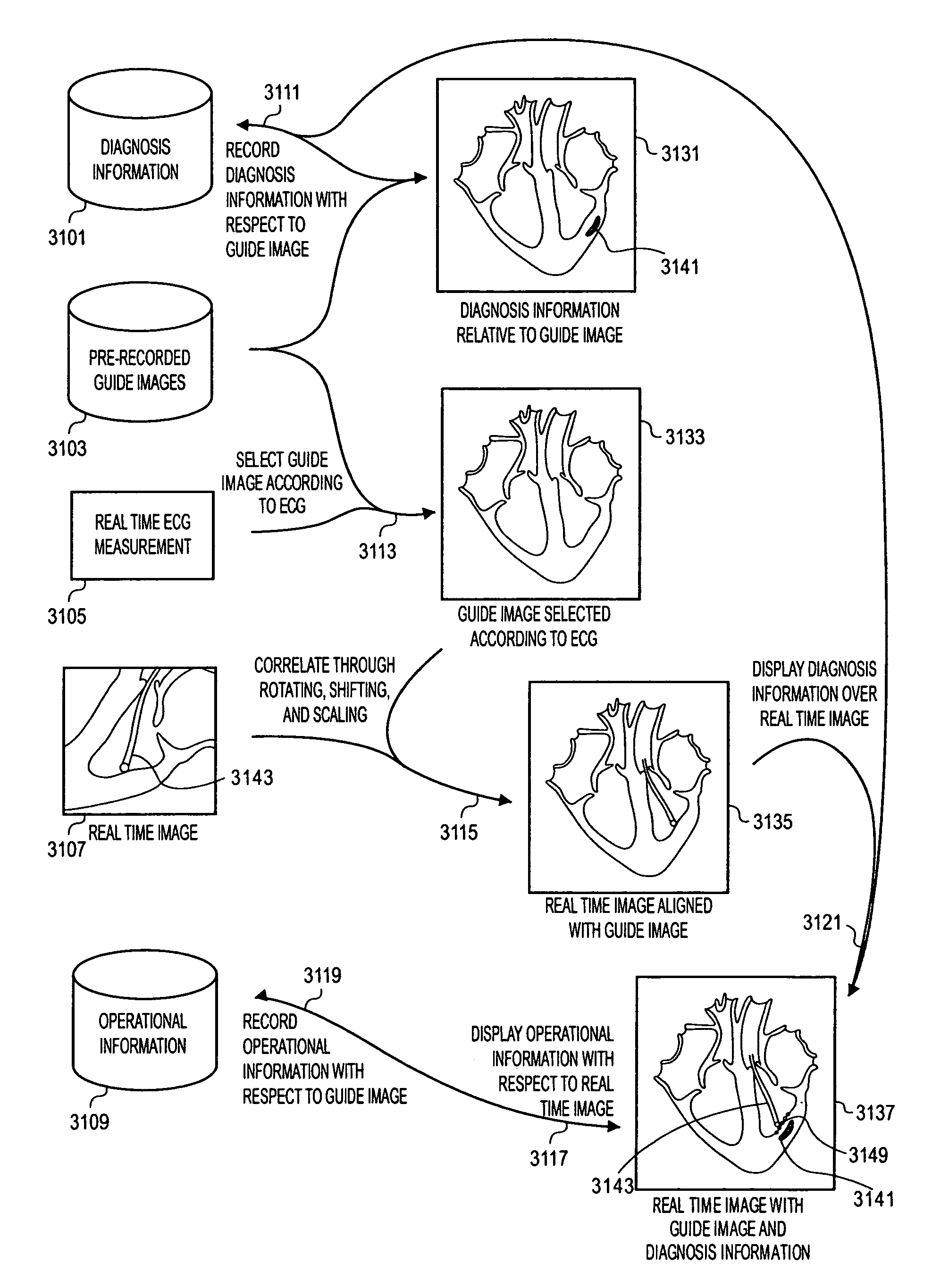
"The present invention relates to a method for aligning and displaying two images, one real-time and one recorded, to provide a larger field of view and better spatial alignment. The method involves determining the parameters of scaling, rotation, and translation to match the two images. The recorded image can include information about the patient, such as blood pressure or heart rate, to select the frames that correspond to the recorded image. The real-time image can show the position of a medical device in relation to the recorded image. The method can also compensate for uncertainties in the position of the anatomy or equipment movement. Overall, the invention improves the spatial alignment of images and provides a larger anatomical context for real-time images."

Problems solved by technology

Attempting to widen the field of view typically introduces noticeable and annoying time delays between the displayed image and the actual condition of the anatomy and / or requires impractically rapid patient and / or equipment position changes.
When attempting to guide a medical device in the anatomy and record locations in the anatomy, real time imaging is desired and these problems become particularly bothersome.
The real time image's limited field of view makes the anatomical context of the real time image or a diagnosis based on tissue image properties and / or their relationship to the adjacent anatomy difficult to interpret.
Thus, the location of the medical device relative to the adjacent anatomy or anatomy landmarks as shown in the real time image can be difficult to determine or time consuming to determine.
Additionally, a tissue diagnosis based on the real time image can be difficult to determine or time consuming to determine.
It is not very practical to expect the patient to hold their breath in a repeatable manner on command or to be able to remain perfectly still on the positioning table during the medical operation and real time imaging.
Therefore, the distances between locations recorded in real time image reference frame and between recorded locations and anatomical structures in real time image reference frame will be uncertain or in error by the amount (and timing) of this uncontrolled patient motion.
This uncertainty or error is undesirable in many situations, such as when attempting to control the spacing of a therapy applied by a medical device using imaging.
These problems are greatly accentuated in conventional ultrasonic based imaging systems, because the field of view of ultrasonic systems is typically much smaller than CT and MR based imaging systems.
Additionally, the image sensing / producing equipment (the imaging probe) of conventional ultrasonic systems is manually positioned and its location and / or orientation is not controlled or sensed by the imaging system.
Without ECG synchronization, the motion of the heart can cause very indistinct / low resolution cardiac images and varying device location information that can't be easily interpreted when images or location data or other data collected from different times in the cardiac cycle are combined.
One difficulty with ultrasound imaging has been visualization anomalies, including artifacts and overly bright images, in the ultrasonic images of catheters.
Such artifacts can provide a misleading and inaccurate impression of the shape and / or location of the catheter within the patient.

Method used


Image

  • Methods and apparatuses for image guided medical procedures
  • Methods and apparatuses for image guided medical procedures
  • Methods and apparatuses for image guided medical procedures

Examples


Embodiment Construction

[0127]The following description and drawings are illustrative of the invention and are not to be construed as limiting the invention. Numerous specific details are described to provide a thorough understanding of the present invention. However, in certain instances, well known or conventional details are not described in order to avoid obscuring the description of the present invention.

[0128]In the present application, the term “image” is not limited to a 2-D image. The term “image” may also generally refer to a set of 3-D volumetric image data, or a 4D imaging data set that includes a series of 3-D volumetric image data paired with another dimension (such as time interval in relation to the cardiac cycle), or other types of imaging data. In the following description, the word “location” refers to the X, Y, and Z points in space or memory address locations, and the word “position” refers to general position information that may include any or all of the 6 degrees of (motion) freedom...


PUM


Abstract

Methods and apparatuses for the image guidance and documentation of medical procedures. One embodiment includes combining small field of view images into a recorded image of with a large field of view and aligning the small field of view real time image with the recorded image through correlation of imaging data. A location and orientation determination system may be used to track the imaging system and provide a starting set of image alignment parameters and / or provide change updates to a set of image alignment parameters, which is then further improved through correlating imaging data. The recorded image may be selected according to real time measurement of a cardiac parameter during an image guided cardiac procedure. Image manipulations planned based on the recorded image can be stored and applied to the real time information. The position of the medical device may be determined and recorded through manipulating a cursor in a 3-D image space shown in two non-parallel views.

Description

[0001]The present patent application relates to co-pending U.S. patent application Ser. No. 10 / 390,065, filed Mar. 14, 2003, entitled “Method and Apparatus for Image Guided Position Tracking During Percutaneous Procedures” by inventor William E. Webler, which is hereby incorporated herein by reference.TECHNOLOGY FIELD[0002]Some embodiments of the present inventions relate to the spatial alignment (which may include scaling) of a real time image / data and a recorded image / data to display a limited field of view real time image in an anatomically correct alignment with the larger field of view recorded image to show the anatomic relationship of the limited field of view image to the overall anatomy image and / or to compensate for undetected motion of the anatomy in the real time image relative to the anatomy in the recorded image. Some embodiments of the present invention relate to imaging and / or tracking of medical instruments relative to the anatomy, and more particularly to the guida...

Claims


Application Information

Patent Timeline
Patent Type & Authority Patents(United States)
IPC IPC(8): A61B8/00
CPCG06T7/0026G06T7/0038G06T19/00G06F19/3437A61B17/3478A61B2017/00247A61B2017/00455A61B2018/00392G06T2210/41G06T2207/30004A61B8/4245A61B8/445A61B8/483G06T7/32G06T7/38G16H50/50
Inventor WEBLER, WILLIAM E.CHOW, MINA
Owner ABBOTT CARDIOVASCULAR
Learn More