
Subunit vaccine for aquaculture

a subunit vaccine and aquaculture technology, applied in the field of aquatic subunit vaccines, can solve the problems of rare fish cell lines, severe threat of ipnv, environmental protection, etc., and achieve the effects of reducing manufacturing costs, improving vaccine purity, and simplifying the mass production process

Active Publication Date: 2013-01-01
SBC VIRBAC LTD
View PDF2 Cites 1 Cited by
  • Summary
  • Abstract
  • Description
  • Claims
  • Application Information

AI Technical Summary

Benefits of technology

[0016]This invention provides an aquatic subunit vaccine prepared by using genetic engineering techniques to fuse an antigenic protein against fish disease with a receptor binding protein, a translocation protein and a signal peptide such that the antigen can be targeted efficiently and precisely to the effector cell as well as bind and then penetrate the plasma membrane barriers. The antigen from the aquatic subunit vaccine can be presented correctly onto the effector cell and then induce fishes an immunity sufficient to resist viral infection. A number of advantages of vaccines created by recombinant DNA technology include simpler process for mass-production, lower cost of manufacturing, higher purity of produced vaccine, and safety of utilizing the vaccine.
[0018]The inventor conjugates the PE protein (containing Domains I and II of the exotoxin A protein, whose amino acid sequence were shown as SEQ ID No: 8) to the N terminus of the fish antigenic protein and the KDEL signal peptide (shown as SEQ ID No: 10) to the C terminus of fish antigenic protein. Therefore, the fish disease-related antigenic protein (thereafter antigenic fusion protein) prepared by fusing PE protein and KDEL signal peptide can be targeted under the action of the PE protein to appropriate cell (i.e. CD8+ T cell) through specific protein-protein interaction between domain I of exotoxin A in the vaccine and receptors on the surface of CD8+ T cell. After the antigenic fusion protein entering the endosome of the CD8+ T cell via endocytosis, it will be processed by protease in the endosome and released into cytoplasm. Then, the KDEL signal peptide will target properly the vaccine protein to bind ER inside the CD8+ T cell for antigen processing and hence induce the effective immunity in fish.

Problems solved by technology

Therefore, IPNV is a severe threat, ecologically and economically, to the aquaculture and sea-farming industries.
However, the fish cell lines are uncommon and the options are rare in addition to its difficulty in development as well as the high cost therefore.
However, protective immunological effects offered by those approaches described above can not satisfy the standard required in the aquaculture industry.
Accordingly, there are a lot of drawbacks associated with conventional aquatic vaccines and improvements thereof have to be improved urgently.
Then, the third domain of truncated exotoxin A enables to inhibit protein synthesis in the cell, resulting in cell death.

Method used


Image

  • Subunit vaccine for aquaculture
  • Subunit vaccine for aquaculture
  • Subunit vaccine for aquaculture

Examples


example 1

The Construction of pET24-PE-VP2-KDEL Expression Vector for Antigenic Fusion Protein

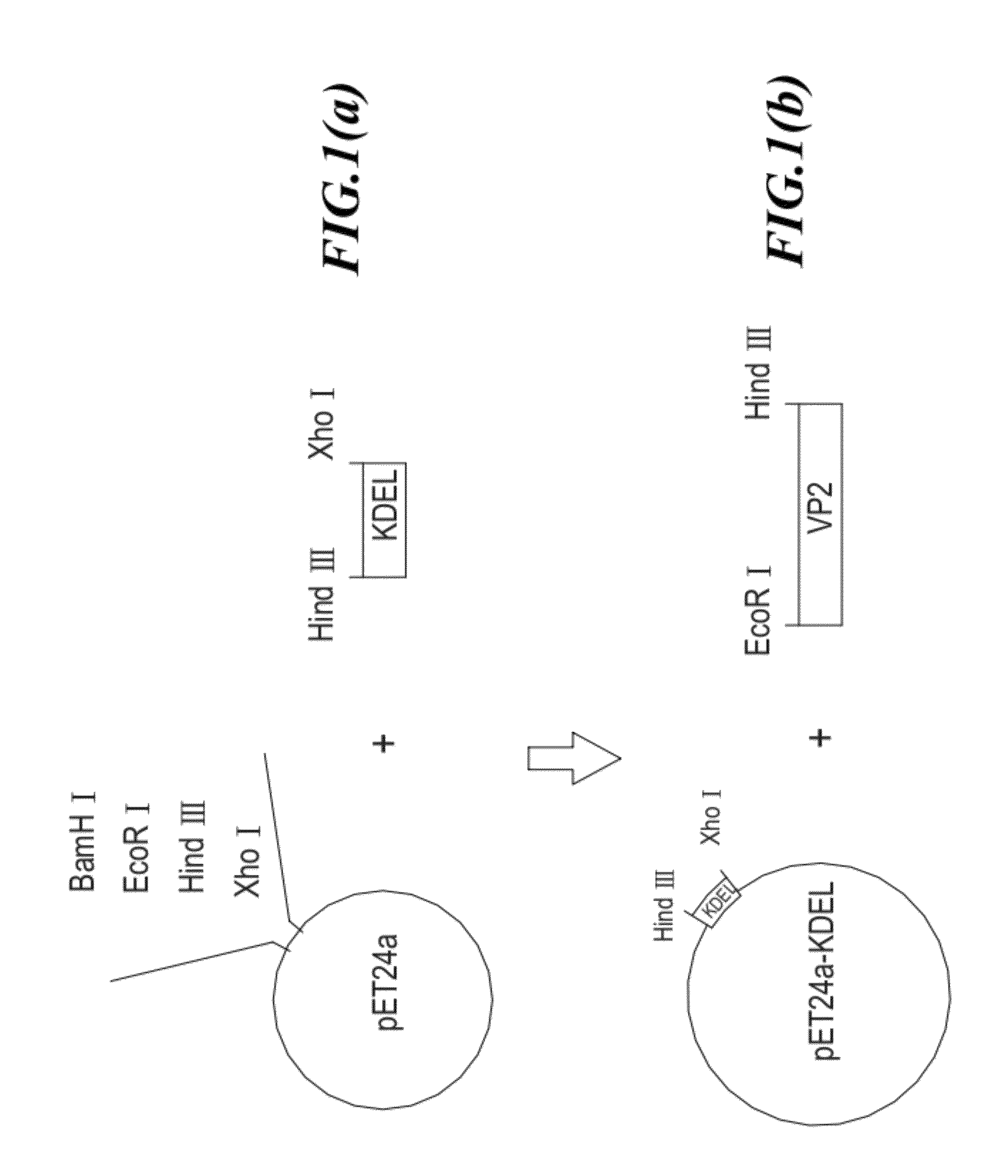
[0033]In this example, a fragment of IPNV VP2 was used as the antigen of the aquatic subunit vaccine. A receptor binding motif and the a translocation protein (PE protein) of exotoxin A were conjugated to the N terminus of VP2, as well as a KDEL signal peptide was conjugated to its C terminus, thereby formed the antigenic fusion protein (PE-VP2-KDEL).

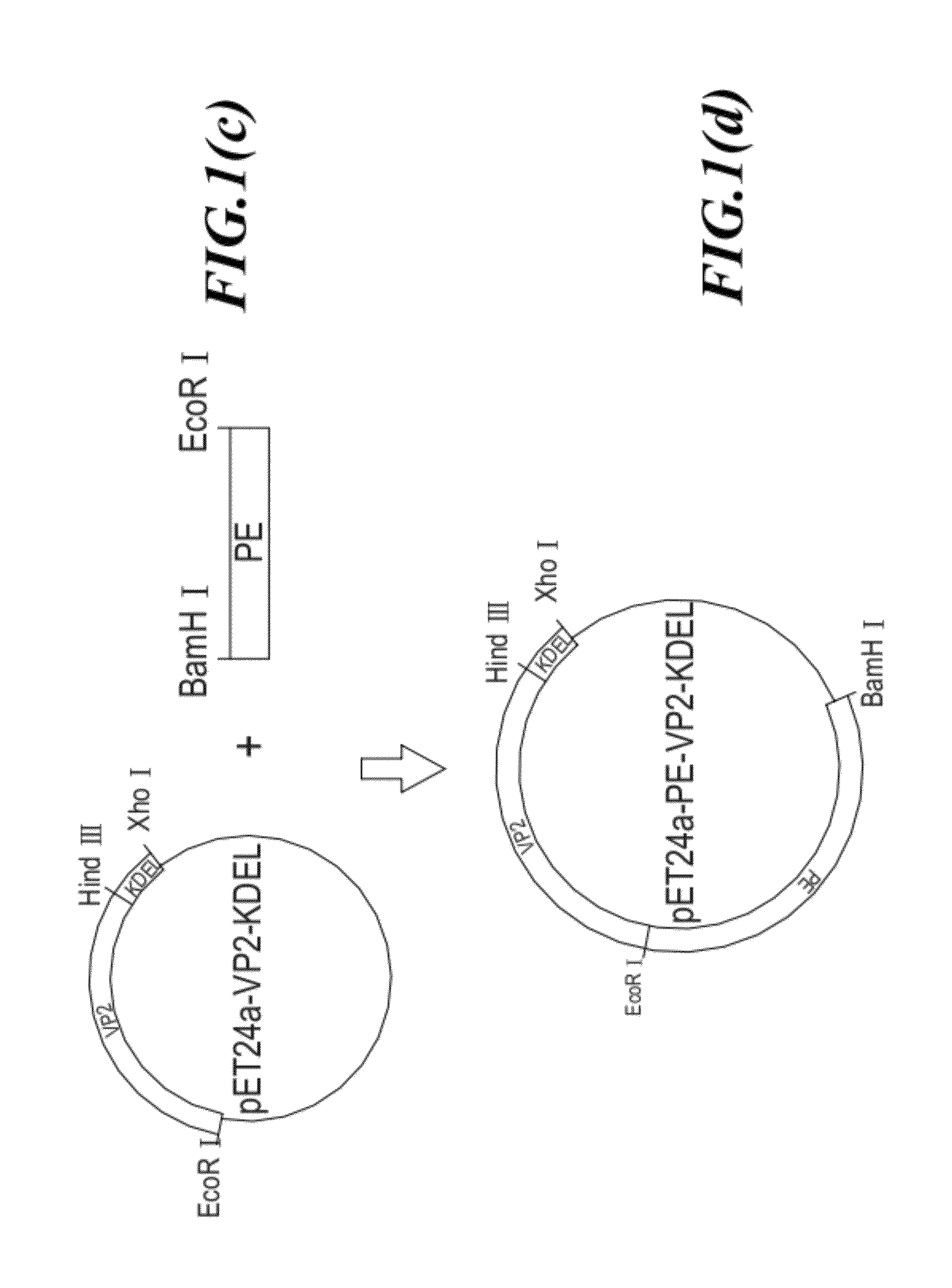
[0034]The antigenic fusion protein (PE-VP2-KDEL) was created by a gene cloning technology comprising cloning DNA sequences encoding respective proteins into an expression vector to form an expression vector pET24a-PE-VP2-KDEL, and then inducing protein expression.

[0035]The strategy for constructing the antigenic fusion protein expression vector (24a-PE-VP2-KDEL) is shown in FIG. 1. First, the DNA sequence encoding KDEL signal peptide is cloned into a plasmid pET24a to form plasmid pET24a-KDEL (as shown in FIGS. 1a and 1b). Next, the DNA sequence encoding...

example 2

Vaccine Preparation

1. The Expression of Antigenic Fusion Protein

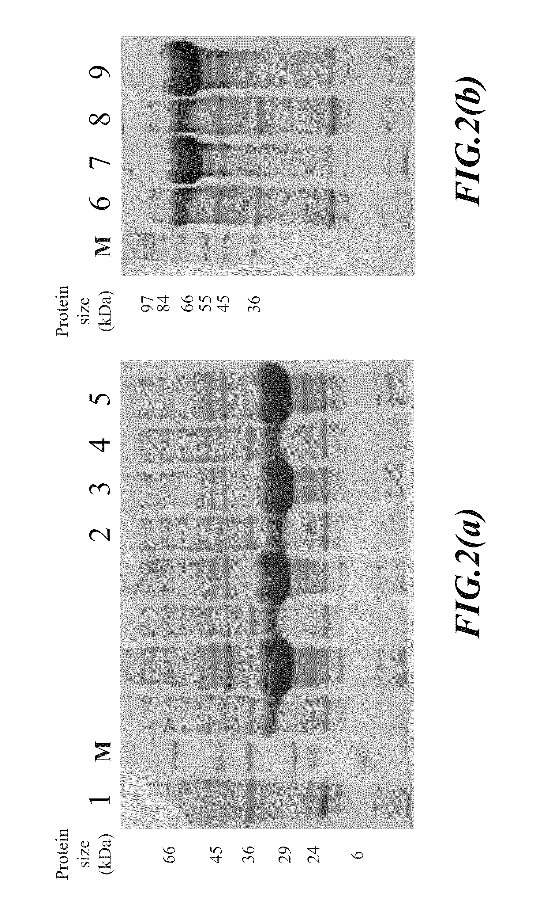
[0053]E. coli BL21 cells transformed as described above with plasmid pET24a-PE-VP2-015-KEDL or plasmid pET24a-PE-VP2-016-KEDL, or E. coli BL21 cells containing plasmid pET24a-PE-VP2-016 or plasmid pET24a-PE-VP2-016 were cultured, respectively, at 37° C. in LB broth containing 50 μg / ml kanamycin overnight. Then, 10 ml of the culture was added into 990 ml fresh LB broth containing 50 μg / ml kanamycin. The diluted cell culture was incubated further at 37° C. by shaking at 250 rpm for 3 hours. Then, 1 ml 1M IPTG (final concentration 1 mM) as an inducer, was added to the culture to induce the expression of the antigenic fusion protein (PE-VP2-015-KEDL whose amino acid sequence was shown as SEQ ID No: 12; or PE-VP2-016-KEDL whose amino acid sequence was shown as SEQ ID No: 14). After IPTG induction, cells were cultured for another three hours and then, harvested by centrifugation at 6,5000×g at 4° C. for 30 minutes. The supern...

example 3

Anti-Viral Test in Salmons

[0057]The basic information about salmons tested was as followed:

[0058]

SpeciesAtlantic salmon (Salmo salar L.)StrainAquagen AS, LR (Low resistance) 06AGHe01SizeAverage 60-90 gVaccination historyNot previously vaccinatedDiseases historyNone

[0059]Culture conditions of salmons in the stage of smoltification:

[0060]

WaterSalinityTemp.QualityTimePhotoperiodFlowDensity0‰8-12° C.Fresh5 weeks12 hours daylight0.8 l / kg fishMax. 40water12 hours darknesspr. min.kg / m30‰8-12° C.FreshFrom week 524 hour daylight0.8 l / kg fishMax. 40waterand onwardspr. min.kg / m3

[0061]There were four groups plus one control group of salmons used in virus resistant test. Each group had thirty salmons. The prepared vaccines were administrated by intraperitoneal injection (i.p.) into the fishes. The dose of the vaccine was 0.1 ml per fish, which corresponded to 0.1 μg of antigenic fusion protein or VP2 viral protein per fish. The virus resistant test was designed as described below:[0062]Group 1: ...


PUM

PropertyMeasurementUnit
pHaaaaaaaaaa
pHaaaaaaaaaa
molecular weightsaaaaaaaaaa

Abstract

An aquatic subunit vaccine comprises an antigenic fusion protein and suitable carrier or adjuvant. The antigenic fusion protein sequence consists from its amino terminus to carboxyl terminus of a receptor binding motif and a translocation domain of Pseudomonas aeruginosa exotoxin A and has the amino acid sequence of SEQ ID No: 8; a viral antigenic protein affecting fish disease; and a signal peptide having the amino acid sequence of SEQ ID No: 10.

Description

BACKGROUND OF THE INVENTION[0001]1. Field of the Invention[0002]The invent relates to aquatic subunit vaccine and in particular, to an aquatic subunit vaccine prepared based upon recombinant DNA technology by fusing a fish antigenic protein with the receptor binding motif and the translocation domain of an exotoxin A, as well as with the signal peptide of KDEL.[0003]2. Description of the Prior Art[0004]Infectious Pancreatic Necrosis (IPN) is a worldwide highly contagious fish disease. Infectious Pancreatic Necrosis Virus (IPNV), the pathogen of IPN, belongs to the family of Birnaviridae. The genome of IPNV consists of two segments of double-stranded RNA (dsRNA), segments A and B. The virion is a non-enveloped icosahedron particle. The length of the A segment gene is 3.2 Kb and codes predominantly for primary virus binding protein (VP2), secondary virus binding protein (VP3), and enzyme protein (VP4).[0005]The major hosts of Infectious Pancreatic Necrosis Virus are salmon and trout. ...

Claims


Application Information

Patent Timeline
Patent Type & Authority Patents(United States)
IPC IPC(8): A61K39/12A61K39/295
CPCA23K1/188A61K39/104A61K39/12A61K2039/552A61K2039/6037C07K2319/02C12N2720/10034C12N2710/00034A23K50/80A61P1/00A61P1/12A61P1/18A61P31/14Y02A40/81A23K50/00A61K39/00C12N15/62
Inventor KUO, TSUNYUNGCHEN, GABRIEL HSUCHUNGWU, CHUNGCHINEVENSEN, OYSTEINSINGH MARJARA, INDERJIT
Owner SBC VIRBAC LTD
Learn More