
Method and apparatus for magnetic resonance guided high intensity focused ultrasound focusing under simultaneous temperature monitoring

a high intensity, focused ultrasound technology, applied in the direction of instruments, measurement devices, acoustic wave reradiation, etc., to achieve the effect of precise localization of the hifu focal point, optimal plane resolution, and acquisition speed

Active Publication Date: 2013-04-23
SIEMENS HEALTHCARE GMBH
View PDF5 Cites 4 Cited by
  • Summary
  • Abstract
  • Description
  • Claims
  • Application Information

AI Technical Summary

Benefits of technology

[0005]An object of the present invention is to provide a method and an apparatus that allows the HIFU focal point to be precisely localized while avoiding, or at least alleviating, the disadvantages of the conventional approaches described above.
[0007]The acquisition speed with which the data representing the aforementioned GRE phase images is increased by using GRE-EPI and / or parallel imaging techniques and / or partial Fourier reconstruction, this further reduces the subsequent heating of the examination subject, because fewer HIFU pulses are activated when magnetic resonance signals are acquired, and / or the magnetic resonance reconstruction matrix size is reduced.

Problems solved by technology

One challenge in MRgHIFU is to provide safe and thermally neutral focusing of the HIFU beam pattern using acoustic radiation force imaging (ARFI).

Method used


Image

  • Method and apparatus for magnetic resonance guided high intensity focused ultrasound focusing under simultaneous temperature monitoring
  • Method and apparatus for magnetic resonance guided high intensity focused ultrasound focusing under simultaneous temperature monitoring
  • Method and apparatus for magnetic resonance guided high intensity focused ultrasound focusing under simultaneous temperature monitoring

Examples


Embodiment Construction

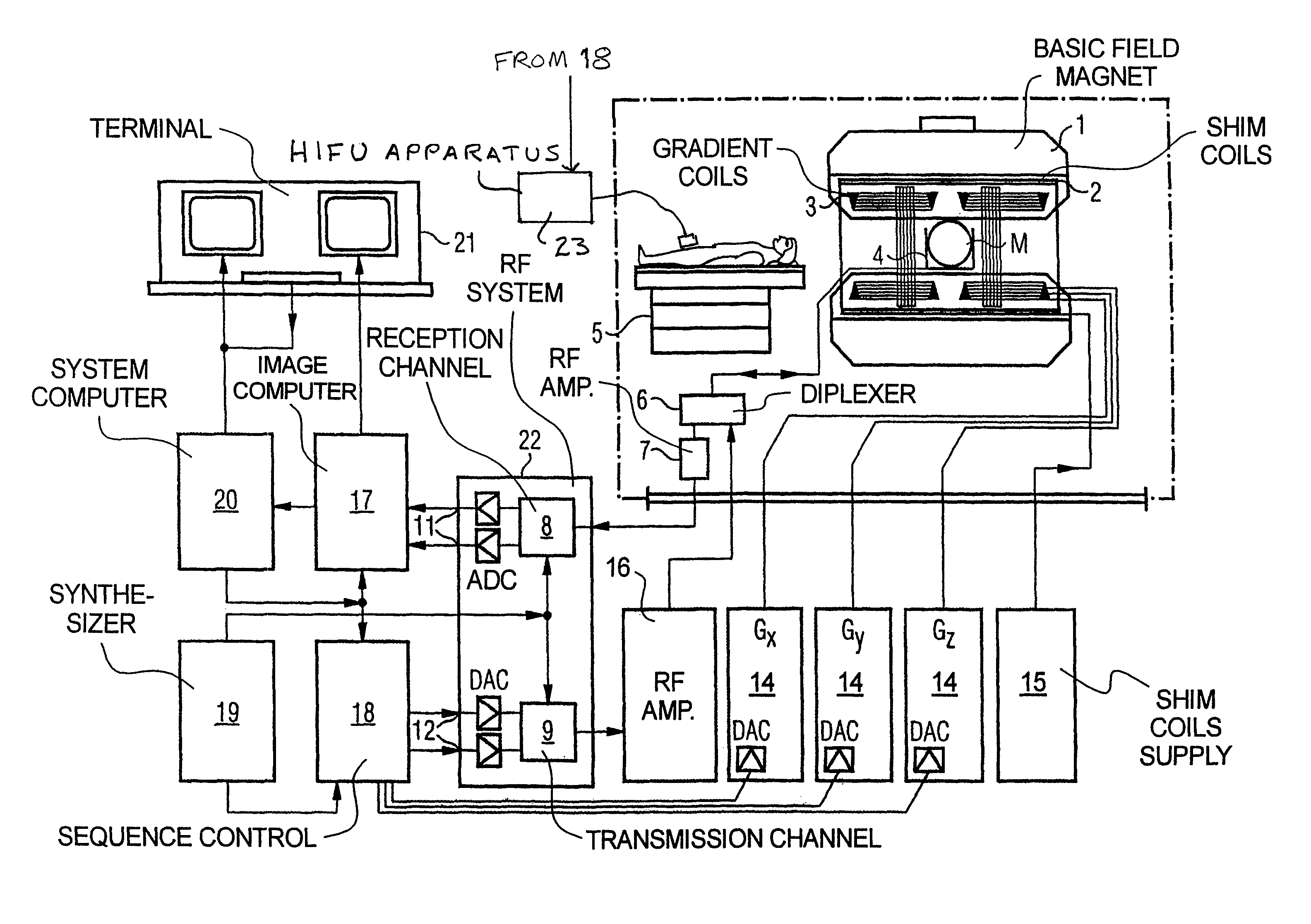
[0011]FIG. 1 is a schematic illustration of a magnetic resonance tomography apparatus operable according to the present invention. The structure of the magnetic resonance tomography apparatus corresponds to the structure of a conventional tomography apparatus, with the differences described below. A basic field magnet 1 generates a temporally constant, strong magnetic field for the polarization or alignment of the nuclear spins in the examination region of a subject such as, for example, a part of a human body to be examined. The high homogeneity of the basic magnetic field required for the magnetic resonance measurement is defined in a spherical measurement volume M into which the parts of the human body to be examined are introduced. For satisfying the homogeneity requirements and, in particular, for eliminating time-invariable influences, shim plates of ferromagnetic material are attached at suitable locations. Time-variable influences are eliminated by shim coils 2 that are driv...


PUM


Abstract

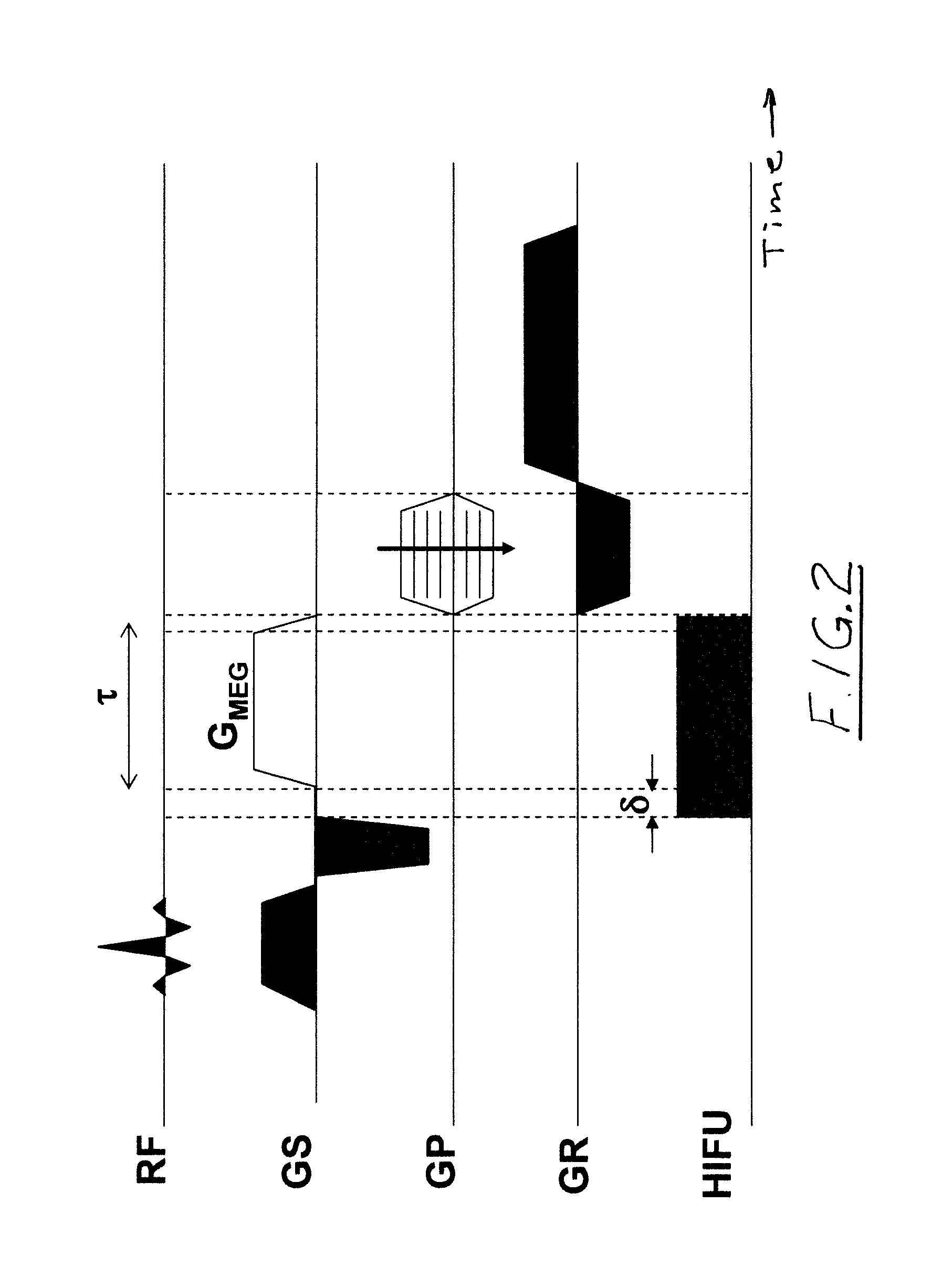
In a method and an apparatus for magnetic resonance guided high intensity focused ultrasound (HIFU), precise localization of the focal point of the HIFU is determined by imaging an examination subject in parallel with GRE sequences that respectively include a positive monopolar gradient pulse and a negative monopolar gradient pulse, that respectively encode the acoustic radiation force (ARF)-induced phase shift that is induced by the simultaneous activation of HIFU during the sequences. A GRE phase image is reconstructed from each acquisition sequence, and a difference image is formed between the two GRE phase images, from which the HIFU focal point is determined. An average image is formed of the two GRE phase images from which PRFS temperature map is determined simultaneously to ARFI map. The use of parallel imaging and the use of partial Fourier reconstruction for reconstructing the GRE phase images allows the data to be acquired sufficiently rapidly so as to minimize the adverse effect of tissue heating that occurs with conventional longer-duration, and repetitious, techniques.

Description

BACKGROUND OF THE INVENTION[0001]1. Field of the Invention[0002]The present invention concerns magnetic resonance guided high intensity focused ultrasound (MRgHIFU), and in particular concerns a method and an apparatus for focusing high intensity focused ultrasound (HIFU).[0003]2. Description of the Prior Art[0004]One challenge in MRgHIFU is to provide safe and thermally neutral focusing of the HIFU beam pattern using acoustic radiation force imaging (ARFI). The radiation force is localized and highly directional (along the main propagation axis of the HIFU beam) while negligible outside the focal zone. This force initiates a tissue displacement correlated to the amplitude of the acoustic field, and thus produces a phase shift that can be encoded in the MR signal using a motion encoding gradient (MEG) (1) Souchon et al. MRM 2008. (2) Plewes D. B. at al. JMRI 1995. (3) McDannold et al. MedPhys Med. Phys. 35(8):3748-58 (2008). In addition, ARFI also provides “stiffness weighted” image...

Claims


Application Information

Patent Timeline
Patent Type & Authority Patents(United States)
IPC IPC(8): G01V3/00
CPCG01R33/4804G01R33/4814G01R33/5616G01R33/5611
Inventor SALOMIR, RARESHYACINTHE, JEAN-NOELMAGALIE, VIALLON
Owner SIEMENS HEALTHCARE GMBH
Learn More