Methods for localized in situ detection of mRNA

a localized detection and in situ technology, applied in the field of rna detection, can solve the problem of no other in situ method available today

Active Publication Date: 2013-10-08
LEICA BIOSYST NEWCASTLE
View PDF19 Cites 140 Cited by
  • Summary
  • Abstract
  • Description
  • Claims
  • Application Information

AI Technical Summary

Benefits of technology

The patent describes methods and compositions that allow for the detection of RNA and single nucleotide variations in RNA. In particular, it allows for the study of differences in the relative expression of two allelic transcripts directly in tissue. This is important for research and diagnostics, as it provides new insights about the human transcriptome. The methods use a modified RT primer that can be linked to amines present on proteins in the cellular matrix, using EDC-mediated conjugation, to help maintain the localization of the RNA relative to other cellular components. The RT primer also includes LNA residues, which confer ribonuclease resistance and enhance hybridization efficiency. In a representative embodiment, the RT primer includes LNA residues every second or third residue.

Problems solved by technology
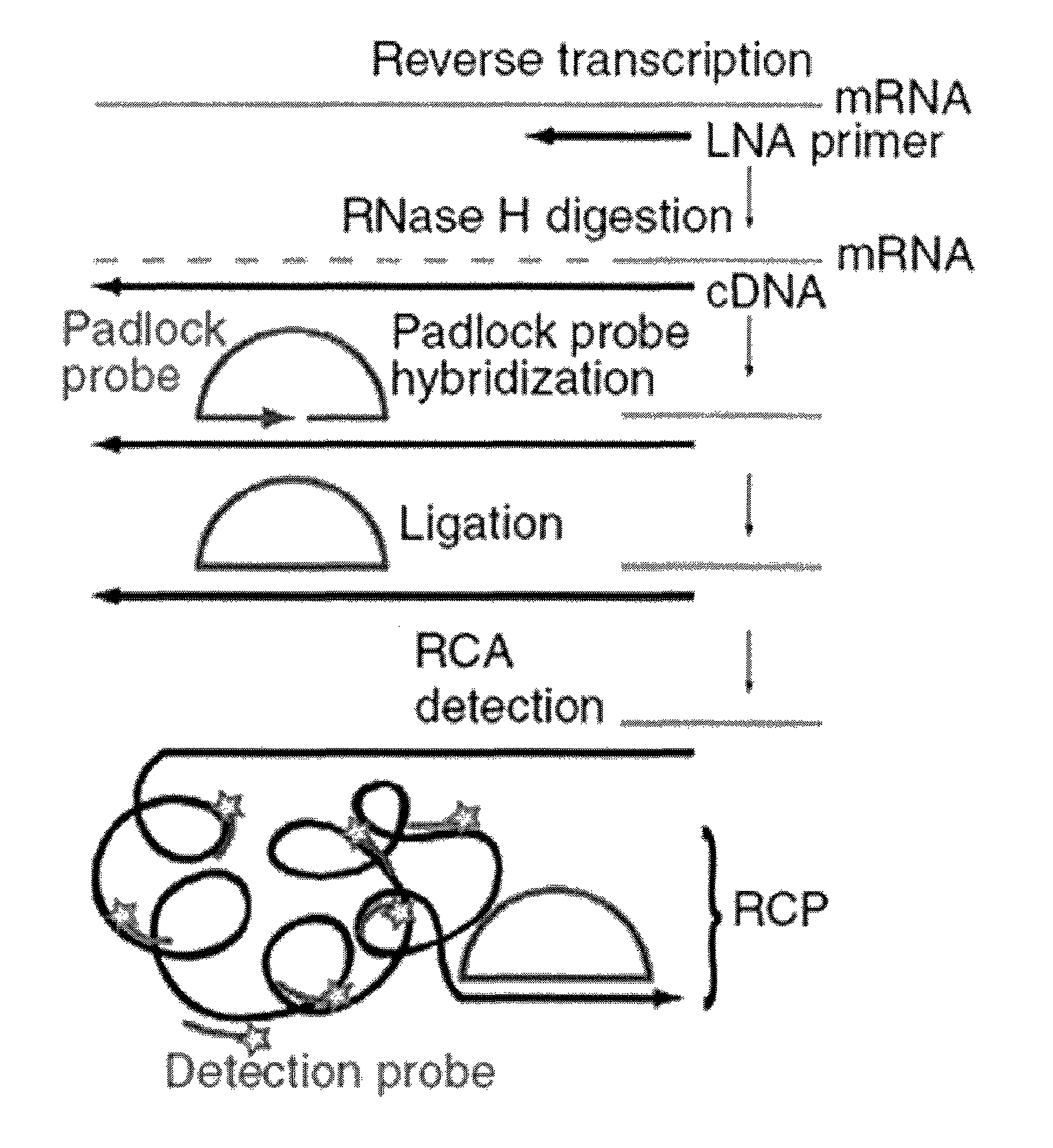
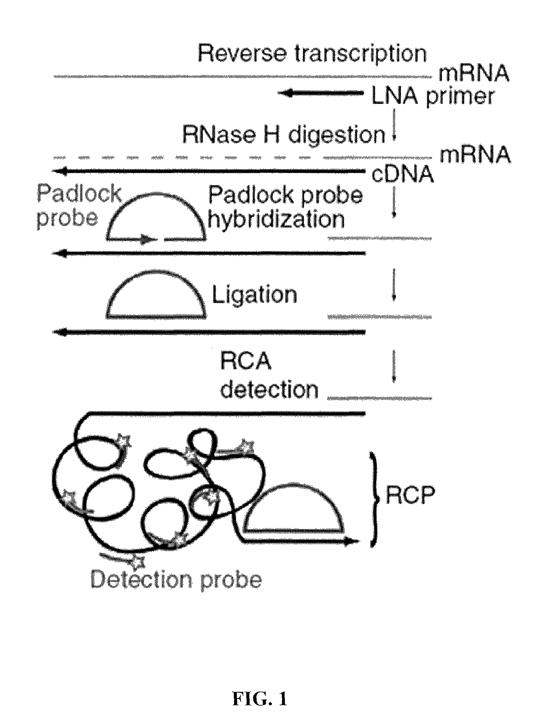
No other in situ method exists today that can perform multiplex detection of expressed single nucleotide sequence variants in RNA.

Method used


Image

  • Methods for localized in situ detection of mRNA
  • Methods for localized in situ detection of mRNA
  • Methods for localized in situ detection of mRNA

Examples


example 1

Detection of β-Actin (ACTB) Transcripts in Cultured Human Cells Using Padlock Probes

[0278]To detect β-actin (ACTB) transcripts in cultured human cells, two different padlock probes were used targeting sequences in the first and last exon, respectively. Many bright, spot-like signals localized to the cytoplasm of cells were visualized, consistent with previous observations of this transcript consistent with previous reports regarding this transcript. The detection efficiency was similar for the two padlock probes, indicating that in this case detection was not highly dependent on target position along the transcript (FIG. 5). In contrast, when reverse transcriptase was omitted from the cDNA synthesis reaction, no signals were detected, verifying that the signals were cDNA dependent (FIG. 5). It was estimated that the overall in situ detection efficiency to be ˜30% of available transcripts, on the basis of a comparison to quantitative PCR (qPCR) data for β-actin mRNA in the GM08402 ce...

example 2

Detection of Single-Nucleotide Variants of Transcripts in Cultured Cells In Situ

[0279]To demonstrate high selectivity of detection, an assay was used to detect of single-nucleotide variants of transcripts in situ. Expressed polymorphisms are rare in β-actin, therefore a single-base difference between the human and mouse β-actin sequences was used as genotyping target. Co-cultured human and mouse fibroblast cells were subjected to in situ genotyping of cDNA using padlock probes PLP-βhum (human) and PLP-βmus (Mus musculus) and target-primed RCA. There was a clear-cut distinction observed between the two subpopulations of cells in the co-culture. The preference for perfectly matched padlock probes at the circularization step ensures distinction between the two targets by the ligase.

example 3

Detection of Single-Nucleotide Variants of Transcripts in Fresh Frozen Tissue In Situ

[0280]To test the method in fixed tissue sections, closely related skeletal muscle α1-actin (Acta1) and cytoplasmic β-actin (Actb) transcripts were targeted in fresh frozen tissue from an E14.5 mouse embryo cross sectioned at the level of the neck. The two actin transcripts were successfully detected in the tissue using padlock probes designed with target sequences differing by a single base. The α1-actin signals were mainly distributed to skeletal muscles, whereas β-actin signals were widely distributed but showed slightly more signals in the non-muscular tissue. The ability to distinguish three transcripts from the same gene family was demonstrated by including a probe specific for the cytoplasmic α1-actin (Acta1) transcript.


PUM


Abstract

The present invention relates to the detection of RNA in a sample of cells. More particularly, the present invention relates to the localized detection of RNA in situ. The method relies on the conversion of RNA to complementary DNA prior to the targeting of the cDNA with a padlock probe(s). The hybridization of the padlock probe(s) relies on the nucleotide sequence of the cDNA which is derived from the corresponding nucleotide sequence of the target RNA. Rolling circle amplification of the subsequently circularized padlock probe produces a rolling circle product which may be detected. Advantageously, this allows the RNA to be detected in situ.

Description

[0001]This application is a continuation of co-pending U.S. patent application Ser. No. 13 / 397,503 filed Feb. 15, 2012, which claims priority to U.S. Provisional Application Ser. No. 61 / 473,662, filed Apr. 8, 2011, and U.S. Provisional Application Ser. No. 61 / 442,921, filed Feb. 15, 2011, both of which are incorporated by reference in their entirety. This application is related to the PCT application No. PCT / IB2012 / 000995 entitled “Method for Localized In Situ Detection of mRNA” by Mats Nilsson Bernitz, Chatarina Larsson, and Ida Grundberg filed on Feb. 15, 2012, which is incorporated by reference in its entirety.BACKGROUND[0002]In some embodiments, the present invention relates to the detection of RNA, especially mRNA, in a sample of cells. More particularly, the present invention relates in particular embodiments to the localized detection of RNA, particularly mRNA, in situ. In certain aspects, the method relies on the conversion of RNA to complementary DNA (cDNA) prior to the tar...

Claims


Application Information

Patent Timeline
Patent Type & Authority Patents(United States)
IPC IPC(8): C12Q1/68C07H21/04
CPCC12Q1/6809C12Q1/6827C12Q1/6841C12Q1/6886C12Q2600/112C12Q2600/156C12Q2600/16C12Q2521/107C12Q2521/327C12Q2525/107C12Q2525/125C12Q2525/161C12Q2525/307C12Q2531/125C12Q2537/143C12Q2543/10
Inventor BERNITZ, MATS NILSSONLARSSON, CHATARINAGRUNDBERG, IDA
Owner LEICA BIOSYST NEWCASTLE
Why Eureka
Try Eureka