
Detection of circulating endothelial cells

a technology of endothelial cells and detection methods, applied in the field of detection of circulating endothelial cells, can solve the problems of high background staining of flow cytometry, cumbersome and time-consuming approach, and hampered clinical testing of these agents

Active Publication Date: 2013-10-15
PHARMA CINQ LLC
View PDF10 Cites 0 Cited by
  • Summary
  • Abstract
  • Description
  • Claims
  • Application Information

AI Technical Summary

Problems solved by technology

But “The clinical testing of these [antiangiogenic] agents is currently hampered, by the lack of surrogate markers for measuring their biological effect and predicting which patients are most likely to benefit.” (Davis D W, et al., 2003, Br J Cancer 89:8-14).
This is a cumbersome and time-consuming approach not readily amenable to automation.
A further problem with flow cytometry et al is the high background staining in flow cytometry.
Excessive data storage capacity is another problem with rare event analysis by flow cytometry (Khan et al., 2004).

Method used


Image

  • Detection of circulating endothelial cells
  • Detection of circulating endothelial cells
  • Detection of circulating endothelial cells

Examples


example 1

[0066]A patient comes into the office and a blood sample is collected in a tube to prevent clotting. mCECs are isolated and then lyzed using a lysis buffer. A ruthenium-labeled antibody against an antigen against mCECs and a biotinylated antibody (also against the antigen from mCECs) is added along with a solution of tripropylamine and magnetic beads with avidin attached. An electric current is applied and electrochemiluminescence (ECL) is detected using an ECL detection device such as one commercially available (BioVeris Corporation or Roche Diagnostics). The signal is proportional to the amount of mCECs per ml found in the circulation.

example 2

[0067]Methods as in Example 1, in which mCECs are isolated or enriched using magnetic beads coated with antibodies against vWF. These cells are lysed using Sigma Lysis Buffer [Sigma CelLytic™-M (Sigma Product Number C 2978, Sigma-Aldrich, Inc., St. Louis, Mo. 63103)]. Cell lysis is performed as per the manufacture's recommendation with the addition of 5 minutes of vigorous vortexing prior to cell debris removal. Cell debris is removed from the cell lysate by centrifugation at 14,000 rpm for 30 minutes in an Eppendorf Centrifuge (Model 5415C). A magnet is used to further deplete the magnetic particles. Ruthenium-labeled antibodies against CD-146 and biotinlyated antibodies against CD-146 are added to the lysate and then magnetic strepavidin beads and a solution containing tripropylamine. An electric current is applied and electrochemiluminescence (ECL) is detected using an ECL detection device such as one commercially available (BioVeris Corporation). The signal is proportional to th...

example 3

[0068]Methods as in example 2, except that the number of CEPs is determined and the magnetic beads used for isolation or enrichment of CEPs are coated with antibodies against CD133 not vWF and the sandwich ECL immunoassay uses antibodies against VEGFR-2 not CD-146.


PUM

PropertyMeasurementUnit
sizesaaaaaaaaaa
sizesaaaaaaaaaa
sizesaaaaaaaaaa

Abstract

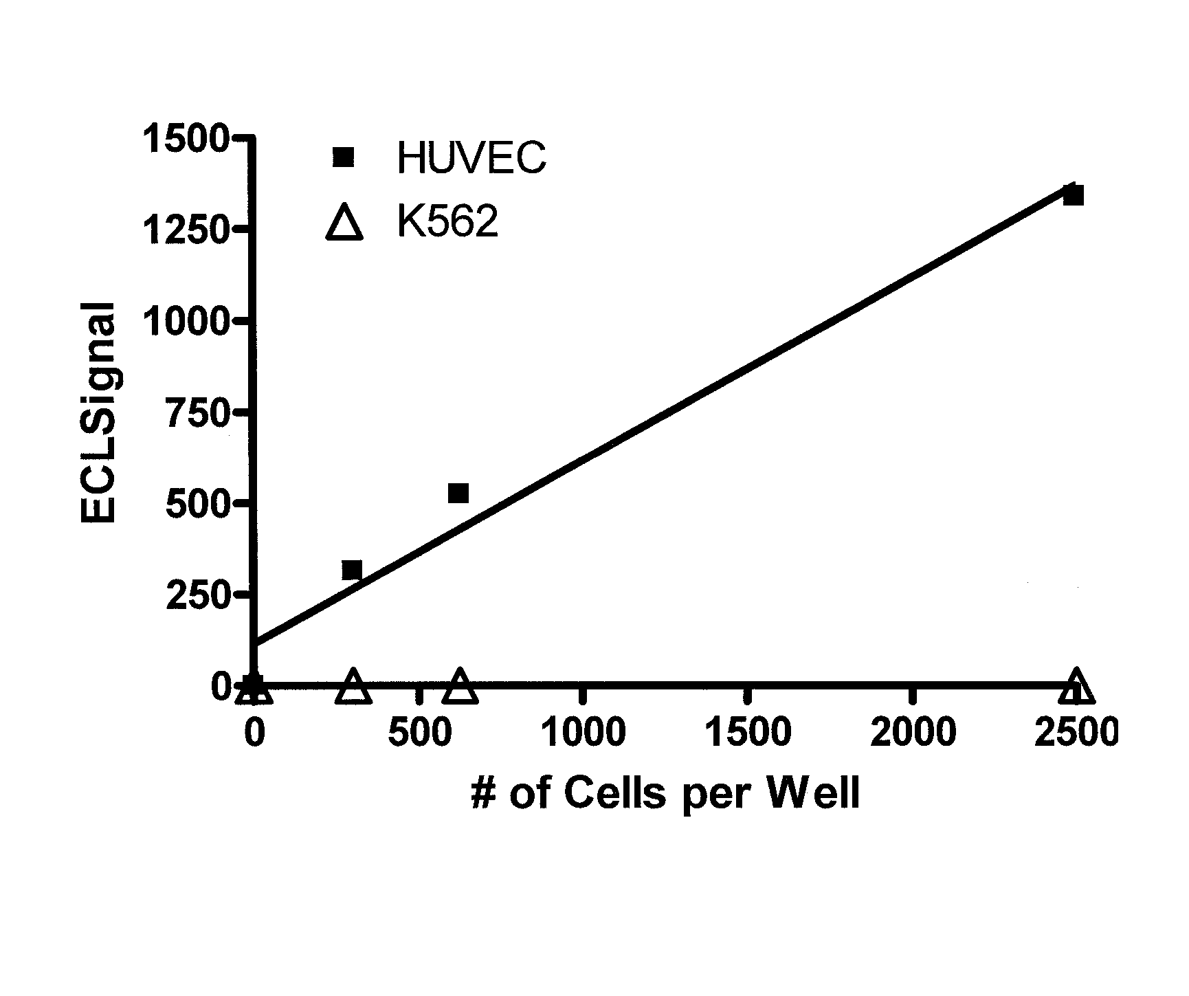
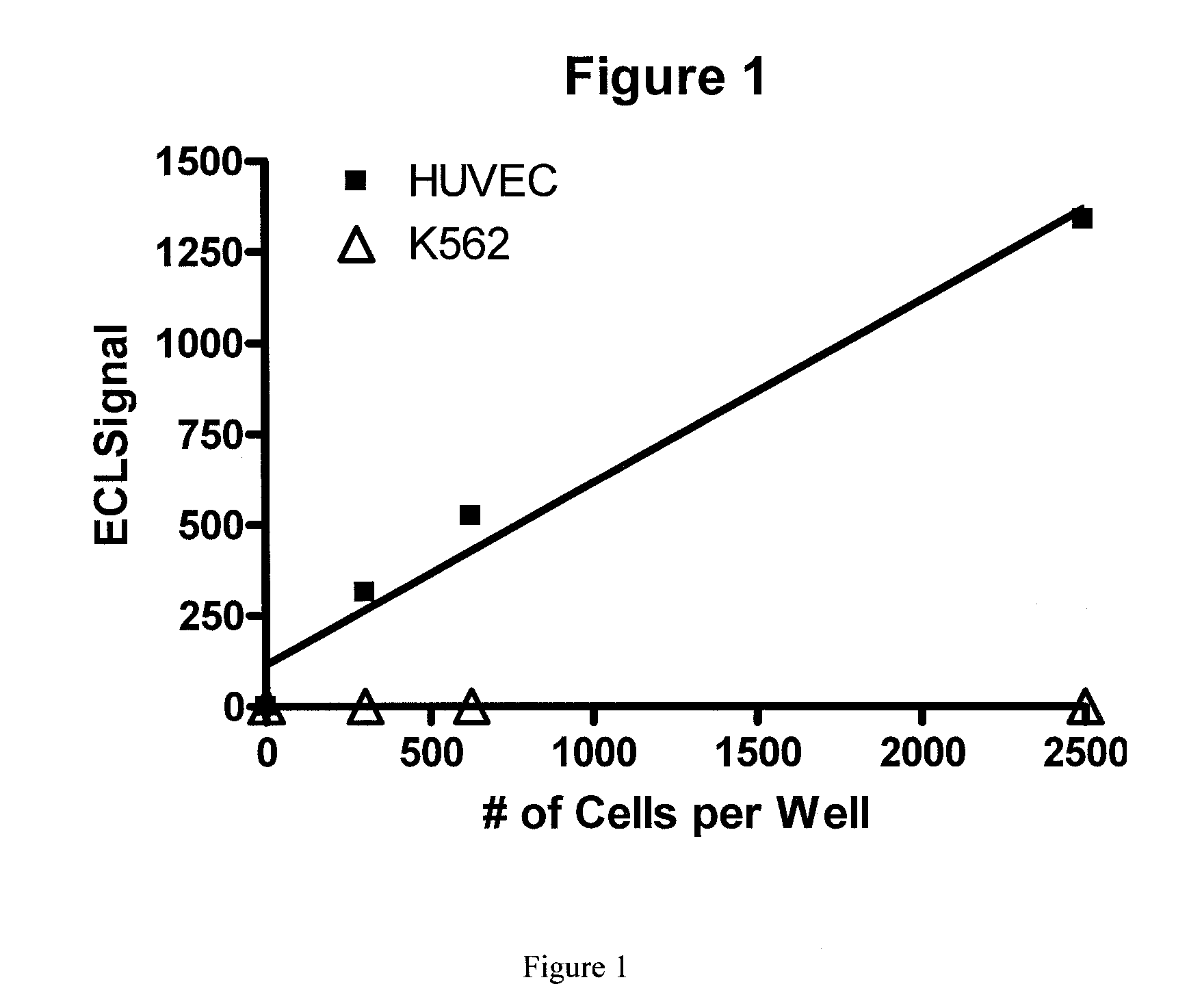
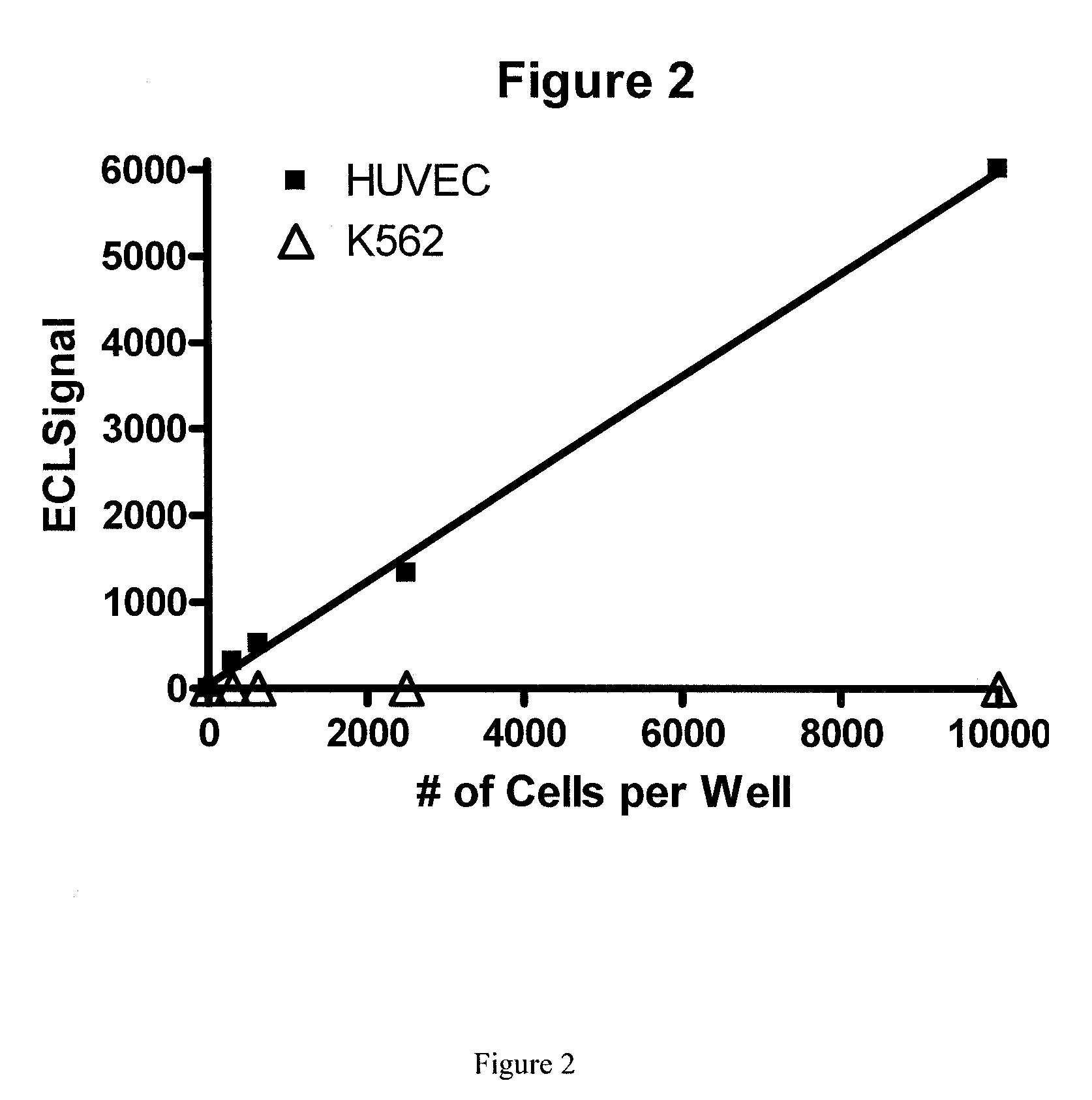
Endothelial cells are detected in a blood sample by enriching the endothelial cells from the blood sample followed by performing on the enriched endothelial cells an immunoassay capable of detecting antigens expressed by the endothelial cells. The immunoassay is capable of detecting antigen expressed from 300 endothelial cells per milliliter of blood. The method can be used for assaying mature circulating endothelial cells or circulating endothelial progenitor cells.

Description

BACKGROUND OF THE INVENTION[0001]There are two distinct populations of circulating endothelial cells: bone marrow derived circulating endothelial progenitors (CEPs) and mature circulating endothelial cells (mCECs) (Beaudry et al., 2005, Clin Cancer Res 11:3514; For additional background see Rosenzweig, 2005; New Engl J Med 353:1055-1057; Khan SS, 2004; Cytometry Part B, Clinical Cytometry 64B: 1-8). Measurement of circulating endothelial cells can act as surrogate markers for biological activity of antiangiogenic agents and used to select patients who will benefit from such therapy for cancer (Beaudry et al., 2005, Clin Cancer Res.). For example, patients with CEPs expressing high levels of VEGFR are excellent candidates for treatment with VEGFR inhibitors such as bevacizumab (Avastin). Also, measurement of mCECs is useful in cancer since an increased level indicates disease progression (Mancuso P et al., 2001, Blood 97:3658-61; Beerepoot L V et al, 2004, Ann Oncol 15:139-45).[0002]...

Claims


Application Information

Patent Timeline
Patent Type & Authority Patents(United States)
IPC IPC(8): A01N1/02G01N1/30G01N33/567G01N1/18C12N5/07C12N5/074C12N5/0789
CPCG01N33/54326G01N33/56966G01N33/582G01N2333/70596Y10T436/25125G01N2333/755G01N2458/30Y10T436/25375Y10T436/101666G01N2333/71
Inventor LORENCE, ROBERT M.
Owner PHARMA CINQ LLC
Learn More