System and method for evaluating tissue

a tissue and quantitative evaluation technology, applied in the field of tissue quantitative evaluation system and method, can solve the problems of high false positive rate, high sensitivity, and inability to accurately detect breast cancer in women with dense breast tissue, and achieve low false positive rate, low probability of cancerous tissue being overlooked, and low sensitivity

Active Publication Date: 2013-10-22
DREXEL UNIV
View PDF117 Cites 17 Cited by
  • Summary
  • Abstract
  • Description
  • Claims
  • Application Information

AI Technical Summary

Benefits of technology

[0108]One advantage of the invention is that it provides significantly less false positive readings than current mammography techniques, thereby minimizing the need to perform numerous unnecessary tissue biopsies for the false positive results of the mammograms.
[0109]Another potential advantage of the present invention is that it has a very high sensitivity indicating that there is a very low likelihood that cancerous tissue would be overlooked using the method of the present invention. This is important because it ensures that the method of the present invention, used alone, may be a reliable cancer screening method.
[0110]The G / E ratio may also be used to identify specific types of malignant or benign tumors. Specifically, a G / E ratio of about 0.5, more preferably about 0.5 to 0.6, may be indicative of hyperplasia, and a G / E ratio of about 0.3, more preferably about 0.2-0.4 combined with an identification of the presence of abnormal tissue obtained from one or more of the individual shear and elastic modulus maps of the sample, may be indicative of carcinoma in situ or benign tumors, such as fibrocystic or fibroadipose. Although healthy tissue may also exhibit G / E ratios about 0.3, cancerous tissue and tumors having a G / E ratio in this range can still be identified by its higher shear and elastic modulus than that of the healthy surrounding tissue. It has been found, for example, that a benign tumor may have a G / E ratio of about 0.3, the same as for healthy tissue, but that the individual measurements of shear modulus and elastic modulus of the benign tumor are typically higher than the individual measurements of shear modulus and elastic modulus for healthy tissue, thereby allowing the prediction of the presence of a benign tumor under these circumstances.
[0111]In the case of carcinoma in situ, it may be necessary to also consider the location of the tissue in question to complete the prediction of whether there is cancerous tissue or not. In this case, the surrounding tissue of, for example, a milk duct, can confine the cancerous tissue, thereby altering the shear and / or elastic moduli of the cancerous tissue in the interfacial area. As a result, the carcinoma in situ will typically exhibit a G / E ratio of about 0.3 due to confinement of the interfacial area by the surrounding tissue. However, individual shear and elastic modulus measurements can again be used to predict the presence of the carcinoma in situ under these circumstances since the individual shear and elastic moduli will differ from that of healthy tissue. Although benign tumors such as fibrocystic tumors also exhibit a higher shear modulus and elastic modulus value than those of the surrounding healthy tissue, inclusion of abnormal tissue as identified by the individual elastic modulus map or shear modulus map that exhibit a G / E about 0.3 would still allow positive predictions of all carcinomas in situ.
[0112]The method of the present invention may further involve the step of artificially increasing the perceived interfacial roughness of a tumor in order to enhance the sensitivity of the indicator for malignancy, invasiveness or tumor type. The perceived interfacial roughness may also be enhanced for examination of tumors located at a significant depth below the tissue surface or tumors which have developed large interlocking networks. In an exemplary embodiment, the perceived interfacial roughness may be increased by increasing the angle of the scan path relative to the interfacial protrusions. For purposes of the present invention, the angle of the scan path refers to the orientation of PEFS 1 relative to the interfacial protrusions when the shear force is applied. Different scan angles are achieved by rotating PEFS 1, shown in FIG. 5(b), about its longitudinal axis, relative to the tissue sample. Preferably, the angle is at least 30 degrees, more preferably, at least 60 degrees and, most preferably, about 90 degrees.
[0113]Upon analyzing and diagnosing the tissue sample, measuring means 2 may be used to express the location of tissue abnormalities in polar coordinates so as to graphically and accurately locate the abnormal tissue within or relative to the tested tissue based on the elastic modulus, shear modulus and / or G / E measurements. Additionally, measuring means 2 may be also be used to quantitatively determine and map the size and depth of the tissue abnormalities. In an exemplary embodiment, the location and dimensions of the abnormal tissue may be displayed on a 2D or 3D map to facilitate surgery.

Problems solved by technology

These sensors, however, require the extraction and destruction of tissue specimens; during operation, since the specimens must be cut to conform with and fit within the sensor.
Mammography is the only FDA approved breast cancer screening technique, which has a typical sensitivity of 85% that decreases to 65% in radiodense breasts.9 However, in these screening processes there is a high incidence of false positives.
Although effective for screening women over 40, mammography is not as effective for screening women who have dense breast tissue.
Additionally, mammography and other tactile imaging tools do not have the ability to probe tumor interface properties.
Although the patent concludes that these measurements may offer the potential for non-invasive breast cancer malignancy screening, it does not disclose a method for determining malignancy, invasiveness or tumor type.

Method used


Image

  • System and method for evaluating tissue
  • System and method for evaluating tissue
  • System and method for evaluating tissue

Examples


example 1

[0130]A study was performed to determine the effectiveness of the PEFS of the present invention to accurately evaluate a set of artificial tissues, which mimic the physical properties of various types of tumors. The PEFS was used to determine whether the artificial tumors embedded in the artificial tissue samples have a rough or branchy interfacial surface, a potential indicator of invasive malignant cancer such as malignant breast cancer, by measuring the elastic modulus (E), shear modulus (G) and determining the G / E ratio for the artificial tissues. It was found that either the elastic modulus or the shear modulus may be used to discern the dimensions of the artificial tumor, and that the shear modulus may further be used to characterize the texture of interface of a tumor with the surrounding tissue. Additionally, when the shear modulus was measured using a scan path substantially perpendicular to the direction of corrugation at the interface of the tissue, a G / E ratio of greater...

example 2

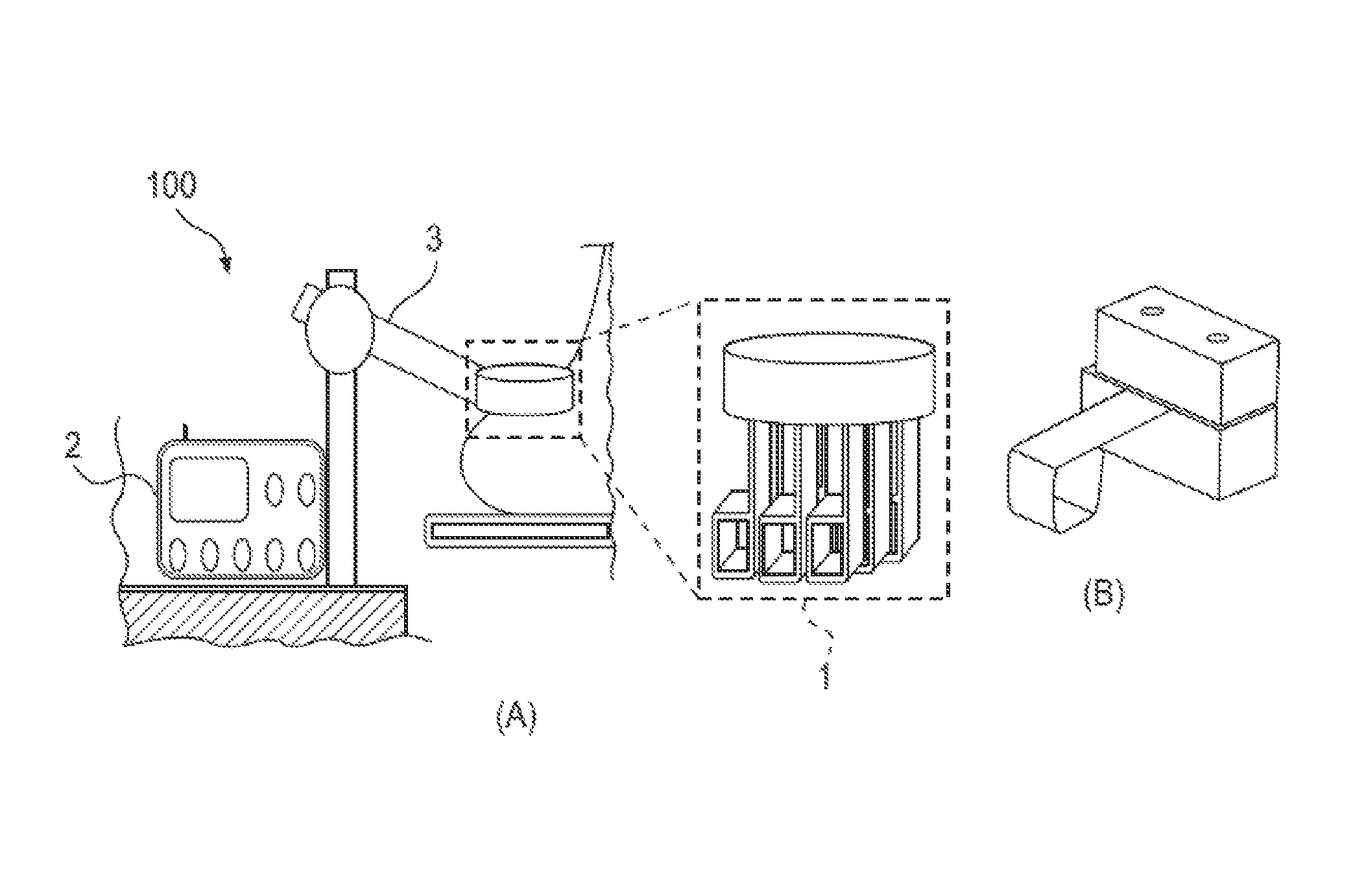
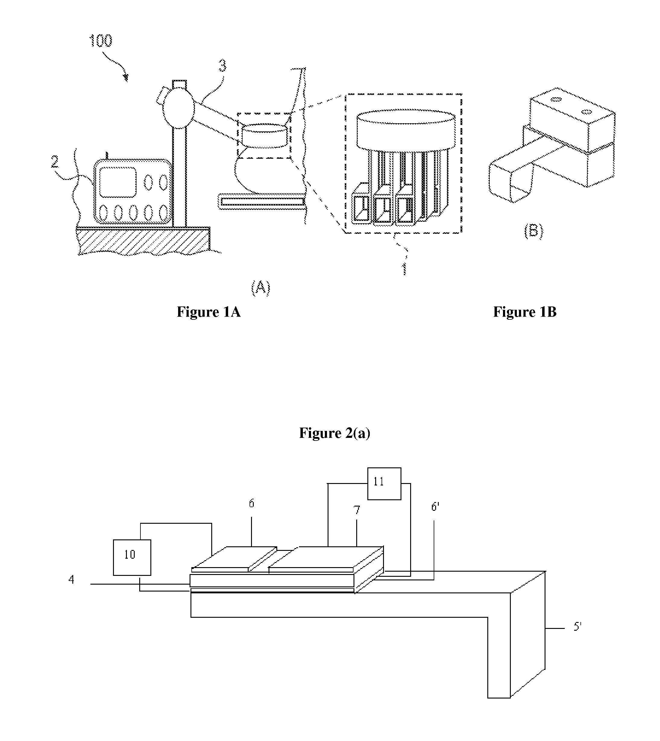
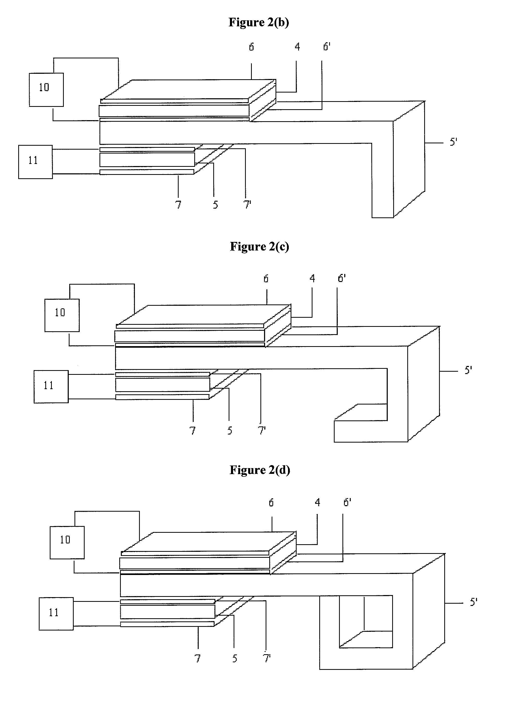
[0151]A PEFS was investigated to determine the depth sensitivity of elastic modulus measurements. The PEFS was fabricated from two piezoelectric layers, namely, a top 127 um thick PZT layer (105-H4E-602, Piezo System, Cambridge, Mass.) that functioned to drive a bottom 127 um thick sensing PZT layer bonded to a 50 um thick stainless steel layer (Alfa Aesar, Ward Hill, Mass.). The stainless steel layer formed a square tip at a distal end of the PEFS that was used to perform compression and shear tests.

[0152]To determine the depth sensitivity of the PEFS, artificial tissue samples were prepared by embedding modeling clay model inclusions, having an elastic modulus of about 80 kPa, in a gelatin matrix, which has an elastic modulus of about 4±1 kPa, at various depths ranging from 2 to 17 mm, as shown in FIG. 13(a). The elastic moduli of the model tissues were then measured by indentation compression tests using three PEFS of various widths, namely 8.6 mm, 6.1 mm and 3.6 mm. The resultan...

example 3

[0153]The depth sensitivity of the PEFS of Example 1, defined as the maximum depth for which it is possible to obtain an accurate measurement, was investigated to determine sensor accuracy and reliability. Specifically, the depth sensitivity of the PEFS was investigated for determining the shear modulus and the G / E ratio of a tissue sample. Similar to previous studies which have confirmed that elastic modulus measurements are accurate to a depth sensitivity of about twice the size of the contact area of the PEFS, the depth limit for shear modulus measurements and the G / E ratio was also found to be about twice the size of the contact area.

[0154]The shear modulus of seven S inclusions embedded in artificial tissue samples and seven R inclusions embedded in artificial tissue samples were investigated. Each inclusion was about 22 mm long and 12 mm wide and was made of C92 modeling clay, which has an elastic modulus that closely mimics that of breast tumors. The inclusions were embedded ...


PUM


Abstract

The present invention provides a sensor system for measuring an elastic modulus and a shear modulus and a method for using the sensor system to evaluate a tissue by determining the presence of and / or characterizing abnormal growths. The method involves applying a set of forces of different magnitudes to one or more locations of tissue, detecting the corresponding displacements due to said applied forces, determining the forces acting on those locations of tissue which are a combination of forces from the applied voltages and the countering forces from tissue deformation, obtaining the elastic modulus and / or shear modulus for a plurality of locations, and determining abnormal growth invasiveness, malignancy or the presence of a tumor from said elastic and / or shear moduli.

Description

CROSS-REFERENCE TO RELATED APPLICATIONS[0001]This application claims priority to and is a non-provisional of U.S. Provisional Patent Application No. 61 / 054,100, filed on May 16, 2008, pursuant 35 U.S.C. 119(e), the entire disclosure of which is incorporated herein by reference.STATEMENT OF GOVERNMENT INTEREST[0002]This invention was made with government support under Contract No. RO1 EB000720 awarded by the National Institutes of Health. The Government has certain rights in the invention.BACKGROUND OF THE INVENTION[0003]1. Field of Invention[0004]The present invention relates to a system and method for quantitatively evaluating a tumor. More particularly, the invention involves using a piezoelectric sensor to detect the existence of, determine the dimensions of, determine the location of, identify the type of, determine the invasiveness of and / or determine the malignancy of a tumor.[0005]2. Brief Description of the Prior Art[0006]The key to successful treatment of cancer lies in ear...

Claims


Application Information

Patent Timeline
Patent Type & Authority Patents(United States)
IPC IPC(8): A61B5/103
CPCA61B5/0053A61B5/4312G01N29/0672A61B5/441A61B8/085A61B8/403A61B8/485A61B5/4244A61B5/4381A61B5/442G01N2291/02475G01N2291/02827
Inventor SHIH, WAN Y.SHIH, WEI-HENGYEGINGIL, HAKKIBROOKS, ARI D.
Owner DREXEL UNIV
Why Eureka
Try Eureka