
Multiparameter perfusion imaging with leakage correction

a multi-parameter perfusion and leakage correction technology, applied in the field of multi-parameter perfusion imaging with leakage correction, can solve the problem that no other technology is able, and achieve the effect of reducing the cumulative dose needed, eliminating the effect of t1 leakage, and accurate and robust blood volume measurements

Active Publication Date: 2014-03-11
IMAGING BIOMETRICS
View PDF6 Cites 4 Cited by
  • Summary
  • Abstract
  • Description
  • Claims
  • Application Information

AI Technical Summary

Benefits of technology

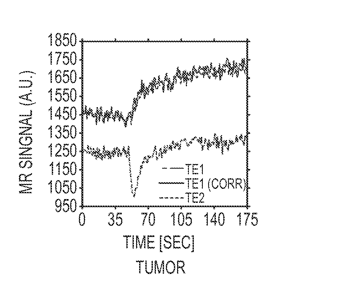
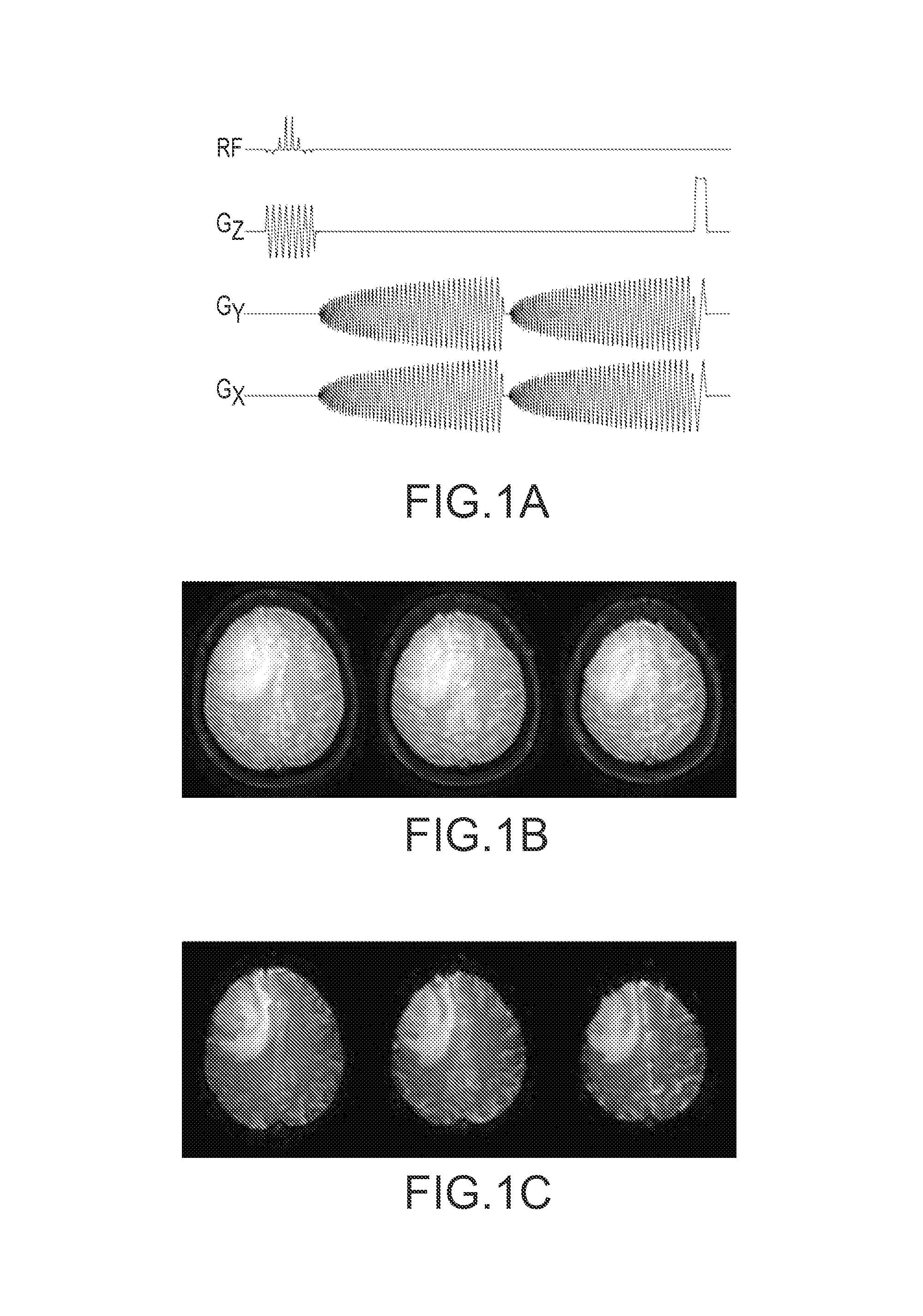
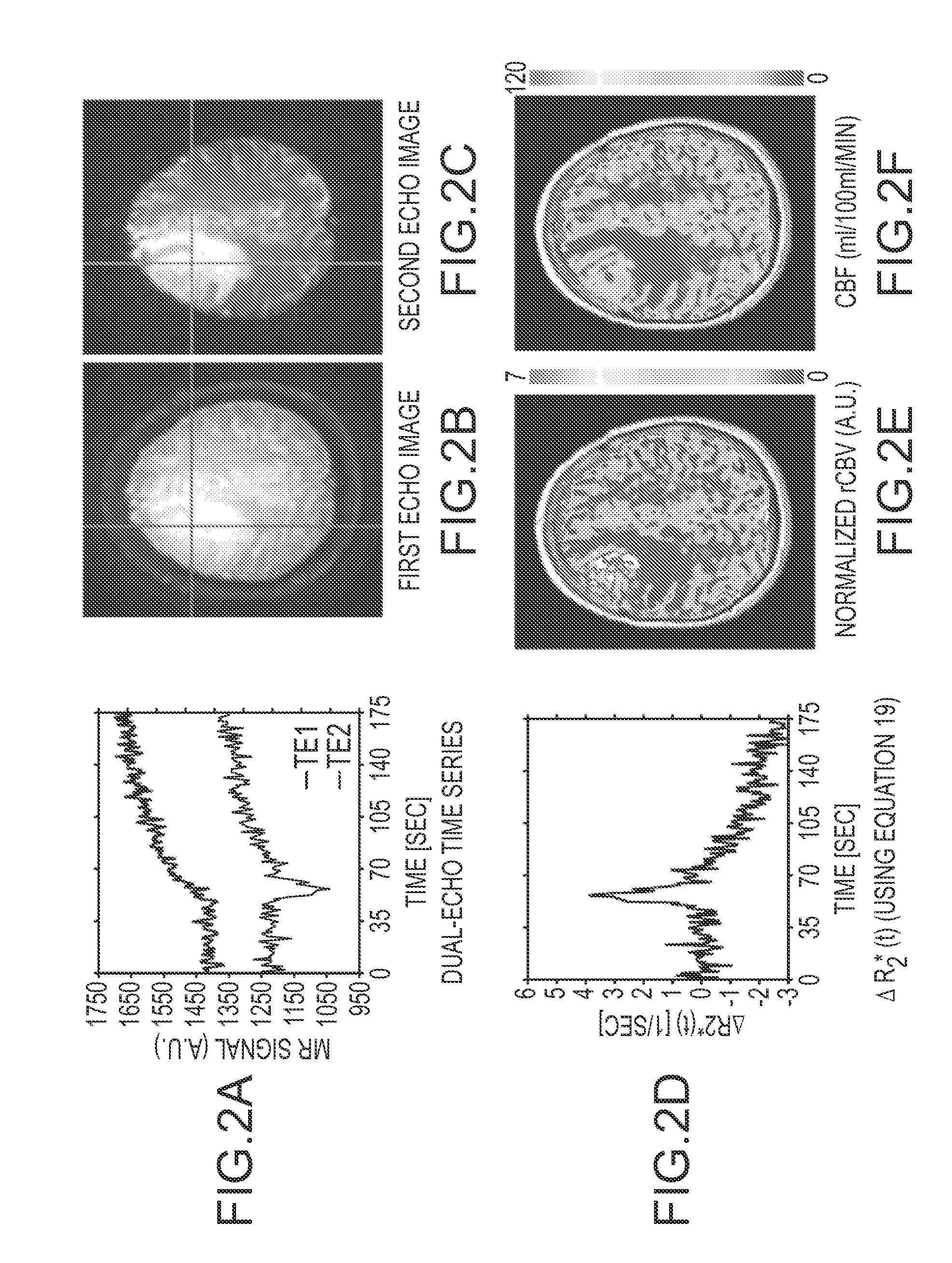
The present invention provides a new and useful method for MRI that combines dual-echo acquisition and single shot SPIRAL acquisition to measure blood volume, flow, and vascular permeability accurately and robustly, even in the absence ofT1 leakage effects. This method can also be used to measure perfusion in various normal and disease states, including but not limited to organ transplants, sickle cell disease, and diabetes. Additionally, it reduces the cumulative dose of Gd contrast agent required for each patient's study by precluding the need for a loading dose.

Problems solved by technology

No other technology enables the collection of both T2 / T2* perfusion data and T1-based permeability data at the same time.

Method used


Image

  • Multiparameter perfusion imaging with leakage correction
  • Multiparameter perfusion imaging with leakage correction
  • Multiparameter perfusion imaging with leakage correction

Examples


Embodiment Construction

[0018]Dynamic Susceptibility Contrast (DSC) MRI and Dynamic Contrast Enhanced (DCE) MRI are two minimally invasive imaging techniques frequently employed to probe the angiogenic activity of brain neoplasms based on estimates of vascularity and vascular permeability. It is well known that gadolinium produces simultaneous T1, 12, and T2* shortening effects in tissue, and these properties are uniquely exploited in DSC- and DCE-MRI. However; several different MRI contrast agents are capable of inducing susceptibility contrast effects when injected. Therefore, several different contrast agents can be used in conjunction with the described invention. Most commonly, paramagnetic agents such as Gd (gadolinium)-chelated contrast agents are used. When Gd(III)-chelated agents are injected quickly, under bolus-like conditions, they induce susceptibility contrast in tissues, notable as a transient decrease in a T2 or T2*-weighted MRI signal. Alternatively, another category of contrast agents can...


PUM


Abstract

A magnetic resonance imaging (MRI) methodology is provided for simultaneous measurement of dynamic susceptibility contrast (DSC) MRI and dynamic contrast enhanced (DCE) MRI perfusion and permeability parameters using a combination of dual echo and spiral acquisition techniques with no contrast agent preload. T1 and T2 / T2* leakage effects are eliminated, thereby permitting accurate measurement of blood volume, blood flow and vascular permeability which are used in evaluating tumor angiogenesis.

Description

CROSS-REFERENCE TO RELATED APPLICATIONS[0001]This application is the National Stage of International Application No. PCT / US2008 / 064597 filed May 22, 2008, which claims the benefit of U.S. Provisional Patent Application Ser. No. 60 / 939,539 filed on May 22, 2007.BACKGROUND OF THE INVENTION[0002]Magnetic resonance imaging (MRI) relies on the relaxation properties of excited hydrogen nuclei in water and lipids to create images. When the target object to be imaged is placed in a uniform magnetic field, the forces in the magnetic field cause the spins of atomic nuclei having a non-zero spin to align in a particular manner with the applied magnetic field. By way of example, hydrogen atoms have a simple spin (1 / 2) and therefore align either parallel or anti-parallel to the magnetic field. A radio frequency pulse (RF) is then applied in a direction perpendicular to the magnetic field and removed. When the RF signal is removed, the atomic nuclei relax. During the relaxation process, the nucle...

Claims


Application Information

Patent Timeline
Patent Type & Authority Patents(United States)
IPC IPC(8): A61B5/05G06K9/00
CPCG01R33/56366G01R33/4824G01R33/5601A61B5/413A61B5/0263
Inventor SCHMAINDA, KATHLEENPAULSON, ERIC S.PRAH, DOUGLAS E.
Owner IMAGING BIOMETRICS
Learn More