Ocular ultrasound based assessment device and related methods

a technology of ocular ultrasound and assessment device, which is applied in the field of ultrasonic devices, can solve the problems of increasing the cost of ct imaging, the lack of alternative providers for positively diagnosing elevated intracranial pressure, and the excessive radiation exposure of ct imaging, etc., and achieves the effects of easy use, low cost, and high accuracy

Active Publication Date: 2014-03-18
DBMEDX
View PDF20 Cites 29 Cited by
  • Summary
  • Abstract
  • Description
  • Claims
  • Application Information

AI Technical Summary

Benefits of technology

[0007]Ocular ultrasonography is a relatively new application but has seen growing acceptance and provides a quick, accurate, well-tolerated, noninvasive tool for evaluating

Problems solved by technology

CT is a generally available but expensive imaging modality and one which exposes the patient to ionizing radiation.
The use of CT in the emergency department has seen a dramatic increase in recent years, causing concern from physicians and payers (e.g., insurers) regarding excessive radiation exposure and cost associated with CT imaging.
However, when faced with a patient suffering sympt

Method used


Image

  • Ocular ultrasound based assessment device and related methods
  • Ocular ultrasound based assessment device and related methods
  • Ocular ultrasound based assessment device and related methods

Examples


Embodiment Construction

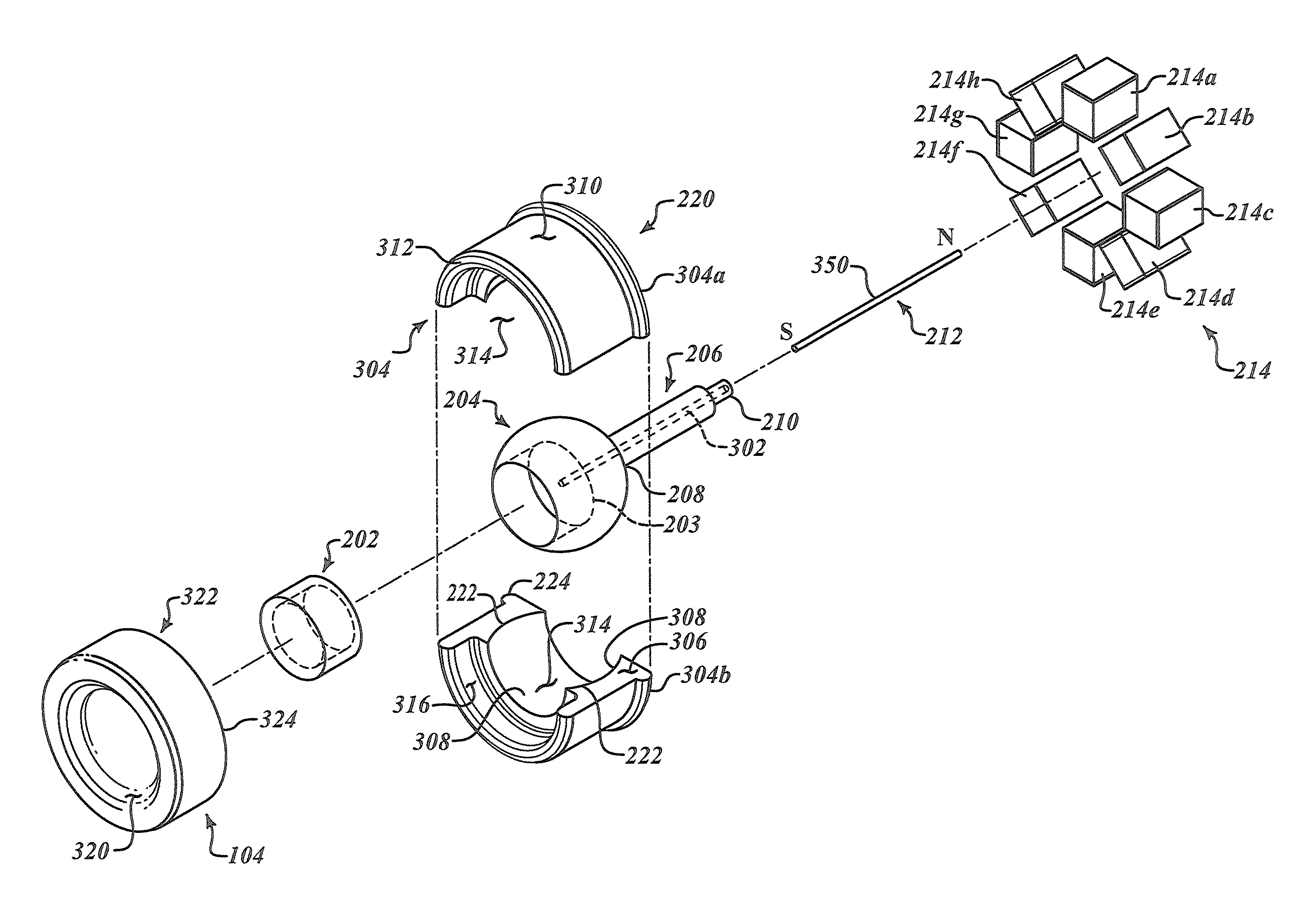
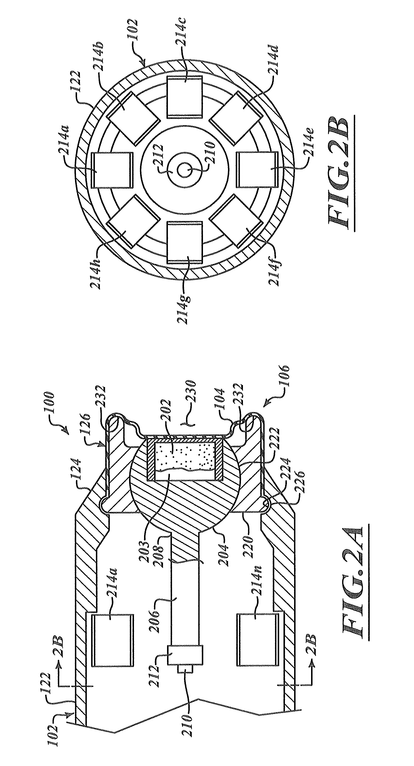
[0052]In the following description, certain specific details are set forth in order to provide a thorough understanding of various disclosed embodiments. However, one skilled in the relevant art will recognize that embodiments may be practiced without one or more of these specific details, or with other methods, components, materials, etc. In other instances, well-known anatomical structures and structures associated with ultrasound systems and transducers have not been shown or described in detail to avoid unnecessarily obscuring descriptions of the embodiments.

[0053]Unless the context requires otherwise, throughout the specification and claims which follow, the word “comprise” and variations thereof, such as, “comprises” and “comprising” are to be construed in an open, inclusive sense, that is as “including, but not limited to.”

[0054]Reference throughout this specification to “one embodiment” or “an embodiment” means that a particular feature, structure or characteristic described...


PUM


Abstract

A three-dimensional ultrasound based assessment device includes a housing having a front end, a back end, and a cavity. An ultrasonic transducer assembly can be mounted to a ball joint which is, in turn, pivotably (pitch, roll, yaw) coupled to a socket proximate the front end. A resilient or elastic sheath retains pieces forming the socket under elastic compression, with the ball pivotally retained therein. A drive assembly positioned proximate a tail or end of a stem that extends from the ball joint may be operated to sweep the ball and ultrasonic transducer. A simple interface provides at least one of a number, text, graphic, color or symbol as a visual indicator indicative of at least one of a measurement of an anatomical structure or a comparison of the measurement of the anatomical structure with a reference, without providing images of the anatomical structure.

Description

BACKGROUND[0001]1. Technical Field[0002]This disclosure generally relates to ultrasound devices, and more particularly to ultrasound devices having dimensional scanning capabilities and methods of using ultrasound devices for diagnostic purposes.[0003]2. Description of the Related Art[0004]Patients presenting at an emergency department often require assessment for elevated intracranial pressure (“EICP”), particularly as a result of head trauma or spontaneous intracranial hemorrhage. These patients often present acutely, and a diagnosis of elevated intracranial pressure suggests a serious pathology that may require rapid intervention. Typically, the diagnosis of elevated intracranial pressure is made using cranial computed tomography (“CT”). CT is a generally available but expensive imaging modality and one which exposes the patient to ionizing radiation. CT imaging typically requires a wait, while CT imaging equipment is made available to a patient, and may even require transport of...

Claims


Application Information

Patent Timeline
IPC IPC(8): A61B8/14
CPCA61B8/4461A61B8/4281A61B8/56A61B8/4218A61B8/14A61B3/00A61B8/08A61B8/10A61B8/4427A61B8/462A61B8/483A61B8/5223G16H50/30
Inventor QUIRK, WILLIAMBARNARD, WILLIAM L.SHINE, DAVID B.
Owner DBMEDX
Why Eureka
Try Eureka